

metricas
Gastroenterología y Hepatología (English Edition) Beaver tail liver
Journal Information
Vol. 47. Issue 8.
Pages 793-924 (October 2024)
Cite
Cite
Share
Download PDF
More article options
Vol. 47. Issue 8.
Pages 793-924 (October 2024)
Image of the month
Full text access

Beaver tail liver

Hígado en cola de castor
Visits
953
David Villaescusa Arenasa,
Corresponding author
davidvillaescusar@gmail.com

Corresponding author.
, Elena Garrido Gómeza, Carlos Martín de Argilaa, Carmen Díaz del Ríob, Claudia Álvarez Varelaa, Carmen García Huertaa, Ana Belén Agarrabeitia Casadoa, Miguel García Gonzáleza, Agustín Albillos Martíneza
a Gastroenterology and Hepatology Department, Hospital Universitario Ramón y Cajal, Instituto Ramón y Cajal de Investigación Sanitaria (IRYCIS), Centro de Investigación Biomédica en Red (CIBEREHD), Universidad de Alcalá, Madrid, Spain
b University Hospital of Fuenlabrada Radiodiagnosis Service, Madrid, Spain
This item has received
Article information
Full Text
Bibliography
Download PDF
Statistics
Figures (3)
fig0005
fig0010
fig0015
Full Text

A 29-year-old woman underwent an abdominal ultrasound due to chronic abdominal pain.

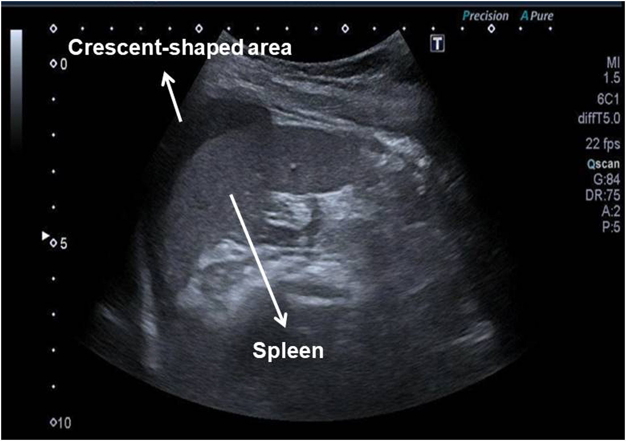
A subdiaphragmatic, hypoechogenic crescent-shaped area with a maximum diameter of 70mm, similar to the characteristic images of splenic hematomas, was observed at the perisplenic level (Fig. 1). The patient had no history of previous abdominal trauma.

Figure 1.

B-mode ultrasound image. Subdiaphragmatic, hypoechogenic crescent-shaped area at the perisplenic level.

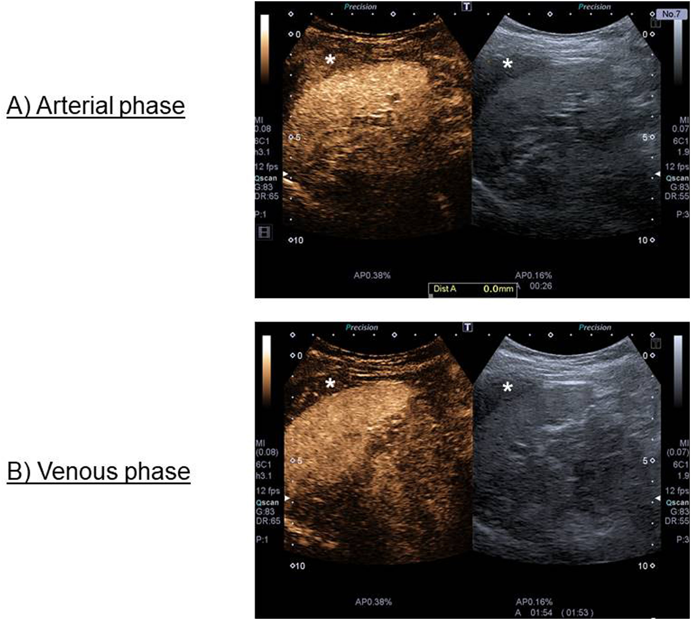
A study with intravenous contrast (SonoVue®) was performed, showing in arterial phase a homogeneous filling of the crescent-shaped formation with less intensity than the splenic parenchyma; in addition, the area showed washout with respect to the splenic surface in very late venous phase (Fig. 2).

Figure 2.

Intravenous contrast (SonoVue®) ultrasound image. (A) Arterial phase: homogeneous filling of the crescent-shaped formation; (B) Late venous phase: washout with respect to the splenic surface of the crescent-shaped formation.

*Area of interest.

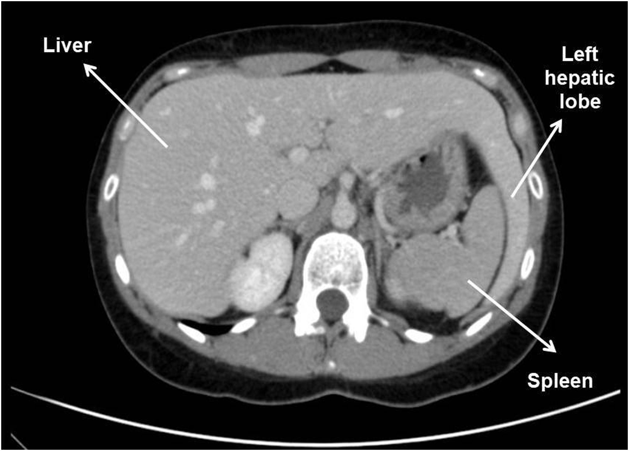
Finally, the dependence of this crescentic image on the hepatic parenchyma was appreciated, being in fact a prolongation of the same (left hepatic lobe) (Fig. 3), confirming by CT scan the variant of normality known as beaver tail liver, a rare hepatic shape with no pathological implications.1,2

Figure 3.

CT scan image. Left lobe extends laterally to contact and enclose the spleen.

It is important to know the possible variants of normality to avoid erroneous interpretations of the imaging tests. Also, the case shows the usefulness of intravenous contrast to increase the characterization capability of B-mode ultrasound.

Funding

There are no financing for this manuscript.

Conflicts of interest

The authors declare they have no conflicts of interest.

References
[1]
M. Haydar Atalar, K. Karakus.
Beaver tail liver.
Abdom Radiol, 43 (2018), pp. 1851-1852
[2]
H. Xiang, J. Han, W.E. Ridley, L.J. Ridley.
Beaver tail liver: anatomic variant.
J Med Imaging Radiat Oncol, 62 (2018), pp. 57
Copyright © 2023. Elsevier España, S.L.U.. All rights reserved
asdasdasd
Article options
Tools
