metricas
Gastroenterología y Hepatología (English Edition) Antithrombotics drugs in Gastrointestinal Endoscopy. Position Statment of the Ca...
Journal Information
Visits
525
Vol. 49. Issue 2.
(February 2026)
Clinical practice guidelines
Full text access

Antithrombotics drugs in Gastrointestinal Endoscopy. Position Statment of the Catalan Society of Gastroenterology

Fármacos antitrombóticos en la endoscopia digestiva. Documento de posicionamiento de la Societat Catalana de Digestologia
Visits
525
Faust Riu Ponsa,b,c,
Corresponding author
friu@hmar.cat

Corresponding author.
, Noèlia Vilaltad, Gemma Ibáñez-Sanze,f, Sergi Quintana-Carbóe, Andrea Calvog, Hugo Uchimah, Pilar García-Iglesiasi, Cristina Romero-Mascarellj, Ermengol Vallèsk, Marco Antonio Álvarezl
a Sevicio Aparato Digestivo, Hospital del Mar, Barcelona, Spain
b Hospital del Mar Research Institute, Barcelona, Spain
c Departament de Medicina i Ciències de la Vida, Universitat Pompeu Fabra, Barcelona, Spain
d Servicio de Hematología y Hemoterapia, Hospital de Sant Pau, Barcelona, Spain
e Servicio de Aparato Digestivo, Hospital Universitari de Bellvitge, L’Hospitalet de Llobregat, Barcelona, Spain
f Grupo de Investigación de Cáncer Colorrectal, Programa ONCOBELL, Institut d'Investigació Biomèdica de Bellvitge (IDIBELL), Hospitalet de Llobregat, Barcelona, Spain
g Servicio de Anestesiología y Reanimación, Hospital Clínic, Barcelona, Spain
h Servicio de Aparato Digestivo, Hospital Germans Trias i Pujol, Badalona, Barcelona, Spain
i Servicio de Aparato Digestivo, Consorci Sanitari d’Anoia. Hospital d’Igualada, Barcelona, Spain
j Servicio de Aparato Digestivo, Hospital de Sant Pau, Barcelona, Spain
k Servicio de Cardiología, Hospital del Mar, Barcelona, Spain
l Servicio de Aparato Digestivo, Althaia, Xarxa Assistencial Universitària de Manresa, Manresa, Barcelona, Spain
Ver más
This item has received
Article information
Abstract
Full Text
Bibliography
Download PDF
Statistics
Figures (1)
fig0005
Tables (21)
Table 1. Stratification of Bleeding Risk and Management of Complications According to Endoscopic Procedure.
Tables
Table 2. Stratification of Thrombotic Risk Following Withdrawal of Clopidogrel, Prasugrel or Ticagrelor.
Tables
Table 3. Stratification of Thrombotic Risk Following Withdrawal of Oral Anticoagulants (Vitamin K Antagonists).
Tables
Table 4. Summary of Laboratory Test Interpretation in Patients Treated with Direct Oral Anticoagulants (DOACs).
Tables
Tables
Tables
Tables
Tables
Tables
Tables
Tables
Tables
Tables
Tables
Tables
Tables
Tables
Tables
Tables
Tables
Tables
Show moreShow less
Additional material (1)
Abstract
Introduction

The use of antiplatelet and anticoagulant drugs is common in patients undergoing elective endoscopic procedures. To minimize both bleeding and thromboembolic risks, an individualized assessment is essential. For this purpose, the Societat Catalana de Digestologia (SCD) commissioned a consensus document aimed at providing practical recommendations based on the best available evidence and expert agreement.

Material and methods

A panel of 10 specialists from the fields of Gastroenterology, Cardiology, Hematology, and Anesthesiology were involved in the development of the document. A non-systematic review of the literature was conducted using a modified Delphi method through online voting. Recommendations were reviewed and discussed during each round until consensus was reached. Recommendations with ≥80% agreement were accepted.
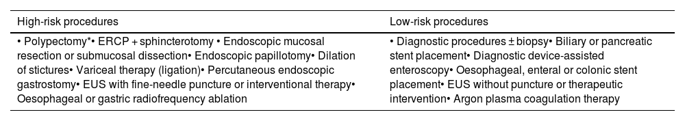
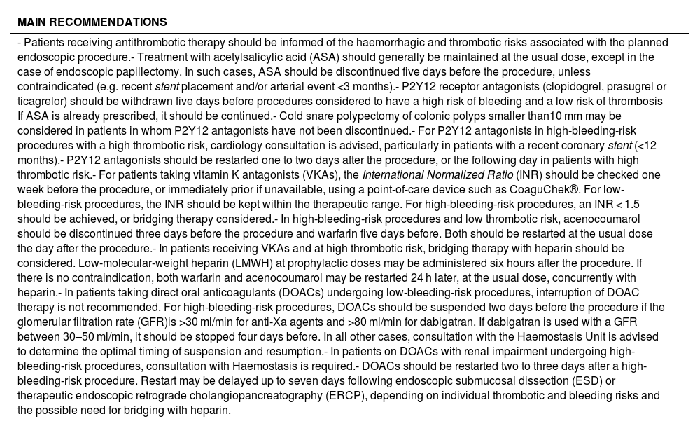
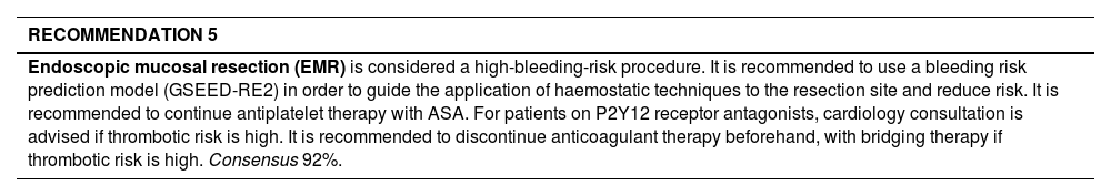
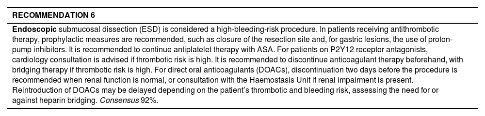
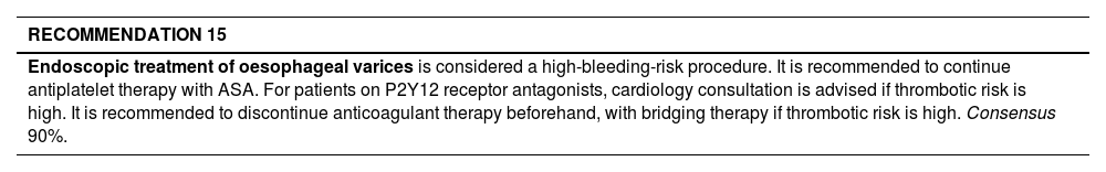
Results

A total of 16 endoscopic procedures were assessed based on their hemorrhagic risk, and tailored management guidelines were defined to different groups of antithrombotic agents. The document includes a management algorithm and stratification tables for bleeding and thrombotic risk.

Conclusion

The recommendations developed by the SCD integrate international evidence and are adapted to the local healthcare context, providing a valuable tool for clinical practice. The importance of multidisciplinary coordination and effective communication with the patient is emphasized to ensure safety throughout the periprocedural period.

Keywords:
Antithrombotics
Antiplatelets agents
Anticoagulants
Direct oral anticoagulant
Gastrointestinal endoscopy
Resumen
Introducción

El uso de fármacos antiagregantes y anticoagulantes es habitual en pacientes que se realizan un procedimiento endoscópico electivo. Para minimizar el riesgo tanto de hemorragia como de trombosis, es fundamental ofrecer una evaluación individualizada. Para ello, la Societat Catalana de Digestología (SCD) encargó un documento de consenso con el objetivo de ofrecer unas recomendaciones prácticas basadas en la mejor evidencia disponible y en consenso de expertos.

Material y métodos

Diez especialistas en Aparato Digestivo, Cardiología, Hematología y Anestesiología participaron en la elaboración del documento. Se llevó a cabo una revisión no sistemática de la literatura y se aplicó el método Delphi modificado con votación telemática. Las recomendaciones se revisaron y comentaron en cada ronda para llegar a un consenso, aceptándose aquellas que alcanzaron un nivel de acuerdo del 80 %.

Resultados

Se evaluaron 16 procedimientos endoscópicos según su riesgo hemorrágico, y se definieron pautas específicas de manejo adaptadas a los distintos grupos de fármacos antitrombóticos. El documento incluye un algoritmo de manejo y tablas de estratificación de riesgo hemorrágico y trombótico.

Conclusión

Las recomendaciones de la SCD integran la evidencia internacional y se adaptan a nuestro entorno sanitario, ofreciendo una herramienta útil para la práctica clínica. Se resalta la necesidad de coordinación multidisciplinaria y de una buena comunicación con el paciente para garantizar la seguridad durante el periodo periprocedimiento.

Palabras clave:
Antitrombótico
Antiagregante plaquetario
Anticoagulantes
Anticoagulante oral de acción directa
Endoscopia gastrointestinal
Full Text
Introduction

The use of antiplatelet and anticoagulant drugs is increasingly common among patients requiring gastrointestinal endoscopy, as these agents are prescribed to prevent arterial and venous thrombotic events However, such treatments may increase the risk of gastrointestinal bleeding, particularly depending on the type of endoscopic procedure and its indication. It is therefore essential to consider previous antithrombotic therapy carefully, and to have clear, evidence-based protocols for periprocedural management to minimise potential complications.

Inadequate management of these drugs may be associated with significant morbidity and mortality, reported to be as high as 2%.1 To reduce these risks, it is recommended that both thrombotic and bleeding risks be assessed individually, according to the type of endoscopic procedure and the patient’s clinical profile. In general, maintaining antithrombotic therapy in patients at high risk of thrombosis is advised, as the consequences of a thrombotic event are usually more severe and irreversible than those of a bleeding episode.

Several clinical guidelines, such as the Cardiovascular Thrombosis Working Group of the Spanish Society of Cardiology guideline, developed in consensus with multiple societies,2 and the European Society of Gastrointestinal Endoscopy (ESGE)3 guideline, provide recommendations for periprocedural management. Nonetheless, their applicability may be limited in certain local clinical settings.

For this reason, the Catalan Society of Gastroenterology Societat Catalana de Digestologia, SCD) has developed this consensus document to provide practical, up-to-date, evidence-based recommendations — agreed upon by experts — for the assessment and management of antithrombotic drugs in patients undergoing elective endoscopic procedures.

Method

This consensus document was prepared by a panel of experts from the SCD, based on the recommendations of the ESGE, the British Society of Gastroenterology (BSG), and the American Society for Gastrointestinal Endoscopy (ASGE). These were updated with the most recent evidence and adapted to the Spanish healthcare context. A non-systematic review of the evidence was conducted, without a structured search protocol or systematic assessment of bias, to identify the relevant literature for each section up to October 2023. The electronic database consulted was MEDLINE (accessed via PubMed). Controversial aspects were discussed online until consensus was reached, and the final version of the document was drafted and reviewed by all contributing experts.

Recommendations were jointly evaluated by all members of the working group, taking into account the limited quality of the available scientific evidence, as most studies were of low methodological robustness. Consensus was reached using a modified Delphi method4 via an online voting platform (Google Forms). Voting was conducted using a five-point Likert scale (1 = completely disagree; 2 = disagree; 3 = neither agree nor disagree; 4 = agree; 5 = completely agree). Ten experts participated, and two voting rounds were carried out to achieve final consensus. A recommendation was considered approved when ≥80% of experts rated it as 4 or 5 on the Likert scale. The first and second rounds were conducted in November and December 2023 respectively (see Appendix A. Supplementary data).

The main recommendations of the working group are summarised in the section Main Recommendations, and the proposed SCD algorithm is presented in Fig. 1.

MAIN RECOMMENDATIONS 
- Patients receiving antithrombotic therapy should be informed of the haemorrhagic and thrombotic risks associated with the planned endoscopic procedure.- Treatment with acetylsalicylic acid (ASA) should generally be maintained at the usual dose, except in the case of endoscopic papillectomy. In such cases, ASA should be discontinued five days before the procedure, unless contraindicated (e.g. recent stent placement and/or arterial event <3 months).- P2Y12 receptor antagonists (clopidogrel, prasugrel or ticagrelor) should be withdrawn five days before procedures considered to have a high risk of bleeding and a low risk of thrombosis If ASA is already prescribed, it should be continued.- Cold snare polypectomy of colonic polyps smaller than10 mm may be considered in patients in whom P2Y12 antagonists have not been discontinued.- For P2Y12 antagonists in high-bleeding-risk procedures with a high thrombotic risk, cardiology consultation is advised, particularly in patients with a recent coronary stent (<12 months).- P2Y12 antagonists should be restarted one to two days after the procedure, or the following day in patients with high thrombotic risk.- For patients taking vitamin K antagonists (VKAs), the International Normalized Ratio (INR) should be checked one week before the procedure, or immediately prior if unavailable, using a point-of-care device such as CoaguChek®. For low-bleeding-risk procedures, the INR should be kept within the therapeutic range. For high-bleeding-risk procedures, an INR < 1.5 should be achieved, or bridging therapy considered.- In high-bleeding-risk procedures and low thrombotic risk, acenocoumarol should be discontinued three days before the procedure and warfarin five days before. Both should be restarted at the usual dose the day after the procedure.- In patients receiving VKAs and at high thrombotic risk, bridging therapy with heparin should be considered. Low-molecular-weight heparin (LMWH) at prophylactic doses may be administered six hours after the procedure. If there is no contraindication, both warfarin and acenocoumarol may be restarted 24 h later, at the usual dose, concurrently with heparin.- In patients taking direct oral anticoagulants (DOACs) undergoing low-bleeding-risk procedures, interruption of DOAC therapy is not recommended. For high-bleeding-risk procedures, DOACs should be suspended two days before the procedure if the glomerular filtration rate (GFR)is >30 ml/min for anti-Xa agents and >80 ml/min for dabigatran. If dabigatran is used with a GFR between 30–50 ml/min, it should be stopped four days before. In all other cases, consultation with the Haemostasis Unit is advised to determine the optimal timing of suspension and resumption.- In patients on DOACs with renal impairment undergoing high-bleeding-risk procedures, consultation with Haemostasis is required.- DOACs should be restarted two to three days after a high-bleeding-risk procedure. Restart may be delayed up to seven days following endoscopic submucosal dissection (ESD) or therapeutic endoscopic retrograde cholangiopancreatography (ERCP), depending on individual thrombotic and bleeding risks and the possible need for bridging with heparin. 

Figure 1.

SCD Algorithm: Antithrombotic drugs in Elective Digestive Endoscopy.

Assessment of bleeding and thrombotic risk

The risk of haemorrhage varies according to the type of endoscopic procedure performed and whether or not therapeutic intervention is required. Bleeding may also require endoscopic treatment for haemostasis in the event of a complication. A procedure is considered to have a high risk of haemorrhage when the probability of clinically significant bleeding is ≥1%5 (Table 1).

Table 1.

Stratification of Bleeding Risk and Management of Complications According to Endoscopic Procedure.

High-risk procedures  Low-risk procedures 
• Polypectomy*• ERCP + sphincterotomy • Endoscopic mucosal resection or submucosal dissection• Endoscopic papillotomy• Dilation of stictures• Variceal therapy (ligation)• Percutaneous endoscopic gastrostomy• EUS with fine-needle puncture or interventional therapy• Oesophageal or gastric radiofrequency ablation  • Diagnostic procedures ± biopsy• Biliary or pancreatic stent placement• Diagnostic device-assisted enteroscopy• Oesophageal, enteral or colonic stent placement• EUS without puncture or therapeutic intervention• Argon plasma coagulation therapy 

ERCP: endoscopic retrograde cholangiopancreatography.

*

In patients on clopidogrel monotherapy, cold-snare polypectomy may be considered for colonic polyps <10 mm.

Two types of haemorrhage related to endoscopic procedures are distinguished: 1) Immediate or intraprocedural bleeding, which may or may not require endoscopic therapy. Most episodes are self-limiting, do not interfere with the procedure, and resolve spontaneously once the intervention is completed. This type of bleeding is only regarded as an adverse event when it disrupts the procedure (e.g. bleeding is controlled but a further procedure is needed to complete the initial intervention) or forces it to be terminated (e.g. need for blood transfusion, hospital admission or observation in a high-dependency unit, emergency surgery or angiography). Clinically significant delayed bleeding, defined as haematemesis, melaena and/or haematochezia associated with a drop in haemoglobin of ≥2 g/dl. This type of bleeding leads to a prolonged hospital stay, readmission, transfusion, or repeat endoscopy (and occasionally surgery or angiography) for haemostasis. It occurs within 24 h and up to 30 days after the procedure.6,7

Conversely, the risk of thrombosis is associated with the temporary interruption of antiplatelet (Table 2) or anticoagulant (Table 3) therapy during the period in which the endoscopic procedure is performed. In these circumstances, patient preferences and joint decision-making with the clinician responsible for cardiovascular management should be taken into account, as they will assess the patient’s individual thromboembolic risk. The consequences of a thrombotic event and its possible sequelae are generally more serious than those of a haemorrhage. From a clinical perspective, it is therefore preferable to accept a certain degree of gastrointestinal bleeding risk rather than provoke a thrombotic event due to inappropriate drug withdrawal.8

Table 2.

Stratification of Thrombotic Risk Following Withdrawal of Clopidogrel, Prasugrel or Ticagrelor.

High thrombotic risk  Low thrombotic risk 
Drug-eluting coronary stent within the first 12 months after placement• Bare-metal coronary stent within the first month after placement  • Ischaemic heart disease without coronary stent• Cerebrovascular disease• Peripheral vascular disease 
Table 3.

Stratification of Thrombotic Risk Following Withdrawal of Oral Anticoagulants (Vitamin K Antagonists).

High thrombotic risk  Low thrombotic risk 
• Mechanical mitral or aortic prosthetic valve• Prosthetic cardiac valve with atrial fibrillation• Atrial fibrillation with mitral stenosis• Atrial fibrillation with CHA₂DS₂-VASc ≥5• Atrial fibrillation with TIA/stroke plus ≥3 of: CHF, hypertension >75 years, diabetes• Within 3 months after DVT/PE or atrial fibrillation with TIA/stroke• Prior venous thromboembolism requiring INR target >3.5• Thrombophilic syndromes (consult haematology)  • Biological heart valve• Atrial fibrillation without high-risk factors (CHA₂DS₂-VASc <5)• >3 months after DVT/PE 

TIA, transient ischaemic attack; DM, diabetes mellitus; CHF, congestive heart failure; HTN, hypertension; DVT, deep vein thrombosis; PE, pulmonary embolism.

In patients with atrial fibrillation, anticoagulant therapy is recommended to prevent thromboembolic complications such as stroke, systemic embolism, and mortality. CHA₂DS₂-VASc score allows for the estimation of thromboembolic risk and the determination of the indication for anticoagulant treatment in men with a score ≥2 and in women with a score ≥3. This scale, which evaluates several cardiovascular risk factors, correlates an increased stroke risk with higher scores9 (see Appendix A. Supplementary data).

A score of ≥5 is considered high thrombotic risk and therefore warrants bridging therapy with heparin before discontinuing anticoagulation.10

A multicentre, prospective observational study conducted in Spain demonstrated that the risk of bleeding related to elective endoscopic procedures was similar for vitamin K antagonists and direct oral anticoagulants (6.2% vs 6.7%), as was the risk of thromboembolic events (1.3% vs 1.5%, respectively). The overall mortality rate was 1.4%, mainly due to thromboembolic events.11

Antiplatelet drugs

Platelets play an essential role in haemostasis following vascular injury, but they are also central to thrombotic processes.

Antiplatelet agents are classified into five main categories:

  • 1

    Acetylsalicylic acid (ASA)

  • 2

    P2Y12 receptor antagonists

  • 3

    Adenosine reuptake inhibitors/phosphodiesterase inhibitors

  • 4

    IIb3 antagonists αβ

  • 5

    Protease-activated receptor 1 (PAR-1) antagonists

Acetylsalicylic acid

ASA acts by blocking the metabolism of arachidonic acid, a precursor of several biologically active molecules including prostaglandin E₂, prostacyclin (PGI₂), and thromboxane A₂ (TxA₂), which regulate platelet activation.12 Many international cardiology societies recommend ASA for secondary prevention in patients with a high cardiovascular risk profile.13

P2Y12 receptor antagonists

In clinical practice, the following are the most widely used: (1) the thienopyridines (ticlopidine, clopidogrel, and prasugrel); and (2) the nucleoside–nucleotide derivatives (ticagrelor and cangrelor).

P2Y12 receptor antagonists have demonstrated efficacy in preventing arterial thrombosis in patients with coronary artery disease, stroke, and peripheral arterial disease.14 Ticagrelor and prasugrel produce a greater reduction in atherothrombotic events compared with clopidogrel, although they are associated with a higher risk of bleeding.15

Phosphodiesterase inhibitors

Cilostazol, an oxyquinoline derivative, inhibits phosphodiesterase type 3 (PDE3) and exerts both antiplatelet and vasodilatory effects.16

Dipyridamole inhibits the uptake of adenosine by red blood cells and exerts antiplatelet activity by stimulating the binding of adenyl cyclase to A₂A and A₂B receptors.

αIIbβ3 antagonists

Drugs targeting the platelet integrin receptor IIb/IIIa (abciximab, eptifibatide, and tirofiban) are used in patients at high risk, such as those with unstable angina or non-ST elevation myocardial infarction, who require percutaneous coronary intervention. However, owing to their high risk of bleeding and potential for thrombocytopenia, their use is now largely restricted to selected cases of patients with reduced coronary flow.17

PAR-1 antagonists

Currently, Vorapaxar® is the only PAR-1 antagonist commercially available.

Anticoagulants

Since the 1940s, vitamin K antagonists (VKAs) have been used in the prevention and treatment of thromboembolic events. They are characterised by a narrow therapeutic margin and high inter- and intra-individual variability, influenced by both genetic and environmental factors.18

The introduction of direct oral anticoagulants (DOACs) sought to overcome the limitations of VKAs. Their effect is based on inhibition of a single coagulation factor, resulting in more predictable pharmacokinetics and pharmacodynamics. This allows fixed dosing and, in general, removes the need for routine monitoring.

Vitamin K antagonists

AVKs exert their anticoagulant activity through the inhibition of subunit 1 of the vitamin K 2,3-epoxide reductase complex (VKORC1) at the hepatocyte level.19

The main VKAs in use are derivatives of 4-hydroxycoumarin (warfarin, acenocoumarol, phenprocoumon) and indandione (fluindione, anisindione, among others).

Pharmacokinetics

The half-life of VKAs varies considerably: approximately 8 h for acenocoumarol, 40 h for warfarin, >60 h for phenprocoumon, and around 31 h for fluindione. Regardless of the half-life, the time it takes for an AVK to reach the anticoagulation threshold is related to the half-life of hypo-fx1-carboxylation of vitamin K-dependent factor, which is 60 h for prothrombin. Consequently, the desired anticoagulation range is usually not reached until five to six days after initiating therapy.18,20

Monitoring

The reagents used to measure the anticoagulant effect of VKAs vary in sensitivity, which led to the adoption of the international normalised ratio (INR) to standardise results. The INR is calculated using the formula:

PT: prothrombin time; s: seconds; ISI: International Sensitivity Index.

The INR is a ratio that compares the patient’s PT with the mean PT of a group of at least 20 healthy individuals, all raised to the power of the ISI. The ISI is specific to each reagent, batch, and instrument.

Direct Oral Anticoagulants (DOACs)

Currently, four DOACs are available, grouped into two mechanisms of action21:

Direct thrombin inhibitor

  • Abigatran (Pradaxa®): a prodrug requiring biotransformation to its active form. It has a short half-life (7–11 h), which may be prolonged in patients with renal impairment.

Direct factor Xa inhibitors

  • Rivaroxaban (Xarelto®): also has a short half-life and is primarily excreted via the kidneys.

  • Apixaban (Eliquis®): has a short half-life, but with less renal excretion than the other DOACs.

  • Edoxaban: similarly has a short half-life, with approximately 50% renal clearance.

Monitoring

The following laboratory tests are available for assessing the effect of DOACs (summarised in Table 4):

  • 1

    Basic haemostasis tests: prothrombin time (PT) and activated partial thromboplastin time (aPTT).

Table 4.

Summary of Laboratory Test Interpretation in Patients Treated with Direct Oral Anticoagulants (DOACs).

  Dabigatran  Rivaroxaban  Apixaban  Edoxaban 
Peak plasma level  2 h after intake  2−4 h after intake  1−4 h after intake  1−2 h after intake 
Trough plasma level  12−24 h after intake  16−24 h after intake  12−24 h after intake  12−24 h after intake 
PT  Not reliable  Prolonged; may indicate bleeding risk  Not reliable  Prolonged, but no known correlation with bleeding risk 
aPTT  With trough level, values ≥2× ULN suggest bleeding risk  Not reliable  Not reliable  Prolonged, but no known correlation with bleeding risk 
dTT  With trough level, >200 ng/mL and/or ≥65 s suggest bleeding risk  Not reliable  Not reliable  Not reliable 
Chromogenic anti-Xa assay  Not reliable  Quantitative; no validated thresholds for bleeding or thrombotic risk  No data  Quantitative; no validated thresholds for bleeding or thrombotic risk 
Ecarin clotting time  With trough level, ≥3× ULN suggests bleeding risk  Not affected  Not affected  Not affected 

PT: prothrombin time; aPTT: activated partial thromboplastin time; TTd: diluted thrombin time.

Adapted from Refs.30–32

PT and aPTT assess the extrinsic and intrinsic coagulation pathways respectively. These tests are not sensitive enough to provide a reliable correlation between drug dose and anticoagulant effect.22,23

  • 2

    Thrombin time (TT)

A functional coagulometric assay that indirectly measures fibrinogen concentration and fibrin formation. A modified version, the diluted thrombin time (dTT) or Hemoclot®, is used for direct thrombin inhibitors. This test is insensitive to direct factor Xa inhibitors.23,24

  • 3

    Anti-Xa activity

Multiple commercial kits are available for measuring anti-Xa activity with low-molecular-weight heparins. For DOACs, additional validation has been required. Rivaroxaban and apixaban show a linear correlation between anti-Xa activity and therapeutic plasma concentrations. Commercially available, drug-specific anti-Xa assays are now in use, though not widely standardised. Tests are available that could be used to measure dabigatran concentrations in plasma; however, only a limited number of laboratories currently perform them.22,24

  • 4

    Ecarin clotting time (ECT)

Used in preclinical studies but not yet standardized.23,25

  • 5

    Thrombin generation tests

Further studies are needed to determine whether these assays yield reproducible and clinically meaningful results.26,27

  • 6

    Thromboelastography

Shows good correlation with prolongation of clotting time for conventional anticoagulants, but limited data are available for DOACs.

  • 7

    HepTest

Not specific for anti-Xa activity, as it can be influenced by the presence of thrombin inhibitors.

  • 8

    Prothrombinase-induced clotting time (PiCT)

Not yet standardised for the monitoring of DOAC effects. Not yet standardised for the monitoring of DOAC effects.28,29

Endoscopic proceduresEndoscopy with or without biopsy, capsule endoscopy, and device-assisted enteroscopyDiagnostic endoscopy with or without biopsy

The risk of haemorrhage associated with endoscopic biopsy is very low, approximately 1‰, and bleeding is usually immediate and controllable endoscopically.33

A randomised study assessing the safety of biopsies in patients receiving dual antiplatelet therapy (ASA and clopidogrel) found no significant bleeding events with either drug, whether the biopsies were taken from the antrum or duodenum.34

Similarly, performing biopsies in patients on antiplatelet or anticoagulant therapy—including DOACs—does not increase bleeding risk. A Japanese multicentre observational study found no cases of bleeding within 30 days after gastroduodenal biopsies in patients who continued antithrombotic therapy, compared with controls. Most of the treated patients were receiving antiplatelet agents, while only 19 of 277 were on DOACs.35

By contrast, an Italian prospective cohort study reported a clinically relevant but statistically non-significant increase in intraprocedural bleeding among patients who had not discontinued DOACs compared with those who had withheld the morning dose (5.2% vs 1.7%; p = 0.23).36

For this reason, the ESGE guideline recommends withholding the morning dose of DOACs on the day of the procedure.3 However, since bleeding from a biopsy occurs during the procedure and does not influence delayed haemorrhage, DOAC interruption may not be strictly necessary, as any bleeding can be managed endoscopically.

RECOMMENDATION 1 
Diagnostic endoscopy with or without biopsy is considered a low-bleeding-risk procedure. It is recommended to maintain single or dual antiplatelet therapy and any anticoagulant treatment. Consensus 94%. 

Capsule endoscopy

Capsule endoscopy is a non-invasive examination that allows visualisation of the entire small intestine. The main complication is capsule retention, which occurs in approximately 2.5% of patients with established Crohn’s disease.37

As a purely diagnostic procedure, it is considered low risk for haemorrhage.

Notably, the use of antithrombotic therapy—whether antiplatelet or anticoagulant—is associated with a higher detection rate of findings in small-bowel capsule studies for obscure gastrointestinal bleeding, as well as an increased risk of rebleeding.38

RECOMMENDATION 2 
Capsule endoscopy is considered a low-bleeding-risk procedure. It is recommended to maintain both antiplatelet and anticoagulant treatment. Consensus 94%. 

Device-assisted enteroscopy

The risk of haemorrhage during enteroscopy is approximately 0.2–0.3%.39 The risk of perforation following double-balloon enteroscopy ranges from 0.1–0.4%, increasing to 1.5% when polypectomy is performed.40 In Crohn’s disease, dilation of small-bowel strictures carries a complication rate of 5.3% (including bleeding, perforation, or the need for surgery).41

A retrospective study of 420 patients undergoing double-balloon enteroscopy reported that 13% were on anticoagulation. The diagnostic yield was 73% and therapeutic intervention was required in 35% of cases. Only procedure duration and advanced age were predictors of diagnostic performance.42

Although considered a low-bleeding-risk technique, in practice more than half of the procedures require therapeutic intervention.41 Therefore, in patients on antithrombotic therapy, enteroscopy should generally be approached as a high-risk procedure.

RECOMMENDATION 3 
Diagnostic enteroscopy is considered a low-bleeding-risk procedure, although in practice most examinations require therapeutic intervention; it should therefore be regarded as high risk from the outset. It is recommended to continue antiplatelet therapy with ASA. For patients on P2Y12 receptor antagonists, cardiology consultation is advised if thrombotic risk is high. It is recommended to discontinue anticoagulant therapy beforehand, with bridging therapy if thrombotic risk is high. Consensus 80%. 

Gastrointestinal polyp resectionPolypectomy

Polypectomy is considered a high-bleeding-risk endoscopic procedure (risk of clinically significant haemorrhage ≥2% within 30 days) when the polyp is ≥10 mm in size. However, reported bleeding rates vary widely between studies (0.07–3%), due to differences in definitions of post-polypectomy bleeding (PPB) — whether early or delayed, and whether minor or major — across publications.

Polyp size is the most important risk factor for PPB, with an odds ratio (OR) of 2.3. In a study of pedunculated polyps, the risk of early or delayed PPB was 3.1% for 98 polyps measuring 10–19 mm, and 15.1% for 66 polyps ≥20 mm (for every1 mm increase in diameter, bleeding risk rose by 9%). Other factors linked to higher risk include age ≥65 years, associated cardiovascular or renal disease, macroscopic morphology (pedunculated or laterally spreading lesions), inadequate bowel preparation, type of electrocautery used, and inadvertent transection of the polyp stalk before application of current.43–45

  • 1

    Polypectomy in patients receiving antiplatelet and/or NSAID therapy

Observational studies in patients undergoing polypectomy while taking ASA have shown that antiplatelet or NSAID use does not increase the risk of PPB.46–48

  • 2

    Polypectomy in patients receiving clopidogrel

A randomised trial comparing continuous clopidogrel with clopidogrel withdrawal seven days before colonoscopic polypectomy (<10 mm) found no significant difference in immediate or delayed PPB (3.8% vs 3.6%), nor in the number of cardiothrombotic events. Most patients were receiving dual antiplatelet therapy with ASA, and the control group’s bleeding rate was higher than that reported in other studies involving non-antiplatelet users.49

A meta-analysis of five studies (two randomised controlled and three case–control) involving 655 patients on continued clopidogrel and 6,620 controls found no significant increase in immediate PPB (relative risk [RR] 1.57; 95% CI 0.98–2.51), but a significant increase in delayed PPB among those who continued clopidogrel (RR 3.10; 95% CI 1.60–5.98). No differences were observed in serious cardiothrombotic events within 30 days post-procedure. Other bleeding-related factors, such as polyp size, site, number, resection technique, or operator experience, were not assessed in that study.50

A multicentre randomised study evaluating cold-snare polypectomy of polyps <10 mm found no increase in delayed bleeding A total of 276 polyps in 107 patients were randomised to two groups: one discontinued clopidogrel seven days before the procedure (continuing ASA and restarting clopidogrel two days after colonoscopy), and one continued clopidogrel or dual therapy. The main limitation is that the sample size was not achieved due to low participation from the centres, so the study had to be stopped. Although recruitment was lower than planned, results indicated that cold-snare polypectomy of polyps <10 mm appears safe during uninterrupted clopidogrel use, with higher rates of intraprocedural bleeding requiring clip application, but fewer delayed haemorrhages.51

These findings suggest that cold-snare polypectomy of polyps <10 mm in patients on clopidogrel monotherapy is generally a safe technique.

Periprocedural decisions regarding clopidogrel withdrawal should always weigh thrombotic versus bleeding risk. In patients with recent percutaneous coronary intervention, coronary stent placement, or acute coronary syndrome, colonoscopy should preferably be deferred.

Alternatively, and in consultation with the prescribing specialist, temporary substitution of clopidogrel with ASA five days before the procedure may be considered.

  • 3

    Polypectomy in patients receiving anticoagulant therapy

Several studies have examined the safety of small-polyp resection in patients continuing anticoagulation. Although colonoscopy may be performed safely, current guidelines consider polypectomy a high-bleeding-risk procedure and recommend temporary discontinuation of anticoagulation. This is due to the difficulty of precisely assessing bleeding risk, which depends on multiple factors, particularly polyp size and INR values.

A retrospective study involving 225 polypectomies of polyps <10 mm (hot or cold snare, with prophylactic clip placement) in 123 patients on continuous warfarin reported a delayed major bleeding rate of 0.8% (one patient requiring transfusion) and minor bleeding in 1.6% (two patients not requiring medical intervention).52

Another retrospective study of 1,177 cold-snare polypectomies of polyps <10 mm compared PPB in patients on antithrombotic therapy (antiplatelet, anticoagulant, or multiple agents) versus controls. Immediate bleeding occurred in 5.4% overall, more frequently in warfarin users, but no differences were observed in delayed bleeding rates (0% in both groups).53

A randomised clinical trial of 70 patients (159 polyps <10 mm) comparing cold-snare and hot-snare polypectomy without warfarin interruption confirmed a lower bleeding rate with cold-snare resection (immediate bleeding 5.7% vs 23.0%; delayed bleeding 0% vs 14%).54

A multicentre randomised study compared cold-snare versus hot-snarepolypectomy in patients on uninterrupted anticoagulation (DOAC or warfarin) versus those receiving LMWH bridging (hot-snare group). Major bleeding occurred in 4.7% of the uninterrupted cold-snare group and 12.0% of the hot-snare/bridging group. Most patients in the latter would not have been eligible for bridging according to current guidelines.55

As for DOACs, there is not as much data available, but a retrospective study compared the risk of post-polypectomy haemorrhage and thromboembolic risk in 218 patients receiving oral anticoagulants (73 DOACs and 145 warfarin) with a control group, adjusted for sex and age, in 218 patients not receiving anticoagulant therapy. The risk of HPP was similar between DOACs and warfarin and significantly higher for both compared with the control group (13.7% vs. 0.9%). Thromboembolic events occurred only in two warfarin patients (one after withdrawal, one with bridging therapy).56 A large Japanese registry (16,977 individuals) comparing high-risk endoscopic procedures showed that DOACs were associated with lower PPB risk than warfarin (9.9% vs 12.0%), with no significant differences in thromboembolic or mortality rates (4.7% vs 5.4%).57 In a retrospective Hong Kong cohort, apixaban was associated with a significantly lower risk of PPB (adjusted hazard ratio 0.39; 95% CI 0.24–0.63) and thromboembolism than warfarin, whereas dabigatran and rivaroxaban were linked to higher risks of both bleeding (HR 2.23 and 2.72, respectively) and thromboembolism compared with apixaban.58

RECOMMENDATION 4 
Polypectomy is considered a high-bleeding-risk procedure. It is recommended to continue antiplatelet therapy with ASA. For patients on P2Y12 receptor antagonists, cardiology consultation is advised in those at high thrombotic risk. Anticoagulant therapy should be discontinued in advance, with bridging if thrombotic risk is high. Exceptionally, cold-snare polypectomy of colonic polyps <10 mm is considered safe during uninterrupted clopidogrel use, but not advisable for patients on DOACs. Consensus 80%. 

Endoscopic Mucosal Resection (EMR)

In endoscopic mucosal resection (EMR) of colorectal lesions, the rate of immediate bleeding ranges from 3 to 11%, while delayed bleeding occurs in approximately 6% of cases.59

Several factors have been identified as increasing the risk of bleeding: proximal colonic location, large lesion size, high patient comorbidity, and the use of antithrombotic therapy.60 The Spanish GSEED-RE2 model developed by the Spanish Endoscopic Resection Group includes these four variables and enables identification of patients at high risk of clinically significant post-EMR bleeding.61

Both antiplatelet62 and anticoagulant therapy, including DOACs,63 have been shown to increase the risk of delayed bleeding after EMR. Moreover, prophylactic clip closure of the mucosal defect following EMR has been reported to be cost-effective in patients on antithrombotic therapy.64

Resection of duodenal lesions carries an even higher complication rate, with delayed bleeding occurring in around 20% of cases and perforation in 13–50%. Consequently, alternative techniques such as underwater EMR or cold-snare polypectomy have been proposed as safer yet equally effective options.65

EMR is therefore considered a high-bleeding-risk procedure, particularly in patients receiving antithrombotic therapy, which further increases this risk.

RECOMMENDATION 5 
Endoscopic mucosal resection (EMR) is considered a high-bleeding-risk procedure. It is recommended to use a bleeding risk prediction model (GSEED-RE2) in order to guide the application of haemostatic techniques to the resection site and reduce risk. It is recommended to continue antiplatelet therapy with ASA. For patients on P2Y12 receptor antagonists, cardiology consultation is advised if thrombotic risk is high. It is recommended to discontinue anticoagulant therapy beforehand, with bridging therapy if thrombotic risk is high. Consensus 92%. 

Endoscopic submucosal dissection

Two meta-analyses and several retrospective studies comparing continuation versus interruption of low-dose ASA found no significant differences in delayed bleeding after gastric ESD.66–74 This was also observed in patients on dual antiplatelet therapy, where ASA alone was continued.67 Moreover, inappropriate interruption of antiplatelet agents was significantly associated with an increased risk of thrombosis.

Continuation of thienopyridine therapy (clopidogrel or prasugrel) or ASA did not increase delayed bleeding in gastric ESD in one retrospective study.71 However, another non-randomised, comparative retrospective study found that continuation of any antithrombotic drug or transition to heparin increased bleeding risk in gastric ESD.72

The risk of bleeding after gastric ESD increases with the number of antiplatelet agents taken, or when antiplatelet and anticoagulant drugs are combined.74 For colorectal ESD, with the exception of ASA monotherapy, antiplatelet agents were identified as independent risk factors for delayed bleeding in a retrospective study.75

In a large national database including 16,977 patients undergoing high-risk endoscopic procedures while receiving oral anticoagulation, both upper and lower gastrointestinal ESD were significantly associated with post-procedural bleeding. The risk of delayed bleeding after ESD at any site was 16% among patients taking warfarin or DOACs, including those receiving heparin bridging therapy.57

A more recent multicentre Japanese retrospective study analysing 34,455 colorectal ESD cases (3% on DOACs, 1.3% on warfarin) found differences in bleeding risk depending on the type of DOAC and compared with warfarin. Dabigatran (18.3%) was associated with a higher risk of delayed bleeding than apixaban (10.08%), edoxaban (7.73%), and rivaroxaban (7.2%). Even rivaroxaban was inferior to warfarin (11.8%). In multivariate analysis, LMWH bridging therapy, rectal location, and procedureduration >55 min were identified as independent risk factors for delayed bleeding in the DOAC group.76

Predictive models for post-ESD bleeding have been proposed to help pre-procedurally identify patients at increased risk. For gastric ESD, the BEST-J score includes variables such as use of antithrombotic therapy, tumour location, and size.77 For colorectal ESD, the Limoges bleeding score includes five factors: age, use of antithrombotic therapy, high comorbidity, tumour size, and location.78

ESD is a high-bleeding-risk procedure. Therefore, in patients taking antiplatelet or anticoagulant drugs, prophylactic measures are recommended, such as closure of the resection site and/or use of proton pump inhibitors in gastric ESD. In patients receiving DOACs, the interval for resuming therapy may be extended to 3–7 days post-procedure, as resumption within 24–48 h doubles the risk of bleeding. This should always be considered only in patients with low thrombotic risk.36,73

RECOMMENDATION 6 
Endoscopic submucosal dissection (ESD) is considered a high-bleeding-risk procedure. In patients receiving antithrombotic therapy, prophylactic measures are recommended, such as closure of the resection site and, for gastric lesions, the use of proton-pump inhibitors. It is recommended to continue antiplatelet therapy with ASA. For patients on P2Y12 receptor antagonists, cardiology consultation is advised if thrombotic risk is high. It is recommended to discontinue anticoagulant therapy beforehand, with bridging therapy if thrombotic risk is high. For direct oral anticoagulants (DOACs), discontinuation two days before the procedure is recommended when renal function is normal, or consultation with the Haemostasis Unit if renal impairment is present. Reintroduction of DOACs may be delayed depending on the patient’s thrombotic and bleeding risk, assessing the need for or against heparin bridging. Consensus 92%. 

Third-Space endoscopy

  • 1

    Peroral Endoscopic Myotomy (POEM)

The use of antithrombotic therapy in patients undergoing peroral endoscopic myotomy (POEM) appears safe, based on data from high-volume centres in Japan and other international institutions.79,80

In an international retrospective case–control study, the risk of clinically significant bleeding after POEM was significantly higher in patients receiving antithrombotic agents than in those who were not (5.6% vs 0.8%). Anticoagulants and clopidogrel were temporarily discontinued in all cases, while ASA was continued in 40.5% of patients without increasing the risk of bleeding.80

RECOMMENDATION 7 
Endoscopic full-thickness resection is considered a high-bleeding-risk procedure. It is recommended to continue antiplatelet therapy with ASA. For patients on P2Y12 receptor antagonists, cardiology consultation is advised if thrombotic risk is high. It is recommended to discontinue anticoagulant therapy beforehand, with bridging therapy if thrombotic risk is high. Consensus 90%. 
  • 2

    Full-thickness Resection

The full-thickness resection device (FTRD) system uses a clip that may be regarded as a haemostatic component, although there are no dedicated studies evaluating the safety of this technique in patients receiving antithrombotic therapy. A review of large case series and a meta-analysis have shown that the bleeding risk associated with this technique is approximately 6%.81,82 Until further evidence becomes available, full-thickness resection should be considered a high-bleeding-risk procedure.

RECOMMENDATION 8 
Endoscopic full-thickness resection is considered a high-bleeding-risk procedure. It is recommended to continue antiplatelet therapy with ASA. For patients on P2Y12 receptor antagonists, cardiology consultation is advised if thrombotic risk is high. It is recommended to discontinue anticoagulant therapy beforehand, with bridging therapy if thrombotic risk is high. Consensus 90%. 

Endoscopic retrograde cholangiopancreatography, endoscopic papillectomy, and diagnostic and therapeutic endoscopic ultrasoundEndoscopic retrograde cholangiopancreatography

Post-ERCP bleeding is considered high (>1%) when sphincterotomy is performed, but low when only balloon dilation or biliary/pancreatic stent placement is carried out without sphincterotomy Bleeding may be immediate—usually self-limited—or delayed, appearing from a few hours up to seven-to-ten days after the procedure. The reported incidence of post-ERCP bleeding ranges from 0.3 to 9.6%,83 most cases being mild,84 with a mortality rate of 0.04%. A meta-analysis of 21 prospective studies, including 16,855 patients, found an overall bleeding rate of 1.3%, mostly moderate (71%), severe in 66 cases (0.39%), and fatal in eight (0.04%); mortality related to bleeding was 3.54% (95% CI 1.08–6.00).85

The ESGE guideline on ERCP-related adverse events classifies patients as high-risk for post-sphincterotomy bleeding if any of the following factors are present: administration of anticoagulants, platelet count <50 × 10⁹/l, liver cirrhosis, chronic renal failure on haemodialysis, intraprocedural bleeding, limited endoscopist experience (<200 ERCPs), or failed cannulation requiring precut sphincterotomy. Risk-reduction strategies include avoiding sphincterotomy before biliary stent placement and using a blended current rather than pure cut current.83

Most studies assessing antithrombotic use in ERCP are retrospective, involve small sample sizes, and have limited statistical power.

  • 1

    ERCP in Patients Receiving Antiplatelet Therapy

A 2018 retrospective study analysed the safety of more than 2,400 ERCPs in patients with and without antiplatelet therapy.86 Bleeding occurred in 0.8% of non-treated patients, 4.7% of those taking ASA, 6.3% of those on other antiplatelets (dipyridamole, cilostazol, or a thienopyridine), and 8.3% of those on multiple agents. Neither the specific antiplatelet drug used, nor the number of days it was withheld prior to the procedure, was significantly associated with post-ERCP bleeding —though the small sample sizes in several subgroups may limit interpretation.

A 2022 meta-analysis of six cohort studies found a modest increase in post-sphincterotomy bleeding among patients on single-agent antiplatelet therapy.87 compared with those not receiving antiplatelets (OR1.53; 95% CI 1.03–2.28; I² = 0%). The number needed to harm—that is, the number of patients who would need to receive single-agent antiplatelet therapy for one additional post-sphincterotomy bleeding event—was 185. However, all included studies had methodological limitations due to small sample sizes. Another 2022 meta-analysis including six observational studies found no significant difference in bleeding risk between patients on dual antiplatelet therapy and those on ASA monotherapy (OR 1.14; 95% CI 0.46–2.81); Overall bleeding risk in dual therapy was about 6%.88 Nevertheless, potential bias cannot be excluded, and further robust evidence is needed to clarify bleeding risk with dual antiplatelet

therapy. Data on P2Y12 receptor antagonists specifically are scarce. Pending larger, adequately powered studies, it seems prudent to exercise caution in patients on antiplatelet therapy undergoing ERCP with sphincterotomy

  • 2

    Especially those on P2Y12 antagonists or dual antiplatelet therapy.

A multicentre retrospective Japanese study compared the risk of post-sphincterotomy bleeding in 149 anticoagulated patients (warfarin or DOACs), either continuing treatment or receiving heparin bridging.89 Heparin substitution was identified as a risk factor for post-procedural bleeding in patients on DOACs. However, no significant differences were found in bleeding rates between patients who continued warfarin and those who received warfarin plus bridging therapy. Furthermore, the rate of post-sphincterotomy bleeding in patients continuing DOACs was significantly lower than in those treated with warfarin (0% vs 16.6%, respectively). These findings are consistent with guideline recommendations to withhold DOACs without heparin bridging for high-bleeding-risk endoscopic procedures.2,3

A single-centre retrospective Spanish study evaluated the incidence of post-ERCP bleeding in 797 procedures performed in 588 patients receiving antiplatelet and/or anticoagulant therapy.90 The overall bleeding incidence was 4.6%, moderate in 60% of cases. Bleeding was more frequent in patients on DOACs compared with those taking acenocoumarol or LMWH (OR 3.63; 95% CI 1.01–12.8). This finding may reflect the higher baseline risk of gastrointestinal bleeding associated with DOACs compared with traditional anticoagulation,91 although it contrasts with two previous studies reporting higher risk with warfarin than with DOACs.89,92

Regarding prophylactic heparin, a retrospective case–control study evaluated bleeding risk in 369 ERCPs with sphincterotomy — 151 with periprocedural LMWH prophylaxis (for venous thromboembolism prevention) and 218 without.93 The overall bleeding rate was 4.6%. No differences were found with the risk of bleeding (3.3 vs. 5.5%, respectively) with no significant difference between the groups (3.3% vs 5.5%, respectively). No differences in 30-day thromboembolic events were observed either. Given these findings and the retrospective design, withdrawal of prophylactic anticoagulation does not appear necessary, as post-ERCP bleeding risk remains low.

A 2018 retrospective study assessed timing of anticoagulant resumption after sphincterotomy in 96 patients94 stratified by: very early (<24 h), early (24–48 h), and delayed (>48 h) resumption. No significant differences were observed in delayed bleeding rates, but there was a marked increase in thromboembolic events in the delayed-resumption group (0%, 0%, and 24%, respectively).

A prospective observational study including 644 ERCPs in patients on antiplatelet or anticoagulant therapy reported six thrombotic events (0.9%) and 63 bleeding events (9.8%) within 30 days post-procedure.95 Periprocedural management of antithrombotic therapy — whether or not consistent with guideline recommendations — was not associated with higher bleeding or thrombotic risk. However, adherence to clinical guidelines remains advisable, as certain comorbidities such as atrial fibrillation, recurrent pulmonary embolism, and rheumatic valvulopathy significantly increase thromboembolic risk.

Given the limited evidence currently available, both ESGE and ASGE guidelines classify ERCP with sphincterotomy as a high-bleeding-risk procedure, while biliary/pancreatic stent placement or balloon dilation without sphincterotomy are considered low-risk.3,96 Accordingly, and in line with the ESGE-specific ERCP guideline, it is recommended to continue antiplatelet or anticoagulant therapy for low-risk procedures such as biliary stent placement without sphincterotomy or cholangioscopy.83 In contrast, for high-bleeding-risk procedures, it is considered safe to continue ASA in all cases — regardless of the underlying thrombotic or bleeding risk — while P2Y12 receptor antagonists and anticoagulants (VKAs and DOACs) should be discontinued. The duration of withdrawal and the need for bridging therapy should be determined by thrombotic risk, following general recommendations. After ERCP, optimal timing for resuming antithrombotic therapy should be based on the balance between post-procedural bleeding and thrombotic risk.

There are currently no data on the safety of mechanical lithotripsy, cholangioscopy, or electrohydraulic lithotripsy in patients receiving antiplatelet or anticoagulant therapy.

RECOMMENDATION 9 
Endoscopic retrograde cholangiopancreatography (ERCP) with sphincterotomy is considered a high-bleeding-risk procedure, while biliary or pancreatic stent placement and balloon dilation without sphincterotomy are classified as low-risk. For low-risk procedures, it is recommended to continue both antiplatelet and anticoagulant therapy. For high-bleeding-risk procedures (sphincterotomy or balloon dilation with sphincterotomy), it is recommended to continue antiplatelet therapy with ASA, to consult cardiology in patients at high thrombotic risk taking P2Y12 receptor antagonists, and to discontinue anticoagulant therapy beforehand, with bridging if thrombotic risk is high. For direct oral anticoagulants (DOACs), discontinuation two days before the procedure is recommended if renal function is normal. The optimal timing for reintroduction of antithrombotic therapy should be determined according to the individual risk of post-procedural bleeding and thrombosis. Consensus 92%. 

Endoscopic papillectomy

Endoscopic papillectomy is the treatment of choice for benign papillary lesions up to 20–30 mm in diameter and ≤20 mm of intraductal extension. It is a technically demanding procedure that requires extensive experience in therapeutic endoscopy. The overall rate of adverse events is approximately 25%, with bleeding being the most frequent (10.6%), usually mild to moderate in severity.97 Most bleeding episodes can be managed conservatively or endoscopically using sclerotherapy, haemostatic clips, or argon plasma coagulation (APC), and do not require blood transfusion. Only in isolated cases is arterial embolisation or surgery necessary.98,99 A retrospective study of 173 patients identified intraprocedural bleeding and lesion size ≥3 cm as independent risk factors for delayed haemorrhage, while clip closure was a protective factor.100

There is no standardised procedural technique. In 2021, an international expert consensus document was published, reaching agreement on 47 of 79 items (59%). It proposed a flow diagram for the diagnostic and staging process, including endoscopic biopsy, imaging assessment, technical aspects of endoscopic resection, strategies to prevent adverse events, and post-procedure follow-up according to histological grade.101

A 2022 retrospective study of 370 patients undergoing endoscopic papillectomy reported a delayed bleeding rate of 14.3%. Multivariate analysis identified anticoagulant use (OR 4.37; 95% CI 2.86–5.95) and intraprocedural bleeding (OR 2.22; 95% CI 1.10–4.40) as independent risk factors for delayed haemorrhage. In the absence of intraprocedural bleeding, anticoagulant use and tumour size remained significant predictors of post-procedural bleeding. No differences were observed, nor an increased risk of bleeding, with antiplatelet therapy.102 To date, no studies have been specifically designed to evaluate the safety of antithrombotic therapy during this procedure.

International guidelines (ESGE, ASGE) define endoscopic papillectomy as a high-bleeding-risk procedure, with the particular recommendation that even ASA should be withheld when the patient’s individual thrombotic risk allows (low level of evidence, weak recommendation). The Japanese Society of Gastrointestinal Endoscopy has recently published a specific guideline for this procedure, recommending withholding ASA for 3–5 days and thienopyridines for 5–7 days before the procedure in patients with low thrombotic risk. However, in patients at high thrombotic risk, if the procedure cannot be postponed, and in agreement with the referring specialist, ASA may be continued or thienopyridines substituted with ASA. Unlike Western guidelines, it also proposes omitting the DOAC on the day of the procedure.3,96,103,104

RECOMMENDATION 10 
Endoscopic papillectomy is considered a high-bleeding-risk procedure. It is suggested to discontinue antiplatelet therapy with ASA five days before the procedure. For patients on P2Y12 receptor antagonists, cardiology consultation is advised if thrombotic risk is high. It is recommended to discontinue anticoagulant therapy beforehand, with bridging therapy if thrombotic risk is high. Consensus 90%. 

Endoscopic Ultrasound (EUS): Diagnostic and Therapeutic

  • 1

    Diagnostic EUS

Endoscopic ultrasound (EUS) with fine-needle aspiration or biopsy (EUS-FNA/FNB) is a minimally invasive technique that enables tissue sampling for diagnosis in a wide range of conditions. Solid pancreatic masses are one of the main indications, with fine-needle aspiration (FNA) and fine-needle biopsy (FNB) showing equivalent efficacy for histopathological diagnosis and comparable safety profiles, as both use needles of similar calibre (19G, 22G, 25G). Other indications include: pancreatic cystic lesions, biliary strictures (particularly distal or extrinsic), subepithelial lesions of the gastrointestinal tract (especially atypical or >2 cm), diffuse mural thickening of the oesophagus, stomach, or rectum, regional or distant staging of selected oesophageal, gastric, or rectal cancers, and evaluation of mediastinal, abdominal, or hepatic masses of unknown origin.105,106

EUS-FNA/FNB is considered a safe procedure, with a very low complication rate — particularly bleeding (<1%). A 2011 meta-analysis of 10,941 EUS-FNA cases reported 107 adverse events (0.98%), mostly mild-to-moderate abdominal pain or pancreatitis; post-procedural bleeding occurred in 14 patients (0.12%).107 A 2020 meta-analysis including 5,330 patients undergoing EUS-FNB for diagnostic or therapeutic purposes found an overall adverse event rate of 0.59%, mainly minor bleeding in solid subepithelial lesions. In multivariate analysis, use of a 22G needle was associated with a lower risk of adverse events and with improved diagnostic accuracy and technical success.108

Another meta-analysis of 5,124 patients specifically evaluated EUS-FNA in pancreatic cystic lesions, finding an overall adverse event rate of 2.66%, most commonly pancreatitis (0.92%) and bleeding (0.69%), with a procedure-related mortality of 0.19%.109

Several studies have evaluated the effect of antithrombotic drugs on EUS-FNA/FNB safety:

  • -

    A prospective controlled study of 214 patients undergoing EUS-FNA/FNB compared bleeding rates between those taking ASA/NSAIDs, LMWH, and no antithrombotic therapy. No bleeding occurred in the ASA/NSAID group, compared with 3.7% in controls and 33.3% among LMWH users — a statistically significant but clinically irrelevant difference.110

  • -

    A retrospective study of 742 patients undergoing EUS-FNA compared the risk of bleeding among four groups of patients: (1) those not receiving antithrombotic therapy, (2) those with complete withdrawal of antithrombotic treatment (antiplatelet or anticoagulant), (3) those who continued antiplatelet therapy with ASA or cilostazol, and (4) those who received heparin bridging therapy (previously on VKA). The proportion of bleeding was 1% (6/611), 0% (0/62), 1.6% (1/61) and 0% (0/8), with no statistically significant differences between the groups. All bleeding events (0.9% overall) occurred intraprocedurally, and only one was severe, requiring haemostatic intervention.111

  • -

    A prospective multicentre Japanese study analysed 85 high-thrombotic-risk patients (out of 2,629 EUS-FNA procedures). ASA was continued in all cases; thienopyridines were withdrawn five days before and replaced with ASA; warfarin and DOACs were discontinued three and two days before, respectively, with intravenous heparin administered until three hours pre-procedure. Two patients (2.4%) experienced bleeding — one on dual antiplatelet therapy (ASA + thienopyridine) and one on warfarin — with no thrombotic events reported.112

  • -

    A retrospective study of 908 patients (114 on antithrombotic therapy) found a significantly higher bleeding rate in antithrombotic users (3.51%) than in non-users (0.25%). The first group was divided into three groups: those who maintained treatment, usually AAS or cilostazol monotherapy; those who discontinued it, whether thienopyridines, warfarin or DOACs (patients at low thromboembolic risk); and those who underwent bridging therapy with LMWH because they were considered at high thromboembolic risk. Globally, significant bleeding occurred in six patients. (0.7%): four in which they received antithrombotic drugs (4/114, 3.51%) and two in which they did not receive it (2/794, 0.25%), with a statistically significant difference. Of the four taking antithrombotics, two were in the treatment maintenance group, one in the interruption group, and one in the heparin replacement group, with no significant differences found between them. All bleeding was minor and was treated conservatively. A single thrombotic event (stroke) was recorded in a patient in whom clopidogrel had been replaced by ASA. Thus, a higher risk of bleeding was observed in patients on antithrombotics, but in no case was it severe regardless of whether the antithrombotic was maintained or not.113

  • -

    A 2023 meta-analysis pooling 12 studies found a global bleeding rate of 2% and major bleeding rate of 0.8% among patients on antithrombotic therapy. Compared with controls, these patients had a higher overall bleeding risk (OR 2.12; 95% CI 1.20–3.83), but there were no significant differences between those who continued versus those who discontinued therapy.114

  • 2

    Therapeutic EUS

Therapeutic EUS is increasingly used, particularly for biliary and pancreatic drainage when ERCP fails or the papilla is inaccessible (e.g. rendezvous, transmural, or antegrade approaches), as well as for EUS-guided creation of endoscopic anastomoses, such as gastroenteric bypasses. The reported rate of post-procedural bleeding after EUS-guided biliary drainage is approximately 4%.115–117

Evidence regarding the safety of antithrombotic therapy in these procedures is limited:

  • -

    A retrospective series of five anticoagulated patients (mostly on heparin) undergoing urgent EUS-guided gallbladder drainage for acute cholecystitis reported no bleeding events.118 A systematic review of similar cases found bleeding rates of 0.65% for transpapillary drainage (0% if no sphincterotomy) and 2.1% for EUS-guided drainage, mostly mild and not attributable to antithrombotic use.119

  • -

    A retrospective case–control study of 195 EUS-guided biliary drainage procedures (41 patients on antiplatelet/anticoagulant therapy and 154 controls) found an overall bleeding rate of 3.6%, with no significant differences between groups and no thromboembolic events. Use of antithrombotic therapy was not an independent risk factor for bleeding (OR 2.96; 95% CI 0.56–14).120 Thus, the incidence of bleeding in patients undergoing EUS biliary drainage and receiving antithrombotics is low, even continuously.

Although current evidence is scarce and occasionally contradictory, the overall bleeding risk reported for EUS-FNA/FNB is below 1%. Nevertheless, all major guidelines classify the technique as high risk for bleeding.3,96,103 Therefore, maintaining ASA therapy in all cases is recommended, while P2Y12 receptor antagonists, VKAs, and DOACs should be discontinued according to general guidelines and thrombotic risk. However, based on the evidence available, continuing antithrombotic therapy in selected high-thrombotic-risk patients may be reasonable, accepting a slight increase in bleeding risk.

Currently, there is still a lack of adequately designed prospective studies to establish firm recommendations. Conversely, diagnostic EUS without puncture should be regarded as a low-bleeding-risk procedure, whereas interventional or therapeutic EUS is considered high risk and should be managed accordingly. That said, studies indicate that in selected cases — depending on thrombotic risk and urgency (e.g. urgent biliary drainage) — performing the procedure without interruption of antithrombotic therapy may be acceptable, with a modest and clinically manageable increase in post-procedural bleeding risk.

RECOMMENDATION 11 
Diagnostic endoscopic ultrasound (EUS) without puncture is considered a low-bleeding-risk procedure. It is recommended to continue antiplatelet therapy, including dual antiplatelet therapy and to continue anticoagulant therapy. It is recommended to continue anticoagulant treatment. Consensus 92%. EUS with fine-needle aspiration or biopsy (FNA/FNB) is considered a high-bleeding-risk procedure. It is recommended to continue antiplatelet therapy with ASA. For patients on P2Y12 receptor antagonists, cardiology consultation is advised if thrombotic risk is high. It is recommended to discontinue anticoagulant therapy beforehand, with bridging therapy if thrombotic risk is high. Consensus 90%.In selected high-thrombotic-risk patients, continuation of antiplatelet or anticoagulant therapy may be considered on an individual basis, accepting a slight increase in bleeding risk. Consensus 80%. Interventional or therapeutic EUS is considered a high-bleeding-risk procedure. It is recommended to withdraw antiplatelet or anticoagulant therapy according to the patient’s thrombotic risk. Exceptionally, depending on thrombotic risk and the urgency of the intervention (e.g. urgent biliary drainage), antithrombotic therapy may be maintained, accepting a slight increase in bleeding risk. Consensus 80%. 

Pneumatic dilation

A review of studies including over 100 patients with benign upper gastrointestinal strictures — whether anastomotic, due to achalasia, post-ESD, eosinophilic oesophagitis, gastric outlet obstruction, or mixed aetiology — shows a bleeding risk well below 1%.121–130 A systematic review and meta-analysis of endoscopic dilation for gastroduodenal strictures in Crohn’s disease found a 2.1% per-procedure bleeding rate.131 A prospective study in 55 patients with oesophageal carcinoma reported no clinically significant bleeding.132 Dilation of benign lower gastrointestinal strictures, whether iatrogenic or inflammatory, also showed no significant bleeding in large prospective or retrospective series (>100 patients).133–137

According to current evidence, pneumatic dilation for achalasia is not usually associated with relevant bleeding, although perforation rates of 3–9% have been reported.138

Overall, dilation of upper or lower gastrointestinal strictures appears to be a low-risk procedure, with the exceptions of intestinal strictures in Crohn’s disease and achalasia dilation. anticoagulant therapy.

According to ESGE guidelines, endoscopic dilation should be regarded as a high-risk procedure in patients taking antithrombotic medication.

RECOMMENDATION 12 
Endoscopic dilation is considered a high-bleeding-risk procedure, particularly in gastrointestinal strictures in Crohn’s disease and in achalasia dilation. It is recommended to continue antiplatelet therapy with ASA. For patients on P2Y12 receptor antagonists, cardiology consultation is advised if thrombotic risk is high. It is recommended to discontinue anticoagulant therapy beforehand, with bridging therapy if thrombotic risk is high. Consensus 88%. 

Gastrointestinal stent placement

The risk of bleeding after endoscopic stent placement is uncertain and difficult to assess because of study heterogeneity regarding stent type and design, anatomical location, and indication (benign vs malignant). Based on current evidence, endoscopic stent placement, regardless of location, is considered a low-bleeding-risk procedure within seven days post-procedure. According to the ESGE review, the bleeding risk of self-expandable metal stents (SEMS) varies by location: Oesophagus: 0% within seven days; delayed bleeding 8–9%. Duodenum: <1% within seven days. Colon: 0–0.5% within seven days.3

Patients on antiplatelet or anticoagulant therapy are assumed to have a higher risk of delayed bleeding, although no specific studies have evaluated stent placement in antithrombotic users.

RECOMMENDATION 13 
Gastrointestinal stent placement is considered a low-bleeding-risk procedure. It is recommended to continue antiplatelet therapy with ASA. It is suggested to maintain single or dual antiplatelet therapy and any anticoagulant treatment. Consensus 86%. 

Percutaneous endoscopic gastrostomy

The risk of bleeding from PEG placement ranges from 1% to 2.5%.139,140Minor bleeding, related to the puncture site or stoma insertion, is typically self-limited. Massive haemorrhage, involving the gastric artery, splenic vein, or mesenteric vein, or resulting in a rectus sheath haematoma, is rare.

PEG appears to be safe in patients on antiplatelet therapy. A meta-analysis of 11 retrospective studies involving 6,233 patients (3,665 on antiplatelet therapy, last dose ≤48 h before the procedure) found the relative risk of bleeding compared with controls to be: ASA: RR 1.43 (95% CI 0.89–2.29) Clopidogrel: RR 1.21 (95% CI 0.48–3.04) Dual antiplatelet therapy: RR 2.13 (95% CI 0.77–5.91).141

A higher bleeding risk has been observed in anticoagulated patients. In Nagata's study, 2.0% with warfarin and 1.2% with DOACs.57

A more recent study of 1,613 PEG procedures, in which 95% of patients continued antithrombotic therapy (antiplatelet and/or anticoagulant), reported only six cases of significant bleeding (0.39%), all in patients receiving LMWH, either alone or with ASA. No significant differences were found between antithrombotic drug types. Procedure-related mortality occurred in five patients (0.31%). These data suggest only a minimal increase in bleeding risk with continued anticoagulation.142

Nevertheless, PEG is classified as a high-bleeding-risk endoscopic procedure, and temporary withdrawal of antithrombotic therapy is considered reasonable.

RECOMMENDATION 14 
Percutaneous endoscopic gastrostomy (PEG) is considered a high-bleeding-risk procedure. Although PEG is generally safe in patients who continue antithrombotic therapy, with only a slight increase in bleeding risk, temporary withdrawal of such therapy appears reasonable. It is recommended to continue antiplatelet therapy with ASA. For patients on P2Y12 receptor antagonists, cardiology consultation is advised if thrombotic risk is high. It is recommended to discontinue anticoagulant therapy beforehand, with bridging therapy if thrombotic risk is high. Consensus 90%. 

Endoscopic treatment of oesophageal varices

Endoscopic variceal ligation (EVL) is indicated for acute variceal bleeding, primary prophylaxis (when pharmacological therapy is contraindicated or not tolerated), and secondary prophylaxis.143 The reported risk of post-ligation bleeding is 2.3–7.3%, usually secondary to ulceration after band sloughing. In the study by Dueñas et al.,144 analysing 521 EV L procedures in 175 patients, the rate of post-ligation bleeding was 4.6%, associated with advanced liver dysfunction or hepatocellular carcinoma, and mortality was 24%. Applying fewer than six bands per session appears to reduce bleeding risk.

There is no evidence on the use of clopidogrel in patients undergoing EVL. In patients receiving VKAs, the risk of bleeding during EVL was not different from that of non-anticoagulated patients.145 A retrospective study of 553 EV L procedures found that LMWH anticoagulation was not significantly associated with post-ligation bleeding, with six cases (3.8% vs 1.6%; p = 0.29) and no difference between heparin users and controls.146 Another retrospective study of 32 cirrhotic patients anticoagulated for portal vein thrombosis who underwent elective EVL reported three cases (9%) of post-ligation bleeding, none leading to haemorrhagic shock or death. Risk factors for bleeding were secondary prophylaxis and previously decompensated cirrhosis.147

Endoscopic variceal ligation is considered a high-bleeding-risk procedure. Given the limited evidence for P2Y12 receptor antagonists and the potential severity of portal-hypertension-related haemorrhage, it is reasonable to withdraw antithrombotic therapy before elective (non-urgent) EVL.

RECOMMENDATION 15 
Endoscopic treatment of oesophageal varices is considered a high-bleeding-risk procedure. It is recommended to continue antiplatelet therapy with ASA. For patients on P2Y12 receptor antagonists, cardiology consultation is advised if thrombotic risk is high. It is recommended to discontinue anticoagulant therapy beforehand, with bridging therapy if thrombotic risk is high. Consensus 90%. 

Ablative therapies

The risk of bleeding after radiofrequency ablation (RFA) in Barrett’s oesophagus is approximately 1%. However, when ablative therapy is combined with EMR of nodular or flat irregular lesions, the risk of adverse events increases four-fold.148

Bleeding risk is also present when RFA is performed for gastric antral vascular ectasia (GAVE). A systematic review of 10 studies (half case series) including 72 patients found 4.2% minor adverse events and no major events; in one study, 2 of 21 patients (9.5%) experienced minor acute bleeding and a superficial ulcer.149

There is no specific evidence regarding argon plasma coagulation (APC) in patients on antithrombotic therapy, aside from limited case series.150

According to European guidelines, ablative therapies—particularly RFA in the oesophagus and stomach—are considered high-bleeding-risk procedures, and both antiplatelet and anticoagulant therapy should be carefully reviewed.3 By contrast, argon plasma coagulation is regarded as a safe, low-bleeding-risk technique.96,151

RECOMMENDATION 16 
Ablative therapy, especially radiofrequency ablation in the oesophagus and stomach, is considered a high-bleeding-risk procedure. It is recommended to continue antiplatelet therapy with ASA. For patients on P2Y12 receptor antagonists, cardiology consultation is advised if thrombotic risk is high. It is recommended to discontinue anticoagulant therapy beforehand, with bridging therapy if thrombotic risk is high. Argon plasma coagulation therapy is considered a low-bleeding-risk procedure. It is recommended to maintain single or dual antiplatelet therapy and any anticoagulant treatment. Consensus 86%. 

Resumption of antithrombotic drugs

There is limited evidence regarding the optimal timing for restarting antithrombotic drugs after endoscopic procedures. Both procedural bleeding risk and individual thrombotic risk should be carefully evaluated for each patient to determine when to restart treatment and to minimise complications. Most clinical guidelines recommend resuming therapy as soon as possible once endoscopic haemostasis has been confirmed, given the thromboembolic risk associated with treatment interruption.

A multicentre Spanish study found no differences in early reintroduction of anticoagulants, either VKAs or DOACs, suggesting that resumption within the first 48 h after the procedure appears safe — though data are not applicable to high-bleeding-risk procedures.11 Conversely, an Italian study cautioned against early resumption of anticoagulants in high-bleeding-risk procedures, as doing so increased the risk of bleeding compared with European guideline recommendations.36

Good clinical practice to enhance endoscopy quality includes providing written and verbal instructions about therapy modification, resumption, and bleeding risks. Including a detailed follow-up plan in the endoscopy report (or in a separate document) specifying the drug, timing, and dosage of resumption helps reduce medical errors, rescheduling, and adverse events.152,153

The following recommendations outline drug-specific and procedure-specific guidance for resuming antithrombotic therapy:

ASA

  • -

    Low-bleeding-risk procedures: It is recommended to continue treatment.

Consensus 100%.

  • -

    High-bleeding-risk procedures: It is recommended to continue treatment. If ASA is prescribed for primary prophylaxis, temporary withdrawal may be considered. With the exception of endoscopic papillectomy, and depending on thrombotic risk, it is recommended to discontinue ASA five days before the procedure (unless contraindicated, e.g. stent placement and/or arterial event <3 months), and to resume 48–72 h post-procedure.

Consensus 92%.

P2Y12 INHIBITORS

  • -

    Low-bleeding-risk procedures: It is recommended to continue treatment, even in cases of dual antiplatelet therapy with ASA.

Consensus 96%.

  • -

    High-bleeding-risk procedures and low thrombotic risk: It is recommended to discontinue P2Y12 inhibitors five days before the procedure, and to resume 24–48 h afterwards. Continue ASA if already prescribed. In the case of ERCP, resumption may be delayed up to seven days if there is a high post-procedural bleeding risk but low thrombotic risk.

Consensus 94%.

  • -

    High-bleeding-risk procedures and high thrombotic risk: Cardiology consultation is recommended. Interruption of the antiplatelet agent may be considered if: a drug-eluting coronary stent was placed >12 months earlier, or a bare-metal stent was placed>1 month earlier. ASA should always be continued if already prescribed. ASA should always be continued if already prescribed. It is recommended to restart the antiplatelet agent 24–48 h post-procedure, or the following day if thrombotic risk is high.

Consensus 98%.

Vitamin K Antagonists (VKAs)

  • -

    Low-bleeding-risk procedures: It is recommended to continue anticoagulant therapy. INR should be checked the week before the procedure or, if not possible, immediately prior using a point-of-care coagulometer (e.g. CoaguChek®). If INR is within the therapeutic range, continue the daily dose. If INR is within range but <5, reduce dosage until stable. If INR >5, postpone the endoscopy.

Consensus 92%.

  • -

    It is recommended to discontinue acenocoumarol three days before or warfarin five days before the procedure, ensuring INR < 1.5. Restart acenocoumarol or warfarin 24 h post-procedure at the usual dose.

Consensus 86%.

  • -

    High-bleeding-risk procedures and high thrombotic risk: It is recommended to discontinue acenocoumarol three days or warfarin five days before the procedure, and to consider bridging therapy with heparin. Start LMWH the day after discontinuation, omitting it on the day of the procedure (last LMWH dose ≥12 h before if prophylactic, ≥24 h if therapeutic). On the same day as the procedure, prophylactic LMWH may be administered six hours post-procedure. If no contraindication exists, resume acenocoumarol or warfarin 24 h after the procedure, overlapping with LMWH until INR returns to therapeutic range.

Consensus 92%.

In high-bleeding-risk ERCP, resumption of anticoagulation may be delayed up to seven days, assessing thrombotic risk in such cases, consultation with the Haemostasis Unit is recommended to assess the need for LMWH bridging therapy.

Consensus 82%.

DIRECT ORAL ANTICOAGULANTS (DOACs)

  • -

    Low-bleeding-risk procedures: It is recommended not to discontinue DOACs. Resume at the usual dose the same evening after the procedure.

Consensus 98%.

  • -

    High-bleeding-risk procedures: It is recommended to withhold DOACs for two days (last dose three days before endoscopy). For dabigatran with creatinine clearance 30–50 mL/min, suspend four days before (last dose five days before). If renal impairment is present, consult the Haemostasis Unit. Resume DOAC therapy 24–48 h post-procedure.

Consensus 86%.

In high-bleeding-risk ESD or ERCP, resumption of anticoagulation may be delayed up to seven days, depending on thrombotic risk; in such cases, consultation with the Haemostasis Unit is recommended to assess the need for LMWH bridging therapy.

Consensus 86%.

Conclusions

In summary, patients undergoing endoscopic procedures while taking antiplatelet and/or anticoagulant drugs require bleeding-risk assessment according to procedure type and individualised peri-procedural management of these drugs. It is important to bear in mind that the level of evidence supporting most recommendations is low or very low. Consequently, clinical decisions should aim to balance the risk of bleeding against the risk of thrombosis to ensure optimal patient safety.

Ethical considerations

This paper corresponds to a non-systematic review based on previously published studies and does not involve the collection of data from patients or human subjects. Therefore, approval by an Ethics Committee and informed consent were not required.

Funding

This document received no specific funding from public, private or non-profit organisations.

Declaration of competing interest

The authors declare no conflicts of interest.

Appendix A
Supplementary data

The following is Supplementary data to this article:

Icono mmc1.doc

References
[1]
Federación Española de A de A.
Manifiesto para el buen control del paciente anticoagulado, Por la seguridad del paciente anticoagulado, (2020),
[2]
D. Vivas, I. Roldán, R. Ferrandis, F. Marín, V. Roldán, A. Tello-Montoliu, et al.
Manejo perioperatorio y periprocedimiento del tratamiento antitrombótico: documento de consenso de SEC, SEDAR, SEACV, SECTCV, AEC, SECPRE, SEPD, SEGO, SEHH, SETH, SEMERGEN, SEMFYC, SEMG, SEMICYUC, SEMI, SEMES, SEPAR, SENEC, SEO, SEPA, SERVEI, SECOT y AEU.
Rev Esp Cardiol., 71 (2018), pp. 553-564
[3]
A.M. Veitch, F. Radaelli, R. Alikhan, J.M. Dumonceau, D. Eaton, J. Jerrome, et al.
Endoscopy in patients on antiplatelet or anticoagulant therapy: British Society of Gastroenterology (BSG) and European Society of Gastrointestinal Endoscopy (ESGE) guideline update.
Gut., 70 (2021), pp. 1611-1628
[4]
R. Boulkedid, H. Abdoul, M. Loustau, O. Sibony, C. Alberti.
Using and reporting the Delphi method for selecting healthcare quality indicators: a systematic review.
PLoS One., (2011), pp. 6
[5]
G.M. Eisen, T.H. Baron, J.A. Dominitz, D.O. Faigel, J.L. Goldstein, J.F. Johanson, et al.
Complications of upper GI endoscopy.
Gastrointest Endosc., 55 (2002), pp. 784-793
[6]
L.M. Wong Kee Song, T.H. Baron.
Endoscopic management of procedure-related bleeding.
Gastrointest Interv., 1 (2012), pp. 43-52
[7]
P.B. Cotton, G.M. Eisen, L. Aabakken, T.H. Baron, M.M. Hutter, B.C. Jacobson, et al.
A lexicon for endoscopic adverse events: report of an ASGE workshop.
Gastrointest Endosc., 71 (2010), pp. 446-454
[8]
S. LaHaye, S. Regpala, S. Lacombe, M. Sharma, S. Gibbens, D. Ball, et al.
Evaluation of patients’ attitudes towards stroke prevention and bleeding risk in atrial fibrillation.
Thromb Haemost., 112 (2014), pp. 465-473
[9]
CatSalut.
Programa d’harmonització farmacoterapèutica. Pautes per a l’harmonització de l’ús d’anticoagulants orals per a la prevenció de l’ictus i l’embòlia sistèmica en pacients amb fibril·lació auricular, Servei Català de la Salut. Departament de Salut, (2018), pp. 76
[10]
D. Keeling, R.C. Tait, H. Watson, British Committee of Standards for Haematology.
Peri-operative management of anticoagulation and antiplatelet therapy.
Br J Haematol., 175 (2016), pp. 602-613
[11]
E. Rodríguez de Santiago, R. Sánchez Aldehuelo, F. Riu Pons, C. Rodríguez Escaja, G. Fernández-Esparrach, Á Cañete-Ruiz, et al.
Endoscopy-related bleeding and thromboembolic events in patients on direct oral anticoagulants or vitamin K antagonists.
Clin Gastroenterol Hepatol., 20 (2022), pp. e380-97
[12]
P. Gresele, H. Deckmyn, G.G. Nenci, J. Vermylen.
Thromboxane synthase inhibitors, thromboxane receptor antagonists and dual blockers in thrombotic disorders.
Trends Pharmacol Sci., 12 (1991), pp. 158-163
[13]
P. Gresele, S. Momi.
Novel approaches to antiplatelet therapy.
Biochem Pharmacol., 206 (2022),
[14]
F. Violi.
CAPRIE trial.
[15]
P. Dobesh, S. Varnado, M. Doyle.
Antiplatelet agents in cardiology: a report on aspirin, clopidogrel, prasugrel, and ticagrelor.
Curr Pharm Des., 22 (2016), pp. 1918-1932
[16]
B. Sun, H. Li, Y. Shakur, J. Hensley, S. Hockman, J. Kambayashi, et al.
Role of phosphodiesterase type 3A and 3B in regulating platelet and cardiac function using subtype-selective knockout mice.
Cell Signal., 19 (2007), pp. 1765-1771
[17]
B. Ibanez, S. James, S. Agewall, M.J. Antunes, C. Bucciarelli-Ducci, H. Bueno, et al.
2017 ESC Guidelines for the management of acute myocardial infarction in patients presenting with ST-segment elevation.
Eur Heart J., 39 (2018), pp. 119-177
[18]
V. Siguret, I. Gouin-Thibault, P. Gaussem, E. Pautas.
Optimizing the use of anticoagulants (Heparins and Oral Anticoagulants) in the elderly.
Drugs and Aging., 30 (2013), pp. 687-699
[19]
D.W. Stafford.
The vitamin K cycle.
J Thromb Haemost., 3 (2005), pp. 1873-1878
[20]
W. Ageno, A.S. Gallus, A. Wittkowsky, M. Crowther, E.M. Hylek, G. Palareti.
Oral anticoagulant therapy - Antithrombotic therapy and prevention of thrombosis, 9th ed: American College of Chest Physicians evidence-based clinical practice guidelines.
Chest, 141 (2012), pp. e44S-e88S
[21]
H. Heidbuchel, P. Verhamme, M. Alings, M. Antz, W. Hacke, J. Oldgren, et al.
European Heart Rhythm Association Practical Guide on the use of new oral anticoagulants in patients with non-valvular atrial fibrillation.
Europace., 15 (2013), pp. 625-651
[22]
M.M. Samama, C. Guinet, L. Le Flem.
Do new oral anticoagulants require laboratory monitoring? the clinician point of view.
Thromb Res., 130 (2012), pp. S88-9
[23]
E.J. Favaloro, G. Lippi.
The new oral anticoagulants and the future of haemostasis laboratory testing.
Biochem Medica., 22 (2012), pp. 329-341
[24]
M.M. Samama, C. Guinet.
Laboratory assessment of new anticoagulants.
Clin Chem Lab Med., 49 (2011), pp. 761-772
[25]
D. Garcia, Y.C. Barrett, E. Ramacciotti, J.I. Weitz.
Laboratory assessment of the anticoagulant effects of the next generation of oral anticoagulants.
J Thromb Haemost., 11 (2013), pp. 245-252
[26]
M.A. Miyares, K. Davis.
Newer oral anticoagulants: a review of laboratory monitoring options and reversal agents in the hemorrhagic patient.
Am J Heal Pharm., 69 (2012), pp. 1473-1484
[27]
G.T. Gerotziafas, I. Elalamy, F. Depasse, E. Perzborn, M.M. Samama.
In vitro inhibition of thrombin generation, after tissue factor pathway activation, by the oral, direct factor Xa inhibitor rivaroxaban.
J Thromb Haemost., 5 (2007), pp. 886-888
[28]
A. Calatzis, D. Peetz, S. Haas, M. Spannagl, K. Rudin, M. Wilmer.
Prothrombinase-induced clotting time assay for determination of the anticoagulant effects of unfractionated and low-molecular-weight heparins, fondaparinux, and thrombin inhibitors.
Am J Clin Pathol., 130 (2008), pp. 446-454
[29]
S. Harder, J. Parisius, B. Picard-Willems.
Monitoring direct FXa-inhibitors and fondaparinux by Prothrombinase-induced Clotting Time (PiCT): Relation to FXa-activity and influence of assay modifications.
Thromb Res., 123 (2008), pp. 396-403
[30]
M.M. Samama, J.L. Martinoli, L. LeFlem, C. Guinet, G. Plu-Bureau, F. Depasse, et al.
Assessment of laboratory assays to measure rivaroxaban - An oral, direct factor Xa inhibitor.
Thromb Haemost., 103 (2010), pp. 815-825
[31]
P.C. Wong, E.J. Crain, B. Xin, R.R. Wexler, P.Y.S. Lam, D.J. Pinto, et al.
Apixaban, an oral, direct and highly selective factor Xa inhibitor: In vitro, antithrombotic and antihemostatic studies.
J Thromb Haemost., 6 (2008), pp. 820-829
[32]
Y.C. Barrett, Z. Wang, C. Frost, A. Shenker.
Clinical laboratory measurement of direct factor Xa inhibitors: Anti-Xa assay is preferable to prothrombin time assay.
Thromb Haemost., 104 (2010), pp. 1263-1271
[33]
A. Parra-Blanco, N. Kaminaga, T. Kojima, Y. Endo, N. Uragami, N. Okawa, et al.
Hemoclipping for postpolypectomy and postbiopsy colonic bleeding.
Gastrointest Endosc., 51 (2000), pp. 37-41
[34]
M.J. Whitson, A.E. Dikman, C. von Althann, S. Sanyal, J.C. Desai, N.D. Bamji, et al.
Is gastroduodenal biopsy safe in patients receiving aspirin and clopidogrel?.
J Clin Gastroenterol., 45 (2011), pp. 228-233
[35]
T. Yuki, S. Ishihara, K. Yashima, K. Kawaguchi, H. Fujishiro, Y. Miyaoka, et al.
Bleeding risk related to upper gastrointestinal endoscopic biopsy in patients receiving antithrombotic therapy: a multicenter prospective observational study.
Curr Ther Res., 84 (2017), pp. 32-36
[36]
F. Radaelli, L. Fuccio, S. Paggi, C. Hassan, A. Repici, E. Rondonotti, et al.
Periendoscopic management of direct oral anticoagulants: a prospective cohort study.
[37]
B. Gonzalez Suarez, A. Berrozpe, J.M. Dedeu, M. Escapa, S. Galter, A. Giordano, et al.
Document de posicionament. Societat Catalana de Digestologia.
Càpsula Endoscòpica, (2022),
[38]
G. Tziatzios, P. Gkolfakis, I.S. Papanikolaou, K. Triantafyllou.
Antithrombotic treatment is associated with small-bowel video capsule endoscopy positive findings in obscure gastrointestinal bleeding: a systematic review and meta-analysis.
Dig Dis Sci., 64 (2019), pp. 15-24
[39]
M.G. Noujaim, A. Parish, D. Raines, S.A. Gross, D. Cave, I. Vance, et al.
Use, yield, and risk of device-assisted enteroscopy in the United States.
J Clin Gastroenterol., 55 (2021), pp. 792-797
[40]
L.B. Gerson, J. Tokar, M. Chiorean, S. Lo, G.A. Decker, D. Cave, et al.
Complications associated with double balloon enteroscopy at nine US centers.
Clin Gastroenterol Hepatol., 7 (2009), pp. 1177-1182.e3
[41]
M. Pennazio, E. Rondonotti, E.J. Despott, X. Dray, M. Keuchel, T. Moreels, et al.
Small-bowel capsule endoscopy and device-assisted enteroscopy for diagnosis and treatment of small-bowel disorders: European Society of Gastrointestinal Endoscopy (ESGE) Guideline – Update 2022.
Endoscopy., 55 (2023), pp. 58-95
[42]
D. Shelnut, O. Sims, J. Zaibaq, H. Oh, K. Venkata, S. Peter.
Predictors for outcomes and readmission rates following double balloon enteroscopy: a tertiary care experience.
Endosc Int Open., 06 (2018), pp. E751-7
[43]
M. Rutter, C. Nickerson, C. Rees, J. Patnick, R. Blanks.
Risk factors for adverse events related to polypectomy in the English Bowel Cancer Screening Programme.
Endoscopy., 46 (2014), pp. 90-97
[44]
M. Sawhney, N. Salfiti, D. Nelson, F. Lederle, J. Bond.
Risk factors for severe delayed postpolypectomy bleeding.
Endoscopy., 40 (2008), pp. 115-119
[45]
H.S. Kim, T.I. Kim, W.H. Kim, Y.-H. Kim, H.J. Kim, S.-K. Yang, et al.
Risk factors for immediate postpolypectomy bleeding of the colon: a multicenter study.
Am J Gastroenterol., 101 (2006), pp. 1333-1341
[46]
A.J. Hui, R.M.Y. Wong, J.Y.L. Ching, L.C.T. Hung, S. Sydney Chung, J.J.Y. Sung.
Risk of colonoscopic polypectomy bleeding with anticoagulants and antiplatelet agents: analysis of 1657 cases.
Gastrointest Endosc., 59 (2004), pp. 44-48
[47]
M.L. Shiffman, M.T. Farrel, Y.S. Yee.
Risk of bleeding after endoscopic biopsy or polypectomy in patients taking aspirin or other NSAIDs.
Gastrointest Endosc., 40 (1994), pp. 458-462
[48]
M. Yousfi, C.J. Gostout, T.H. Baron, J.L. Hernandez, R. Keate, D.E. Fleischer, et al.
Postpolypectomy lower gastrointestinal bleeding: potential role of aspirin.
Am J Gastroenterol., 99 (2004), pp. 1785-1789
[49]
F.K.L. Chan, M.H. Kyaw, J.C. Hsiang, B.Y. Suen, K.M. Kee, Y.K. Tse, et al.
Risk of postpolypectomy bleeding with uninterrupted clopidogrel therapy in an industry-independent, double-blind, randomized trial.
Gastroenterology., 156 (2019), pp. 918-925.e1
[50]
D. Li, X. Chang, X. Fang, J. Wang, Z. Yu, C. Wei, et al.
Colonoscopic post‑polypectomy bleeding in patients on uninterruptedclopidogrel therapy: a systematic review and meta‑analysis.
Exp Ther Med., 19 (2020), pp. 3211-3218
[51]
S. Ket, D. Tjandra, D.G. Hewett, A.O. Kheir, A.J. Metz, A. Moss, et al.
Cold snare polypectomy of colorectal polyps ≤10 mm on clopidogrel: Australian and New Zealand randomized controlled trial.
Endosc Int Open., 10 (2022), pp. E745-52
[52]
S. Friedland, D. Sedehi, R. Soetikno.
Colonoscopic polypectomy in anticoagulated patients.
World J Gastroenterol., 15 (2009),
[53]
J. Arimoto, H. Chiba, K. Ashikari, R. Fukui, H. Anan, J. Tachikawa, et al.
Safety of cold snare polypectomy in patients receiving treatment with antithrombotic agents.
Dig Dis Sci., 64 (2019), pp. 3247-3255
[54]
A. Horiuchi, Y. Nakayama, M. Kajiyama, N. Tanaka, K. Sano, D.Y. Graham.
Removal of small colorectal polyps in anticoagulated patients: a prospective randomized comparison of cold snare and conventional polypectomy.
Gastrointest Endosc., 79 (2014), pp. 417-423
[55]
Y. Takeuchi, K. Mabe, Y. Shimodate, S. Yoshii, S. Yamada, M. Iwatate, et al.
Continuous Anticoagulation and Cold Snare Polypectomy Versus Heparin Bridging and Hot Snare Polypectomy in Patients on Anticoagulants With Subcentimeter Polyps.
(2019),
[56]
N. Yanagisawa, N. Nagata, K. Watanabe, T. Iida, M. Hamada, S. Kobayashi, et al.
Post-polypectomy bleeding and thromboembolism risks associated with warfarin vs direct oral anticoagulants.
World J Gastroenterol., 24 (2018), pp. 1540-1549
[57]
N. Nagata, H. Yasunaga, H. Matsui, K. Fushimi, K. Watanabe, J. Akiyama, et al.
Therapeutic endoscopy-related GI bleeding and thromboembolic events in patients using warfarin or direct oral anticoagulants: results from a large nationwide database analysis.
Gut., 67 (2018), pp. 1805-1812
[58]
L.H. Lau, C.L. Guo, T.C. Yip, J.W. Mak, S.H. Wong, K.L. Lam, et al.
Risks of post-colonoscopic polypectomy bleeding and thromboembolism with warfarin and direct oral anticoagulants: a population-based analysis.
[59]
N.G. Burgess, A.J. Metz, S.J. Williams, R. Singh, W. Tam, L.F. Hourigan, et al.
Risk factors for intraprocedural and clinically significant delayed bleeding after wide-field endoscopic mucosal resection of large colonic lesions.
Clin Gastroenterol Hepatol., 12 (2014), pp. 651-661.e3
[60]
E. Albéniz, M. Fraile, B. Ibáñez, P. Alonso-Aguirre, D. Martínez-Ares, S. Soto, et al.
A scoring system to determine risk of delayed bleeding after endoscopic mucosal resection of large colorectal lesions.
Clin Gastroenterol Hepatol., 14 (2016), pp. 1140-1147
[61]
E. Albéniz, A.Z. Gimeno-García, M. Fraile, B. Ibáñez, C. Guarner-Argente, P. Alonso-Aguirre, et al.
Clinical validation of risk scoring systems to predict risk of delayed bleeding after EMR of large colorectal lesions.
Gastrointest Endosc., 91 (2020), pp. 868-878.e3
[62]
F. Pigò, H. Bertani, G. Grande, F. Abate, S. Vavassori, R.L. Conigliaro.
Post-polypectomy bleeding after colonoscopy on uninterrupted aspirin/non steroideal antiflammatory drugs: Systematic review and meta-analysis.
Dig Liver Dis., 50 (2018), pp. 20-26
[63]
H. Harada, Y. Miyaoka, T. Yuki, T. Iwaki, G. Maeda, R. Nakahara, et al.
Postpolypectomy bleeding of colorectal polyps in patients with continuous warfarin and short-term interruption of direct oral anticoagulants.
Gastrointest Endosc., 93 (2021), pp. 691-698
[64]
N.D. Parikh, K. Zanocco, R.N. Keswani, A.J. Gawron.
A cost-efficacy decision analysis of prophylactic clip placement after endoscopic removal of large polyps.
Clin Gastroenterol Hepatol., 11 (2013), pp. 1319-1324
[65]
M. Kato, T. Kanai, N. Yahagi.
Endoscopic resection of superficial non‐ampullary duodenal epithelial tumor.
DEN Open., 2 (2022),
[66]
V. Jaruvongvanich, T. Sempokuya, K. Wijarnpreecha, P. Ungprasert.
Continued versus interrupted aspirin use and bleeding risk after endoscopic submucosal dissection of gastric neoplasms: a meta-analysis.
Ann Gastroenterol., 31 (2018), pp. 344-349
[67]
H. Harada, S. Suehiro, D. Murakami, R. Nakahara, T. Nagasaka, T. Ujihara, et al.
Feasibility of gastric endoscopic submucosal dissection with continuous low-dose aspirin for patients receiving dual antiplatelet therapy.
World J Gastroenterol., 25 (2019), pp. 457-468
[68]
Y. Horikawa, H. Mizutamari, N. Mimori, Y. Kato, M. Sawaguchi, S. Fushimi, et al.
Effect of continued administration of low-dose aspirin for intraoperative bleeding control in gastric endoscopic submucosal dissection.
Digestion., 100 (2019), pp. 139-146
[69]
K. Igarashi, K. Takizawa, N. Kakushima, M. Tanaka, N. Kawata, M. Yoshida, et al.
Should antithrombotic therapy be stopped in patients undergoing gastric endoscopic submucosal dissection?.
Surg Endosc., 31 (2017), pp. 1746-1753
[70]
Y. Kono, Y. Obayashi, Y. Baba, H. Sakae, T. Gotoda, K. Miura, et al.
Postoperative bleeding risk after gastric endoscopic submucosal dissection during antithrombotic drug therapy.
J Gastroenterol Hepatol., 33 (2018), pp. 453-460
[71]
S. Oh, S.G. Kim, J. Kim, J.M. Choi, J.H. Lim, H.-J. Yang, et al.
Continuous use of thienopyridine may be as safe as low-dose aspirin in endoscopic resection of gastric tumors.
Gut Liver., 12 (2018), pp. 393-401
[72]
S. So, J.Y. Ahn, N. Kim, H.K. Na, K.W. Jung, J.H. Lee, et al.
Comparison of the effects of antithrombotic therapy on delayed bleeding after gastric endoscopic resection: a propensity score–matched case-control study.
Gastrointest Endosc., 89 (2019), pp. 277-285.e2
[73]
J. Dong, K. Wei, J. Deng, X. Zhou, X. Huang, M. Deng, et al.
Effects of antithrombotic therapy on bleeding after endoscopic submucosal dissection.
Gastrointest Endosc., 86 (2017), pp. 807-816
[74]
H. Tomida, T. Yoshio, K. Igarashi, Y. Morita, I. Oda, T. Inoue, et al.
Influence of anticoagulants on the risk of delayed bleeding after gastric endoscopic submucosal dissection: a multicenter retrospective study.
Gastric Cancer., 24 (2021), pp. 179-189
[75]
M. Seo, E.M. Song, J.W. Cho, Y.J. Lee, B.-I. Lee, J.S. Kim, et al.
A risk-scoring model for the prediction of delayed bleeding after colorectal endoscopic submucosal dissection.
Gastrointest Endosc., 89 (2019), pp. 990-998.e2
[76]
N. Yoshida, Y. Hayashi, D. Togo, S. Oka, K. Takada, S. Fukunaga, et al.
An analysis of delayed bleeding in cases of colorectal endoscopic submucosal dissection due to types of direct oral anticoagulants in Japan.
Clin Gastroenterol Hepatol., (2023),
[77]
W. Hatta, Y. Tsuji, T. Yoshio, N. Kakushima, S. Hoteya, H. Doyama, et al.
Prediction model of bleeding after endoscopic submucosal dissection for early gastric cancer: BEST-J score.
[78]
J. Albouys, S. Montori Pina, S. Boukechiche, E. Albéniz, G. Vidal, R. Legros, et al.
Risk of delayed bleeding after colorectal endoscopic submucosal dissection: the Limoges bleeding score.
Endoscopy., (2023),
[79]
Y. Shimamura, H. Sato, M. Onimaru, S. Tanaka, H. Shiwaku, J. Shiota, et al.
Per‐oral endoscopic myotomy in patients with antithrombotic agents: a large‐scale multicenter study in Japan.
J Gastroenterol Hepatol., 37 (2022), pp. 480-488
[80]
E. Rodríguez de Santiago, Y. Shimamura, M. Pioche, N. Eleftheriadis, E. Albéniz, R. Bechara, et al.
Safety and effectiveness of peroral endoscopic myotomy in patients on antiplatelet or anticoagulant therapy: an international multicenter case-control study.
Gastrointest Endosc., 93 (2021), pp. 839-849
[81]
L.W. Zwager, J. Mueller, B. Stritzke, N.S.M. Montazeri, K. Caca, E. Dekker, et al.
Adverse events of endoscopic full-thickness resection: results from the German and Dutch nationwide colorectal FTRD registry.
Gastrointest Endosc., 97 (2023), pp. 780-789.e4
[82]
R.D. Dolan, A.N. Bazarbashi, T.R. McCarty, C.C. Thompson, H. Aihara.
Endoscopic full-thickness resection of colorectal lesions: a systematic review and meta-analysis.
Gastrointest Endosc., 95 (2022), pp. 216-224.e18
[83]
J.-M. Dumonceau, C. Kapral, L. Aabakken, I.S. Papanikolaou, A. Tringali, G. Vanbiervliet, et al.
ERCP-related adverse events: European Society of Gastrointestinal Endoscopy (ESGE) Guideline.
Endoscopy., 52 (2020), pp. 127-149
[84]
F. Alberca de las Parras, J. Egea Valenzuela, F. Carballo Álvarez.
Bleeding risk in endoscopic retrograde cholangiopancreatography. Impact of the use of antithrombotic drugs.
Rev Española Enfermedades Dig., 109 (2017), pp. 202-210
[85]
A. Andriulli, S. Loperfido, G. Napolitano, G. Niro, M.R. Valvano, F. Spirito, et al.
Incidence rates of post-ERCP complications: a systematic survey of prospective studies.
Am J Gastroenterol., 102 (2007), pp. 1781-1788
[86]
H.-C. Oh, I.I. El Hajj, J.J. Easler, J. Watkins, E.L. Fogel, L. McHenry, et al.
Post-ERCP bleeding in the era of multiple antiplatelet agents.
Gut Liver., 12 (2018), pp. 214-218
[87]
A. Zakko, L. Zakko, A.A. Grimshaw, L. Laine.
Antiplatelet monotherapy is associated with an increased risk of bleeding after endoscopic sphincterotomy.
Dig Dis Sci., 67 (2022), pp. 4161-4169
[88]
A. Bhurwal, H. Mutneja, A. Goel, V. Bansal, A. Patel, B. Brahmbhatt, et al.
No significant difference in post-ERCP bleeding rates between dual antiplatelet agents and aspirin alone.
J Clin Gastroenterol., 56 (2022), pp. 546-551
[89]
S. Muro, H. Kato, E. Ishida, T. Ueki, M. Fujii, R. Harada, et al.
Comparison of anticoagulants and risk factors for bleeding following endoscopic sphincterotomy among anticoagulant users: Results from a large multicenter retrospective study.
J Gastroenterol Hepatol., 35 (2020), pp. 37-42
[90]
E. Parras Castañera, P. Rodríguez López, A. Álvarez, F. Muñoz Núñez, F. Geijo Martínez, A. Velasco Guardado.
Predictive factors for post-ERCP bleeding. Influence of direct oral anticoagulants.
Rev Esp enfermedades Dig., 113 (2021), pp. 591-596
[91]
I.L. Holster, V.E. Valkhoff, E.J. Kuipers, E.T.T.L. Tjwa.
New oral anticoagulants increase risk for gastrointestinal bleeding: a systematic review and meta-analysis.
Gastroenterology., 145 (2013), pp. 105-112.e15
[92]
T.H. Lee, Y.K. Jung, S.H. Park.
Preparation of high-risk patients and the choice of guidewire for a successful endoscopic retrograde cholangiopancreatography procedure.
Clin Endosc., 47 (2014), pp. 334-340
[93]
T. Krill, D. Li, A. Sood, M. Baliss, R. Abrol, J. Zaibaq, et al.
Pharmacologic venous thromboembolism prophylaxis is not associated with post sphincterotomy bleeding after endoscopic retrograde cholangiopancreatography.
Dig Liver Dis., 53 (2021), pp. 766-771
[94]
W.H. Paik, S.H. Lee, D.W. Ahn, J.B. Jeong, J.W. Kang, J.H. Son, et al.
Optimal time of resuming anticoagulant after endoscopic sphincterotomy in patients at risk for thromboembolism: a retrospective cohort study.
Surg Endosc., 32 (2018), pp. 3902-3908
[95]
R.M. Alejandre-Altamirano, J. Castro-Rodríguez, M. Pleguezuelo-Navarro, L.L. Casáis-Juanena, F.J. Serrano-Ruiz, A.M. Martínez-Rodríguez, et al.
Risk of thromboembolic events in relation to the management of anticoagulant and antiagregant therapy in patients subjected to endoscopic retrograde cholangiopancreatography.
Gastroenterol Hepatol., 46 (2023), pp. 83-91
[96]
R.D. Acosta, N.S. Abraham, V. Chandrasekhara, K.V. Chathadi, D.S. Early, M.A. Eloubeidi, et al.
The management of antithrombotic agents for patients undergoing GI endoscopy.
Gastrointest Endosc., 83 (2016), pp. 3-16
[97]
S.T. de Campos, M.J. Bruno.
Endoscopic papillectomy.
Gastrointest Endosc Clin N Am., 32 (2022), pp. 545-562
[98]
S.-L. Li, W. Li, J. Yin, Z.-K. Wang.
Endoscopic papillectomy for ampullary adenomatous lesions: a literature review.
World J Gastrointest Oncol., 13 (2021), pp. 1466-1474
[99]
M. Spadaccini, A. Fugazza, L. Frazzoni, M.Di Leo, F. Auriemma, S. Carrara, et al.
Endoscopic papillectomy for neoplastic ampullary lesions: a systematic review with pooled analysis.
United Eur Gastroenterol J., 8 (2020), pp. 44-51
[100]
T.-T. Cui, N.-L. Chai, F.-C. Cai, M.-Y. Li, X.-D. Wang, N.-J. Xiao, et al.
Analysis of risk factors for post-endoscopic papillectomy bleeding.
Scand J Gastroenterol., 57 (2022), pp. 1367-1373
[101]
J.A. Fritzsche, P. Fockens, M. Barthet, M.J. Bruno, D.L. Carr-Locke, G. Costamagna, et al.
Expert consensus on endoscopic papillectomy using a Delphi process.
Gastrointest Endosc., 94 (2021), pp. 760-773.e18
[102]
C. Meunier, A. Lisotti, V. Gupta, A.-I. Lemaistre, F. Fumex, R. Gincul, et al.
Oral anticoagulants but not antiplatelet agents increase the risk of delayed bleeding after endoscopic papillectomy: a large study in a tertiary referral center.
Surg Endosc., 36 (2022), pp. 7376-7384
[103]
N.S. Abraham, A.N. Barkun, B.G. Sauer, J. Douketis, L. Laine, P.A. Noseworthy, et al.
American College of Gastroenterology-Canadian Association of Gastroenterology Clinical Practice Guideline: Management of Anticoagulants and Antiplatelets During Acute Gastrointestinal Bleeding and the Periendoscopic Period.
Am J Gastroenterol., 117 (2022), pp. 542-558
[104]
T. Itoi, S. Ryozawa, A. Katanuma, H. Kawashima, E. Iwasaki, S. Hashimoto, et al.
Clinical practice guidelines for endoscopic papillectomy.
Dig Endosc., 34 (2022), pp. 394-411
[105]
J.-M. Dumonceau, P. Deprez, C. Jenssen, J. Iglesias-Garcia, A. Larghi, G. Vanbiervliet, et al.
Indications, results, and clinical impact of endoscopic ultrasound (EUS)-guided sampling in gastroenterology: European Society of Gastrointestinal Endoscopy (ESGE) Clinical Guideline – Updated January 2017.
Endoscopy., 49 (2017), pp. 695-714
[106]
R.E. Pouw, M. Barret, K. Biermann, R. Bisschops, L. Czakó, K.B. Gecse, et al.
Endoscopic tissue sampling – Part 1: upper gastrointestinal and hepatopancreatobiliary tracts. European Society of Gastrointestinal Endoscopy (ESGE) Guideline.
Endoscopy., 53 (2021), pp. 1174-1188
[107]
K.-X. Wang, Q.-W. Ben, Z.-D. Jin, Y.-Q. Du, D.-W. Zou, Z. Liao, et al.
Assessment of morbidity and mortality associated with EUS-guided FNA: a systematic review.
Gastrointest Endosc., 73 (2011), pp. 283-290
[108]
D. Li, J. Wang, M. Yang, F. Xiong, D. Zhang, Z. Xu, et al.
Factors associated with diagnostic accuracy, technical success and adverse events of endoscopic ultrasound‐guided fine‐needle biopsy: a systematic review and meta‐analysis.
J Gastroenterol Hepatol., 35 (2020), pp. 1264-1276
[109]
H. Zhu, F. Jiang, J. Zhu, Y. Du, Z. Jin, Z. Li.
Assessment of morbidity and mortality associated with endoscopic ultrasound‐guided fine‐needle aspiration for pancreatic cystic lesions: a systematic review and meta‐analysis.
Dig Endosc., 29 (2017), pp. 667-675
[110]
C. Kien-Fong Vu, F. Chang, L. Doig, J. Meenan.
A prospective control study of the safety and cellular yield of EUS-guided FNA or Trucut biopsy in patients taking aspirin, nonsteroidal anti-inflammatory drugs, or prophylactic low molecular weight heparin.
Gastrointest Endosc., 63 (2006), pp. 808-813
[111]
T. Inoue, F. Okumura, H. Sano, Y. Kobayashi, N. Ishii, Y. Suzuki, et al.
Bleeding risk of endoscopic ultrasound‐guided fine‐needle aspiration in patients undergoing antithrombotic therapy.
Dig Endosc., 29 (2017), pp. 91-96
[112]
K. Kawakubo, K. Yane, K. Eto, H. Ishiwatari, N. Ehira, S. Haba, et al.
A prospective multicenter study evaluating bleeding risk after endoscopic ultrasound-guided fine needle aspiration in patients prescribed antithrombotic agents.
Gut Liver., 12 (2018), pp. 353-359
[113]
P. Polmanee, K. Hara, N. Mizuno, S. Hijioka, T. Kuwahara, N. Okuno, et al.
Outcomes of EUS-FNA in patients receiving antithrombotic therapy.
Endosc Int open, 7 (2019), pp. E15-25
[114]
S. Giri, S. Afzalpurkar, P. Anirvan, S. Angadi, S. Kasturi, J. Varghese, et al.
Risk of bleeding with endoscopic ultrasound-guided tissue acquisition in patients on antithrombotic therapy: a systematic review and meta-analysis.
Dig Dis Sci., 68 (2023), pp. 1950-1958
[115]
M. Rimbaş, A. Larghi.
Endoscopic ultrasonography-guided techniques for accessing and draining the biliary system and the pancreatic duct.
Gastrointest Endosc Clin N Am., 27 (2017), pp. 681-705
[116]
S.W. van der Merwe, R.L.J. van Wanrooij, M. Bronswijk, S. Everett, S. Lakhtakia, M. Rimbas, et al.
Therapeutic endoscopic ultrasound: European Society of Gastrointestinal Endoscopy (ESGE) Guideline.
Endoscopy., 54 (2022), pp. 185-205
[117]
K. Wang, J. Zhu, L. Xing, Y. Wang, Z. Jin, Z. Li.
Assessment of efficacy and safety of EUS-guided biliary drainage: a systematic review.
Gastrointest Endosc., 83 (2016), pp. 1218-1227
[118]
C.F. Vozzo, C.R. Simons-Linares, M. Abou Saleh, T. Stevens, P. Chahal.
Safety of EUS-guided gallbladder drainage using a lumen-apposing metal stent in patients requiring anticoagulation.
VideoGIE., 5 (2020), pp. 500-503.e1
[119]
R. Sagami, K. Hayasaka, H. Nishikiori, H. Harada, Y. Amano.
Current status in the treatment of acute cholecystitis patients receiving antithrombotic therapy: is endoscopic drainage feasible? A systematic review.
Clin Endosc., 53 (2020), pp. 176-188
[120]
T. Ogura, N. Nishioka, S. Ueno, T. Yamada, M. Yamada, K. Ueshima, et al.
Antiplatelet and/or anticoagulant treatment does not increase hemorrhagic adverse events during EUS-guided biliary drainage.
Gastrointest Endosc., 92 (2020), pp. 659-666
[121]
P.R. Harvey, B. Coupland, J. Mytton, F. Evison, P. Patel, N.J. Trudgill.
Outcomes of pneumatic dilatation and Heller’s myotomy for achalasia in England between 2005 and 2016.
Gut., 68 (2019), pp. 1146-1151
[122]
L. Qi, W. He, J. Yang, Y. Gao, J. Chen.
Endoscopic balloon dilation and submucosal injection of triamcinolone acetonide in the treatment of esophageal stricture: a single‑center retrospective study.
Exp Ther Med., 16 (2018), pp. 5248-5252
[123]
B.D. Vermeulen, M. de Zwart, J. Sijben, E. Soons, L. van der Weerd, D. Arese, et al.
Risk factors and clinical outcomes of endoscopic dilation in benign esophageal strictures: a long-term follow-up study.
Gastrointest Endosc., 91 (2020), pp. 1058-1066
[124]
E. Pope, M. Mansour, M. Berseneva, C. Liy-Wong, J. Salas, I. Fuentes, et al.
Outcomes and predictors for re-stenosis of esophageal stricture in epidermolysis bullosa: a multicenter cohort study.
J Pediatr Gastroenterol Nutr., 71 (2020), pp. 310-314
[125]
M. Dougherty, T.M. Runge, S. Eluri, E.S. Dellon.
Esophageal dilation with either bougie or balloon technique as a treatment for eosinophilic esophagitis: a systematic review and meta-analysis.
Gastrointest Endosc., 86 (2017), pp. 581-591.e3
[126]
T.M. Runge, S. Eluri, C.C. Cotton, C.M. Burk, J.T. Woosley, N.J. Shaheen, et al.
Outcomes of esophageal dilation in eosinophilic esophagitis: safety, efficacy, and persistence of the fibrostenotic phenotype.
Am J Gastroenterol., 111 (2016), pp. 206-213
[127]
R. Kochhar, S. Malik, P. Gupta, Y.R. Reddy, N. Dhaka, S.K. Sinha, et al.
Etiological spectrum and response to endoscopic balloon dilation in patients with benign gastric outlet obstruction.
Gastrointest Endosc., 88 (2018), pp. 899-908
[128]
A.J. Baumann, L.K. Mramba, R.B. Hawkins, A.M. Carpenter, M.S. Fleisher, A.L. Ayzengart, et al.
Endoscopic dilation of bariatric RNY anastomotic strictures: a systematic review and meta-analysis.
Obes Surg., 28 (2018), pp. 4053-4063
[129]
Y. Kishida, N. Kakushima, N. Kawata, M. Tanaka, K. Takizawa, K. Imai, et al.
Complications of endoscopic dilation for esophageal stenosis after endoscopic submucosal dissection of superficial esophageal cancer.
Surg Endosc., 29 (2015), pp. 2953-2959
[130]
E.E. van Halsema, I.C. Noordzij, M.I. van Berge Henegouwen, P. Fockens, J.J. Bergman, J.E. van Hooft.
Endoscopic dilation of benign esophageal anastomotic strictures over 16 mm has a longer lasting effect.
Surg Endosc, 31 (2017), pp. 1871-1881
[131]
D. Bettenworth, A. Gustavsson, A. Atreja, R. Lopez, C. Tysk, G. van Assche, et al.
A pooled analysis of efficacy, safety, and long-term outcome of endoscopic balloon dilation therapy for patients with stricturing Crohnʼs disease.
Inflamm Bowel Dis., 23 (2017), pp. 133-142
[132]
J.C. Molina, E. Goudie, C. Pollock, V. Menezes, P. Ferraro, E. Lafontaine, et al.
Balloon dilation for endosonographic staging in esophageal cancer: a phase 1 clinical trial.
Ann Thorac Surg., 111 (2021), pp. 1150-1155
[133]
N. Lan, J.J. Wu, X.R. Wu, L.T. Hull, B. Shen.
Endoscopic treatment of pouch inlet and afferent limb strictures: stricturotomy vs. balloon dilation.
Surg Endosc., 35 (2021), pp. 1722-1733
[134]
X. Andújar, C. Loras, B. González, M. Socarras, V. Sanchiz, M. Boscà, et al.
Efficacy and safety of endoscopic balloon dilation in inflammatory bowel disease: results of the large multicenter study of the ENEIDA registry.
Surg Endosc., 34 (2020), pp. 1112-1122
[135]
N. Lan, B. Shen.
Endoscopic stricturotomy versus balloon dilation in the treatment of anastomotic strictures in Crohn’s disease.
Inflamm Bowel Dis., 24 (2018), pp. 897-907
[136]
N. Lan, L. Stocchi, J.H. Ashburn, T.L. Hull, S.R. Steele, C.P. Delaney, et al.
Outcomes of endoscopic balloon dilation vs surgical resection for primary ileocolic strictures in patients with Crohn’s disease.
Clin Gastroenterol Hepatol., 16 (2018), pp. 1260-1267
[137]
M. Biraima, M. Adamina, R. Jost, S. Breitenstein, C. Soll.
Long-term results of endoscopic balloon dilation for treatment of colorectal anastomotic stenosis.
Surg Endosc., 30 (2016), pp. 4432-4437
[138]
E.-H. Metman, J.-P. Lagasse, L. D’Alteroche, L. Picon, B. Scotto, J.-P. Barbieux.
Risk factors for immediate complications after progressive pneumatic dilation for achalasia.
Am J Gastroenterol., 94 (1999), pp. 1179-1185
[139]
A. Anderloni, M. Di Leo, F. Barzaghi, R. Semeraro, G. Meucci, R. Marino, et al.
Complications and early mortality in percutaneous endoscopic gastrostomy placement in lombardy: a multicenter prospective cohort study.
Dig Liver Dis., 51 (2019), pp. 1380-1387
[140]
R.J. Sealock, K. Munot.
Common gastrostomy feeding tube complications and troubleshooting.
Clin Gastroenterol Hepatol., 16 (2018), pp. 1864-1869
[141]
A.J. Lucendo, T. Sánchez-Casanueva, O. Redondo, J.M. Tenias, Á Arias.
Risk of bleeding in patients undergoing percutaneous endoscopic gastrotrostomy (PEG) tube insertion under antiplatelet therapy: a systematic review with a meta-analysis.
Rev Esp Enfermedades Dig., 107 (2015), pp. 128-136
[142]
N. Thosani, S. Rashtak, B.S. Kannadath, A. Munjal, M. Meriwether, E.D. Yoon, et al.
Bleeding risk and mortality associated with uninterrupted antithrombotic therapy during percutaneous endoscopic gastrostomy tube placement.
Am J Gastroenterol., 116 (2021), pp. 1868-1875
[143]
R. de Franchis, J. Bosch, G. Garcia-Tsao, T. Reiberger, C. Ripoll, J.G. Abraldes, et al.
Baveno VII – Renewing consensus in portal hypertension.
J Hepatol., 76 (2022), pp. 959-974
[144]
E. Dueñas, A. Cachero, A. Amador, R. Rota, S. Salord, J. Gornals, et al.
Ulcer bleeding after band ligation of esophageal varices: risk factors and prognosis.
Dig Liver Dis., 52 (2020), pp. 79-83
[145]
M. Guillaume, C. Christol, A. Plessier, M. Corbic, J.M. Péron, A. Sommet, et al.
Bleeding risk of variceal band ligation in extrahepatic portal vein obstruction is not increased by oral anticoagulation.
Eur J Gastroenterol Hepatol., 30 (2018), pp. 563-568
[146]
M. Bianchini, G. Cavani, A. Bonaccorso, L. Turco, F. Vizzutti, A. Sartini, et al.
Low molecular weight heparin does not increase bleeding and mortality post‐endoscopic variceal band ligation in cirrhotic patients.
Liver Int., 38 (2018), pp. 1253-1262
[147]
S. Ponthus, L. Spahr, A. Casini, T. Berney, J.-L. Frossard, P. Majno, et al.
Safety of variceal band ligation in patients with cirrhosis and portal vein thrombosis treated with anticoagulant therapy: a retrospective study.
Eur J Gastroenterol Hepatol., 32 (2020), pp. 395-400
[148]
B.J. Qumseya, S. Wani, M. Desai, A. Qumseya, P. Bain, P. Sharma, et al.
Adverse events after radiofrequency ablation in patients with barrett’s esophagus: a systematic review and meta-analysis.
Clin Gastroenterol Hepatol., 14 (2016), pp. 1086-1095.e6
[149]
M. Maida, S. Camilleri, M. Manganaro, S. Garufi, G. Scarpulla.
Radiofrequency ablation for treatment of refractory gastric antral vascular ectasia: a systematic review of the literature.
Gastroenterol Res Pract., 2017 (2017), pp. 1-5
[150]
E. Hortelano, A. Gómez-Iturriaga, R. Ortiz-De-Zárate, M. Zaballa, Á Barturen, F. Casquero, et al.
Is argon plasma coagulation an effective and safe treatment option for patients with chronic radiation proctitis after high doses of radiotherapy?.
Rev Esp Enfermedades Dig., 106 (2014), pp. 165-170
[151]
F.K.L. Chan, K.-L. Goh, N. Reddy, K. Fujimoto, K.Y. Ho, S. Hokimoto, et al.
Management of patients on antithrombotic agents undergoing emergency and elective endoscopy: joint Asian Pacific Association of Gastroenterology (APAGE) and Asian Pacific Society for Digestive Endoscopy (APSDE) practice guidelines.
Gut., (2018),
[152]
R.N. Keswani, S.D. Crockett, A.H. Calderwood.
AGA clinical practice update on strategies to improve quality of screening and surveillance colonoscopy: expert review.
Gastroenterology., 161 (2021), pp. 701-711
[153]
B. O’Connell, A. Boyd, D. Kothari, N. Miller, J. Cornejo, B. Sullivan.
Improving documentation of anticoagulation and antiplatelet recommendations after outpatient endoscopy.
BMJ Open Qual., 11 (2022),
Copyright © 2025. The Author(s)
Download PDF