


metricas
Clínica e Investigación en Arteriosclerosis (English Edition) Identification of new therapeutic targets related to endoplasmic reticulum stres...
Journal Information
Vol. 37. Issue 4.
(July - August 2025)
Cite
Cite
Share
Download PDF
More article options
Visits
905
Vol. 37. Issue 4.
(July - August 2025)
Original article
Full text access

Identification of new therapeutic targets related to endoplasmic reticulum stress and mitochondrial dysfunction to reduce the risk of rupture in degenerative ascending aortic aneurysm

Identificación de nuevas dianas terapéuticas relacionadas con el estrés del retículo endoplasmático y con la disfunción mitocondrial para reducir el riesgo de rotura en el aneurisma de aorta ascendente degenerativo
Visits
905
Rafael Almendra-Peguerosa, Antonio J. Barros-Membrillab, Elvira Pérez-Marlascac, Josep Julvea,d, José Martinez-Gonzáleze,f, Cristina Rodrigueza,e, María Galánc,e,
Corresponding author
maria.galana@urjc.es

Corresponding author.
a Institut de Recerca Sant Pau (IR SANT PAU), Barcelona, Spain
b Hospital de la Santa Creu i Sant Pau, Barcelona, Spain
c Facultad de Ciencias Básicas de la Salud, Universidad Rey Juan Carlos, Alcorcón, Madrid, Spain
d CIBER de Diabetes y Enfermedades Metabólicas Asociadas, CIBERDEM, Instituto Carlos III (ISCIII), Madrid, Spain
e CIBER de Enfermedades Cardiovasculares, CIBERCV, Instituto Carlos III (ISCIII), Madrid, Spain
f Instituto de Investigaciones Biomédicas de Barcelona-Consejo Superior de Investigaciones Científicas (IIBB-CSIC), Barcelona, Spain
This item has received
Article information
Abstract
Full Text
Bibliography
Download PDF
Statistics
Figures (5)
fig0005
fig0010
fig0015
fig0020
fig0025
Tables (1)
Table 1. Data of patients included in the RNA sequencing analysis.
Tables
Additional material (1)
Abstract
Background

Ascending Thoracic Aortic Aneurysm (ATAA) is a progressive dilation of the aorta that can be complicated by its dissection leading to death in 80–90% of the patients. When associated with ageing and atherosclerosis, the outcome is worse and reconstructive surgery is the only effective therapy. Our objective was to characterise differential expressed genes (DEG) involved in endoplasmic reticulum (ER) and mitochondria dysfunction in patients with degenerative ATAA.

Methods

a transcriptomic analysis was performed by RNA sequencing using RNA isolated from ATAA of patients classified as degenerative (n = 13) and multi-organ healthy donors (n = 6). DEGs related to ER stress and mitochondrial dysfunction were identified with the DESeq2 package. Enriched pathway (Reactome) and protein interaction (PPI) analysis was performed with the clusterProfiles package. PPI of the selected DEGs was analysed based on the string database and visualised by Cytoscape software.

Results

histology revealed a complete disorganisation of the extracellular matrix (ECM) and cell loss in the aortic wall of ATAA patients where the upregulation of 15 DEGs and the downregulation of 13 DEGs that encode proteins related to ER stress (ATF4, EIF2AK3, HSPA5, ERN1, SEL1L), mitochondrial dysfunction (DNML1, IMMT, MT-CO3, MT-CYB, MT ND2, TIMM17B, MT-ERF1, TOMM5) and ECM was detected. The results of GO term and enriched pathway analysis indicated that these DEGs are mainly enriched in pathways related to aortic diseases.

Conclusions

our data show that proteins related to mitochondrial dysfunction and ER stress might be therapeutic targets for the treatment of ATAA.

Keywords:
Ascending thoracic aortic aneurysm
Atherosclerosis
Transcriptome
Mitochondria
Endoplasmic reticulum
Abbreviations:
ATAA
DEG
ECM
EFD
EFFL
EFT
ER
LSMCN
MD
MEMA
MLC
MMP
NOX NADPH
PPI
ROS
Resumen
Antecedentes

El aneurisma de aorta torácica ascendente (AATA) es una dilatación progresiva de la aorta que puede complicarse con su disección causando la muerte del 80-90 % de los pacientes. Cuando se asocia con el envejecimiento y la aterosclerosis, su pronóstico es peor siendo la cirugía reconstructiva la única terapia disponible. Nuestro objetivo es caracterizar el perfil de genes expresados diferencialmente (GED) implicados en el estrés del retículo endoplásmico y la disfunción mitocondrial en pacientes con AATA degenerativo.

Métodos

Mediante secuenciación de ARN utilizando ARN extraído de AATA de pacientes degenerativos (n = 13) y donantes multiorgánicos sanos (n = 6) identificamos GED relacionados con el estrés del RE y la disfunción mitocondrial con el paquete DESeq2. El análisis de vías enriquecidas (Reactome) y de interacción de proteínas se realizó con el paquete clusterProfiles y se analizó con la base de datos String y el software Cytoscape.

Resultados

La histología reveló una desorganización completa de la matriz extracelular y pérdida celular en la pared aórtica de los pacientes donde detectamos una regulación positiva de 15 GED y negativa de 13 GED relacionados con el estrés del RE (ATF4, EIF2AK3, HSPA5, ERN1, SEL1 L), la disfunción mitocondrial (DNML1, IMMT, MT-CO3, MT-CYB, MT ND2, TIMM17B, MT-ERF1, TOMM5) y la remodelación de la matriz extracelular. Los resultados del análisis del término GO y de las vías enriquecidas confirmaron la relación de estos GED con enfermedades de la aorta.

Conclusiones

Nuestros datos demuestran que las proteínas relacionadas con la disfunción mitocondrial y el estrés del RE podrían postularse como dianas terapéuticas para tratar el AATA.

Palabras clave:
Aneurisma de aorta ascendente
Aterosclerosis
Transcriptoma
Mitocondria
Retículo endoplasmático
Full Text
Introduction

An aortic aneurysm is a localised, permanent dilation of the aorta that can affect both the thoracic aorta (TAA) and the abdominal region (AAA).1 As the disease progresses, the aortic wall gradually weakens and dilates. This process that can eventually lead to aortic rupture, which is the most serious complication of the disease and is associated with a mortality rate of 50%–80%.2 Indeed, aortic aneurysm is the fifteenth leading cause of death in people over 55 and the nineteenth leading cause of death overall.2 Ascending thoracic aortic aneurysms (ATAA) or descending aortic aneurysms and AAA are distinct pathological entities with different risk factors.3,4

Specifically, TAA can grow asymptomatically until they rupture, resulting in a mortality rate of over 90%. The true incidence and prevalence of this condition is uncertain, and epidemiological data are scarce and dispersed.5–7 However, it is estimated that TAA has an incidence of 5.3 per 100,000 people/year and a prevalence of .16%.5,6 The incidence of TAA is similar between men and women, but it is diagnosed a decade earlier in men (at 60 years of age versus 70 in women). Depending on their location, TAA are classified as aortic root aneurysms or ATAA, which are the most common (≈60%), followed by descending aortic aneurysms (≈35%) and aortic arch aneurysms (<10%).3,4

ATAA affects 1% of the general population, and in these patients, the aorta increases in diameter by an average of .14 cm per year.5,6 It is a silent and potentially fatal condition due to dissection of the aortic wall.8–10 Around 22% of patients with acute aortic syndrome die at home before receiving medical attention, while the hospital mortality rate is 34%.5,7 Currently, surgery is the only therapeutic strategy capable of limiting the incidence of aortic dissection, so an effective drug therapy to reduce the progression and risk of aneurysm rupture is urgently needed. This has been hindered by a lack of knowledge about the association between local biomechanical properties and the tissue, cellular, and molecular changes involved in the pathogenesis of ATAA.11

Unlike descending TAA, ATAA has been shown to be mainly triggered by genetic causes.6,12 Atherosclerosis is not considered a main cause of ATAA, as patients with ascending aortic dissection do not usually show signs of atherosclerosis or calcification, and the disease is not associated with atherosclerotic risk factors.13–16 However, a group of patients with ATAA has been identified that is associated with atherosclerosis and ageing. These patients are classified as having degenerative atherosclerotic aortic disease, and no genetic causes have been found in this group. The life expectancy of patients with TAA can be improved if early on the disease is diagnosed, surgically treated, and blood pressure is adequately controlled.9,10,17 Identifying the mechanisms underlying ATAA could improve both our understanding of this disease and help to stratify patients according to their risk of ATAA rupture, and identify new therapeutic targets to counteract mechanosensitive matrix remodelling.

Recently, studies conducted by our group and other authors have demonstrated the involvement of endoplasmic reticulum (ER) stress and mitochondrial dysfunction in the pathophysiology of AAA in humans, as well as in the formation of aneurysms in patients with Marfan syndrome.18–22 In fact, there is a close interrelationship between ER stress, mitochondrial dysfunction, and the generation of reactive oxygen species (ROS), with the latter playing a key role in cardiovascular diseases.23–25

ER dysfunction can be exacerbated by mitochondrial dysfunction, causing changes in cell behaviour that lead to energy depletion and ultimately cell death. This eventually contributes to the development of aneurysmal disease.26–29 However, it is unclear whether these processes play a significant role in ATAA. This study aims to characterise the contribution of ER stress and mitochondrial dysfunction to the formation of ATAA in elderly patients with atherosclerotic aortic disease. The ultimate goal is to identify new therapeutic targets to improve the management of these patients. To this end, we performed a transcriptomic analysis to identify genes associated with ER stress and mitochondrial dysfunction that are potentially involved in the disease, and characterised their functional relationship with proteins involved in extracellular matrix (ECM) remodelling and reactive oxygen species production.

MethodologyPatient samples

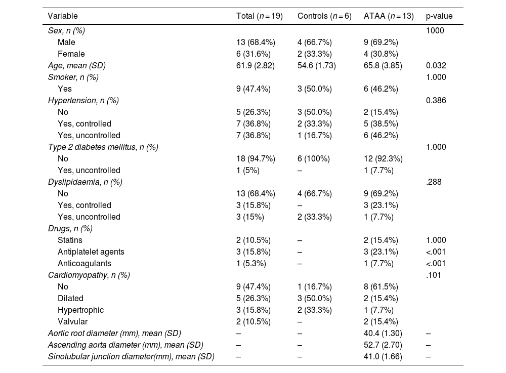
The study population consisted of 19 subjects, including patients with ATAA and multi-organ donors. Samples were obtained during elective or emergency aortic root surgery due to ATAA dissection. Clinical data from the patients included in the study were collected while respecting their anonymity. Samples of human ascending aortic aneurysm were provided by the Functional Aortic Pathology Unit of the Cardiology Department at the Hospital de la Santa Creu i Sant Pau (HSCSP, Barcelona) from patients with aortic atherosclerosis (aged 58–83 years) who underwent corrective surgery for ascending aortic aneurysm. The characteristics of these patients are shown in Table 1.

Table 1.

Data of patients included in the RNA sequencing analysis.

Variable  Total (n = 19)  Controls (n = 6)  ATAA (n = 13)  p-value 
Sex, n (%)        1000
Male  13 (68.4%)  4 (66.7%)  9 (69.2%) 
Female  6 (31.6%)  2 (33.3%)  4 (30.8%) 
Age, mean (SD)  61.9 (2.82)  54.6 (1.73)  65.8 (3.85)  0.032 
Smoker, n (%)        1.000
Yes  9 (47.4%)  3 (50.0%)  6 (46.2%) 
Hypertension, n (%)        0.386
No  5 (26.3%)  3 (50.0%)  2 (15.4%) 
Yes, controlled  7 (36.8%)  2 (33.3%)  5 (38.5%) 
Yes, uncontrolled  7 (36.8%)  1 (16.7%)  6 (46.2%) 
Type 2 diabetes mellitus, n (%)        1.000
No  18 (94.7%)  6 (100%)  12 (92.3%) 
Yes, uncontrolled  1 (5%)  –  1 (7.7%) 
Dyslipidaemia, n (%)        .288
No  13 (68.4%)  4 (66.7%)  9 (69.2%) 
Yes, controlled  3 (15.8%)  –  3 (23.1%) 
Yes, uncontrolled  3 (15%)  2 (33.3%)  1 (7.7%) 
Drugs, n (%)         
Statins  2 (10.5%)  –  2 (15.4%)  1.000 
Antiplatelet agents  3 (15.8%)  –  3 (23.1%)  <.001 
Anticoagulants  1 (5.3%)  –  1 (7.7%)  <.001 
Cardiomyopathy, n (%)        .101
No  9 (47.4%)  1 (16.7%)  8 (61.5%) 
Dilated  5 (26.3%)  3 (50.0%)  2 (15.4%) 
Hypertrophic  3 (15.8%)  2 (33.3%)  1 (7.7%) 
Valvular  2 (10.5%)  –  2 (15.4%) 
Aortic root diameter (mm), mean (SD)  –  –  40.4 (1.30)  – 
Ascending aorta diameter (mm), mean (SD)  –  –  52.7 (2.70)  – 
Sinotubular junction diameter(mm), mean (SD)  –  –  41.0 (1.66)  – 

Categorical variables were compared using Fisher’s exact test. Continuous variables were compared using Student’s t-test.

AATA: ascending thoracic aortic aneurysm; SD: standard deviation.

The study was approved by the Ethics and Clinical Research Committee of the HSCSP, Barcelona (ref. IIBSP-MAR-2019-08), and all samples collected are part of collection C.0005273, which is registered at the Instituto de Salud Carlos III.

The ascending aortas from multi-organ donors (healthy controls [HC]) showed no signs of atherosclerosis and/or aneurysm in the aorta and were obtained from the Barcelona tissue bank for use in research, with approval from the institution’s ethics committee. This work was conducted in accordance with the Declaration of Helsinki. The participation of patients and the collection of samples from control subjects were subject to informed consent from the patients or their legal representatives. Tissue samples were collected and stored at −80 °C for further studies. Some of the tissue was immediately fixed in paraformaldehyde and kept at room temperature for 48 h before being embedded in paraffin for histological characterisation of the samples.

RNA isolation from human tissue, quality control, library construction, and sequencing

Whole-genome analysis was performed by RNA sequencing (Novogene) to identify genes differentially expressed between the aorta of healthy controls and patients with ATAA; whole-genome analysis was performed by RNA sequencing (Novogene). Total RNA was extracted from 25 tissue samples from the ascending aorta that are representative of the pathological condition (ATAA, n = 18) and from multi-organ donors taken as healthy controls (HC, n = 7) using the RNeasy Fibrous Mini Kit (Qiagen, Venlo, Netherlands), in accordance with the manufacturer's instructions. Total RNA was determined and quantified using a NanoDrop 1000 spectrophotometer (Thermo Scientific), and its integrity and quality were assessed using an Agilent 2100 bioanalyser (Aligent Technologies, Santa Clara, CA, USA). For RNA sequencing, samples that did not meet the required RNA integrity values were excluded from the assay, which ultimately included 13 ATAA and 6 HC. Total RNA was used as starting material to synthesise complementary DNA (cDNA) libraries using the Illumina Stranded mRNA Prep kit based on the poly-A selection method, following the manufacturer's protocol (Illumina, San Diego, CA, USA). Briefly, messenger RNA was purified from total RNA using poly-T oligo-attached magnetic beads. After fragmentation, first-strand cDNA was synthesised using random hexamer primers. This was followed by second-strand cDNA synthesis using dUTP for the directional library or dTTP for the non-directional library. Finally, they were simultaneously sequenced using Illumina NextSeq 500 sequencing system with Illumina NextSeq 500/550 high output kit v2.5 (150 cycles, 2 × 74 bp paired end).

Paired sequence files in FASTQ format were analysed using the BaseSpace Sequence Hub platform using Dragen RNA Pipeline v3.7.5 (Illumina). The reference genome used was UCSC hg19. The original data file from the high output sequencing platforms (Illumina) was transformed into sequenced reads using CASAVA base calling. The raw data were stored in FASTQ(fq) format files, which contain read sequences and corresponding base quality.

The limma, DEseq2, and countToFPKM packages of the statistical programme R version 4.4.0 (R Foundation for Statistical Computing, Vienna, Austria. Available at: http://www.r-project.org/) were used to normalise the RNA-seq reads and obtain the fragments per kilobase of transcript per million mapped reads (FPKM).

Gene expression analysis

Based on the FPKM calculated for each sample and a comparison of the ATAA and CS study groups, transcriptomic analysis data was used to quantify the gene expression of genes related to ECM remodelling, encoding metalloproteinases, (MMP1, MMP2, and MMP9), elastin (ELN), fibrillin 1 (FBN1), and fibulin 5 (FBLN5), as well as collagens (COL1A1, COL3A1, COL5A1, COL7A1, COL24A1, and COL26A1). The expression of genes related to endoplasmic reticulum stress was quantified: activating transcription factor-4 (ATF), which encodes transcription factor 4; EIF2AK3, which encodes protein kinase R-like endoplasmic reticulum kinase (PERK); HSPA5, which encodes the molecular chaperone GRP78/BIP; ERN1, which encodes inositol-requiring enzyme 1 (IRE1); SEL1L, which encodes protein sel-1 homologue 1; and oxidative stress-related genes: NOX2 and NOX4. Finally, the expression of genes involved in mitochondrial dysfunction was quantified: DNML1, which encodes dynamin-1-related protein 1 (DRP1); IMMT, which encodes the mitofilin protein; MFF, which encodes the mitochondrial fission factor; MT-CO3, which encodes subunit 3 of cytochrome C oxidase; MT-CYB, which encodes cytochrome B; MT ND2, which encodes subunit 2 of the enzyme NADH dehydrogenase; TIMM17B, which encodes mitochondrial inner membrane translocase 17B; MT-ERF1, which encodes mitochondrial transcription termination factor 1; and TOMM5, which encodes mitochondrial outer membrane translocase 5.

Protein–protein interaction analysis

Protein-protein interaction (PPI) analysis of genes related to ECM remodelling processes, endoplasmic reticulum stress, oxidative stress and mitochondrial dysfunction was performed using the String database, with a minimum interaction score of >.4, considered statistically significant with a p-value of <.05 and a false discovery rate (FDR) of 5%; the results were visualised using Cytoscape software (v3.7.2).

Enrichment analysis: over-representation analysis in disease Gene network and online mendelian inheritance in Man

Genes related to ECM remodelling processes, endoplasmic reticulum stress, oxidative stress, and mitochondrial dysfunction with differential gene expression were annotated for enrichment analysis Over Representing Analysis (ORA) with the clusterProfiler package of the statistical programme R and the WEB-based Gene SeT AnaLysis Toolkit (WebGestalt) with the enrichment categories of the Disease Gene Network (DisGeNET) and Online Mendelian Inheritance in Man (OMIM) databases using the Benjamini & Hochberg method and with an FDR value of <.05.

Histological analysis

The tissue samples were fixed with 4% paraformaldehyde and embedded in paraffin. Histological characterisation of human aorta samples was performed using Masson's trichrome and Picosirius red stains, while the integrity of elastic fibres was assessed by orcein staining using a commercial kit (Casa Álvarez, Madrid, Spain), as previously described.30,31 To this end, the sections were deparaffinised in xylene, rehydrated using a series of alcohols of decreasing concentration, and immersed in distilled water for 5 min. After the various staining procedures, the samples were dehydrated by washing them in 100° alcohol (3 min), followed by two 3-minute washes in xylene. Finally, the slides were mounted with DPX mounting medium (Casa Álvarez, Spain). The samples were scanned with a Pannoramic Midi I Automatic brightfield scan (3DHISTECH) scanner for viewing and capturing representative images with CaseViewer 2.3 software.

Statistical analysis

Statistical analysis was performed using R software, version 4.3.3 (R Foundation for Statistical Computing, Vienna, Austria. Available at: http://www.r-project.org/) and GraphPad Prism software, version 9.2.20 (La Jolla, CA, USA). P-values ≤ .05 were considered significant using the two-tailed statistical test. In addition, the normal distribution of continuous variables was verified using the Shapiro-Wilk test.

The demographic and clinical characteristics of the participants were expressed as mean ± SEM if they were continuous variables. Categorical variables were presented as frequencies and compared using Fisher's exact test. Gene expression results are expressed as median and interquartile range, and differences between groups (ATAA patients versus HC) were assessed using the Mann-Whitney rank sum test. The number (n) of samples is indicated in the figure captions.

Results

The mean age of the patients with AATA was 66 years, 10 years older than that of the control subjects. Most suffered from hypertension (84.56%), but not dyslipidaemia (30.7%), and only 38.5% were receiving antihypertensive treatment. Around 50% of the patients involved in the study were being treated with cholesterol-lowering drugs such as statins, antiplatelet agents, and anticoagulants, and were smokers (Table 1). The ascending aorta was considered dilated if the diameter was 45 mm or more, which is the current threshold for recommending aortic surgery.

Histopathology of the vascular wall in patients with ascending thoracic aortic aneurysm

The morphology of the vessel wall was studied using Masson's trichrome, orcein, and Picrosirius red stains. The aortic wall was histologically analysed in a standardised manner using the classification system described in the consensus statement on surgical pathology of the aorta.32–34

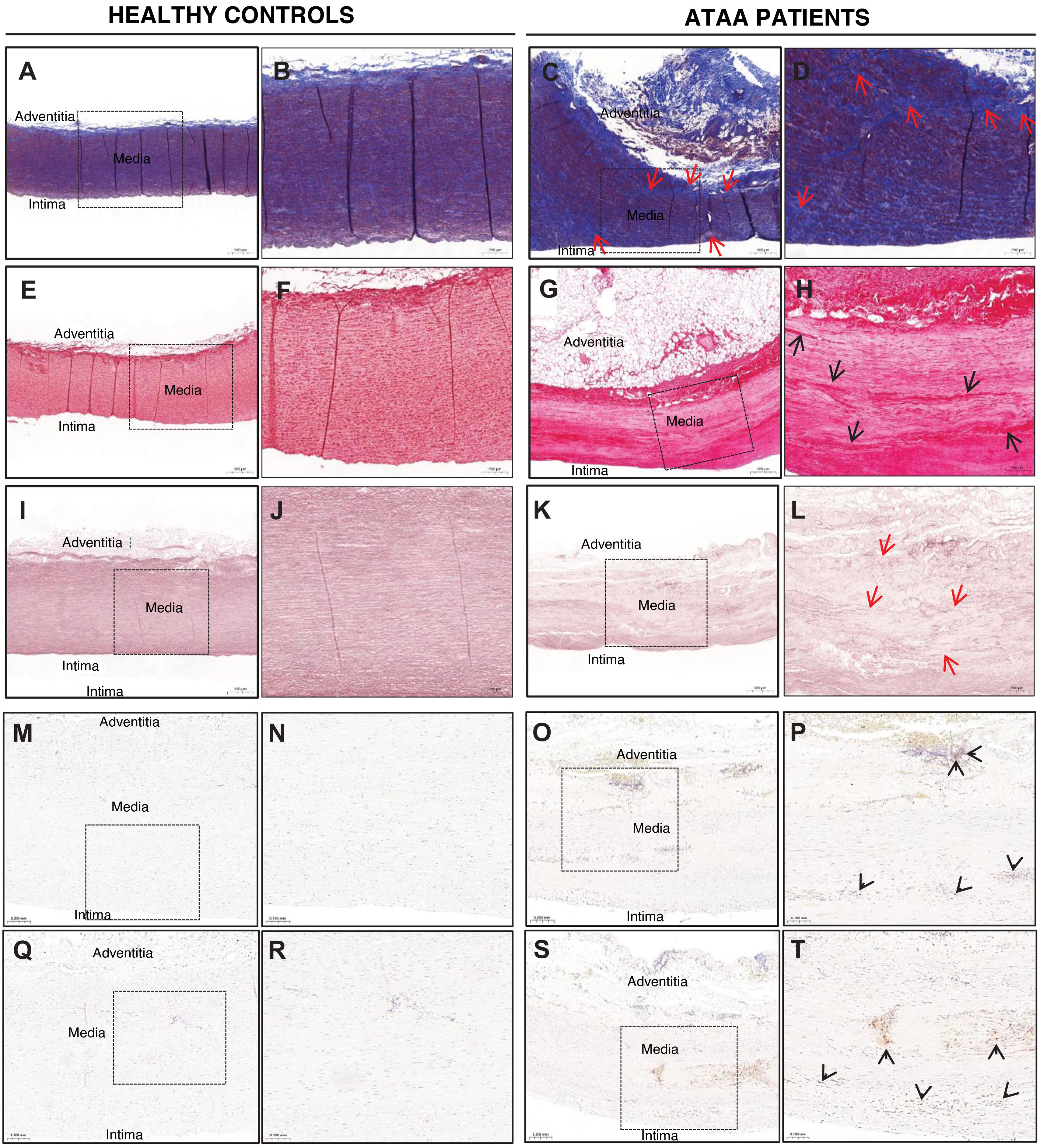
Medial layer degeneration (MLD) was the most common histopathological feature found in ATAA caused by decomposition of the aortic wall tissue and/or fragmentation of the elastic fibres (Fig. 1). Thus, in sections of the aneurysm samples, we identified loss of smooth muscle cell nuclei (LSMCN), middle lobe collapse (MLC), medial fibrosis (MF), and mucoid extracellular matrix accumulation (MEMA) using Masson's trichrome staining (Fig. 1A–D). MEMA can, in turn, be divided into two types: intralamellar (I-MEMA) and translamellar (T-MEMA). In I-MEMA, the increase in MEMA does not alter the arrangement of the lamellar units, unlike T-MEMA.33Fig. 1C and D showed severe T-MEMA, and middle lobe collapse (indicated by arrows) was observed in the ATAA wall of some patients). In turn, both Masson's trichrome and Picrosirius red staining showed an increase in intralamellar collagen, which is seen as an expansion within the intralamellar space (Fig. 1C and D, indicated with arrows) causing scarring alterations in the arrangement of the lamellar units and an increase in translamellar collagen (Fig. 1G and H, indicated by arrows). This collagen is disorganised and unstructured rather than forming bundles of wavy fibres, in comparison with the wall of the healthy donor aortas, where collagen deposition is lower and exhibits a normal distribution (Fig. 1E and F). Orcein staining revealed elastic fibre fragmentation and/or loss (EFFL), elastic fibre thinning (EFT) and elastic fibre disorganisation (EFD) in the aneurysm wall of the patients but not in the aortic wall of the healthy donors (Fig. 1I–L).

Figure 1.

Cross-sectional histological sections of the ascending aorta wall from control subjects and from the dilated ascending aorta wall of patients with ATAA.

A–D. Representative areas of sections stained with Masson’s trichrome are shown, where smooth muscle cells and fibrin are shown in red, while collagen is stained blue. Areas with loss of smooth muscle cell nuclei are indicated with red arrows. B and D show that the intima is significantly thicker in the ascending aorta wall of control subjects. E–H. Representative images of sections stained with Picrosirius red showing increased collagen deposition in the media and adventitia of the aortas of patients. I–L. Sections stained with orcein. Note the fragmentation observed in patients with ATAA (red arrows). M–T. Representative images of immunohistochemistry using the CD3 marker to identify T lymphocytes (M–P) and the CD68 marker to identify macrophages (Q–T). Areas where marker-positive cells are found are indicated by arrowheads.

ATAA, n = 6; and healthy controls, n = 4. Bar: 500 µm (A, E, I, C, G, K); 200 µm (B, F, J, D, H, L, M, O, Q, S); and 100 µm (N, P, R, T).

Finally, we evaluated the content of immune cells infiltrating the ATAA of the patients and the aortic wall of controls. Most of the immune cells present in the aneurysm wall were identified as T cells (CD3) and macrophages (CD68), as shown in Fig. 1M–T.

Expression pattern of genes involved in endoplasmic reticulum stress, mitochondrial dysfunction, and extracellular matrix remodelling

Transcriptomic analysis was performed to analyse the expression of genes normalised by fragments per kilobase of transcripts per million mapped fragments (FPKM), of genes involved in ER stress, mitochondrial dysfunction, and mitophagy, and in the synthesis, maintenance, and destruction of the ECM.

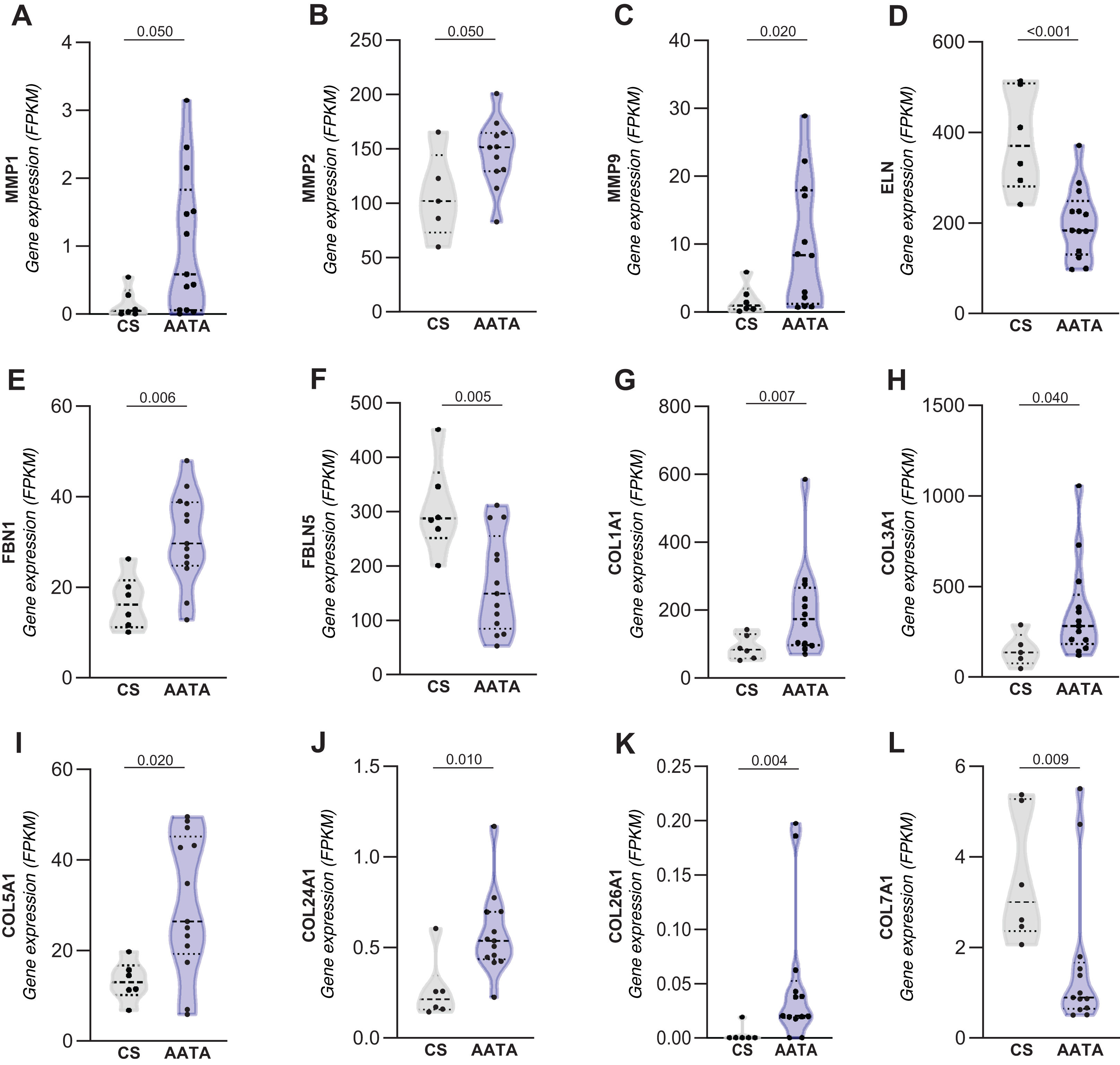
Thus, among the differentially expressed genes involved in ECM remodelling, significant increases were found in MMPs 1, 2, and 9; COL1A1, COL3A1, COL5A1, COL24A1, and COL26A1, which encode the alpha chain of different types of collagen, and FBN1, an essential structural protein that acts as a support for the deposition of elastin (Fig. 2). In contrast, the expression of ELN, FBLN5, which promotes interaction between microfibrils and elastin fibres in the arterial wall, and COL7A1 was significantly decreased (Fig. 2).

Figure 2.

Gene expression of markers related to ECM remodelling process in patients’ ascending thoracic aortic aneurysm (ATAA).

A–F. Expression of genes encoding different metalloproteinases and essential proteins that form and maintain the integrity of elastin fibres. G–L. Expression of genes encoding different subtypes of proteins that form collagen fibres. The studies were performed on samples of the ascending aorta from patients with ATAA (n = 13) and healthy controls ([HC] n = 6). Data are expressed as median and quartile ranges; Wilcoxon rank sum test was used to evaluate differences between groups. A p-value of <.05 was considered statistically significant.

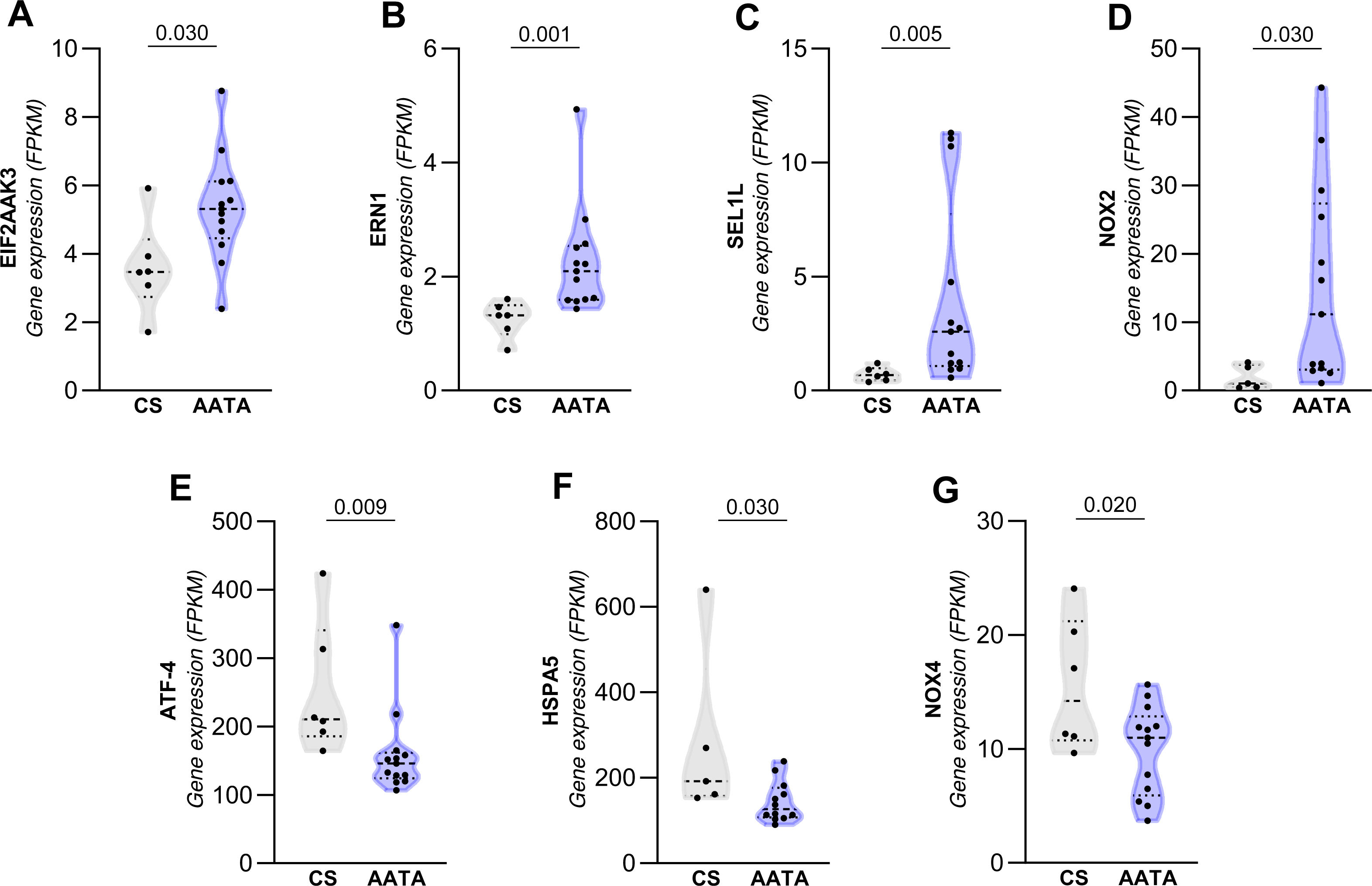
In terms of the differentially expressed genes involved in endoplasmic reticulum stress and those related to the NADPH oxidase enzyme complex, significant increases were found in the EIF2AK3, ERN1, SEL1L, and CYBB genes (Fig. 3). These genes encode the proteins PERK, IRE1, and SEL1L, which are part of the response to the accumulation of misfolded proteins, and for the beta chain of cytochrome B-245 (NOX2), the terminal component of the respiratory chain that transfers electrons from cytoplasmic NADPH through the plasma membrane to molecular oxygen outside. On the other hand, the expression of the ATF4, HSPA5, and CYBB genes, which encode other components of the response to the accumulation of misfolded proteins, such as the transcription factor ATF4, the molecular chaperone GRP78/BIP and the catalytic subunit of the NADPH oxidase complex, NOX4, was found to be significantly decreased, indicating differential regulation of the different pathways involved in the response to misfolded proteins and of the different isoforms of NADPH oxidase (Fig. 3). Linear regression analysis was performed to determine whether age influenced the differential expression of genes involved in endoplasmic reticulum stress and oxidative stress. A positive association was identified with the ERN1, SEL1L, and NOX2 genes (Appendix B Table 1 of the Supplementary material) while, according to logistic regression analysis, only the expression of the EIF2AK3 gene interacts with age and the presence of ATAA (Appendix B Table 2 of the Supplementary material).

Figure 3.

Differential gene expression of endoplasmic reticulum stress (ERS) markers and the catalytic subunit of the NADPH oxidase complex in patients with ascending thoracic aortic aneurysm.

A-D. The EIF2AK3, ERN1, SEL1L, and NOX2 genes were found to be significantly increased in the patient group, while the expression of the genes ATF-4, HSPA5, and NOX4 (E-G) was found to be decreased. The studies were performed on ascending aorta samples from patients with ATAA (n = 13) and healthy controls (n = 6). The Wilcoxon rank sum test was used to evaluate differences between the two groups. A p-value <.05 was considered statistically significant.

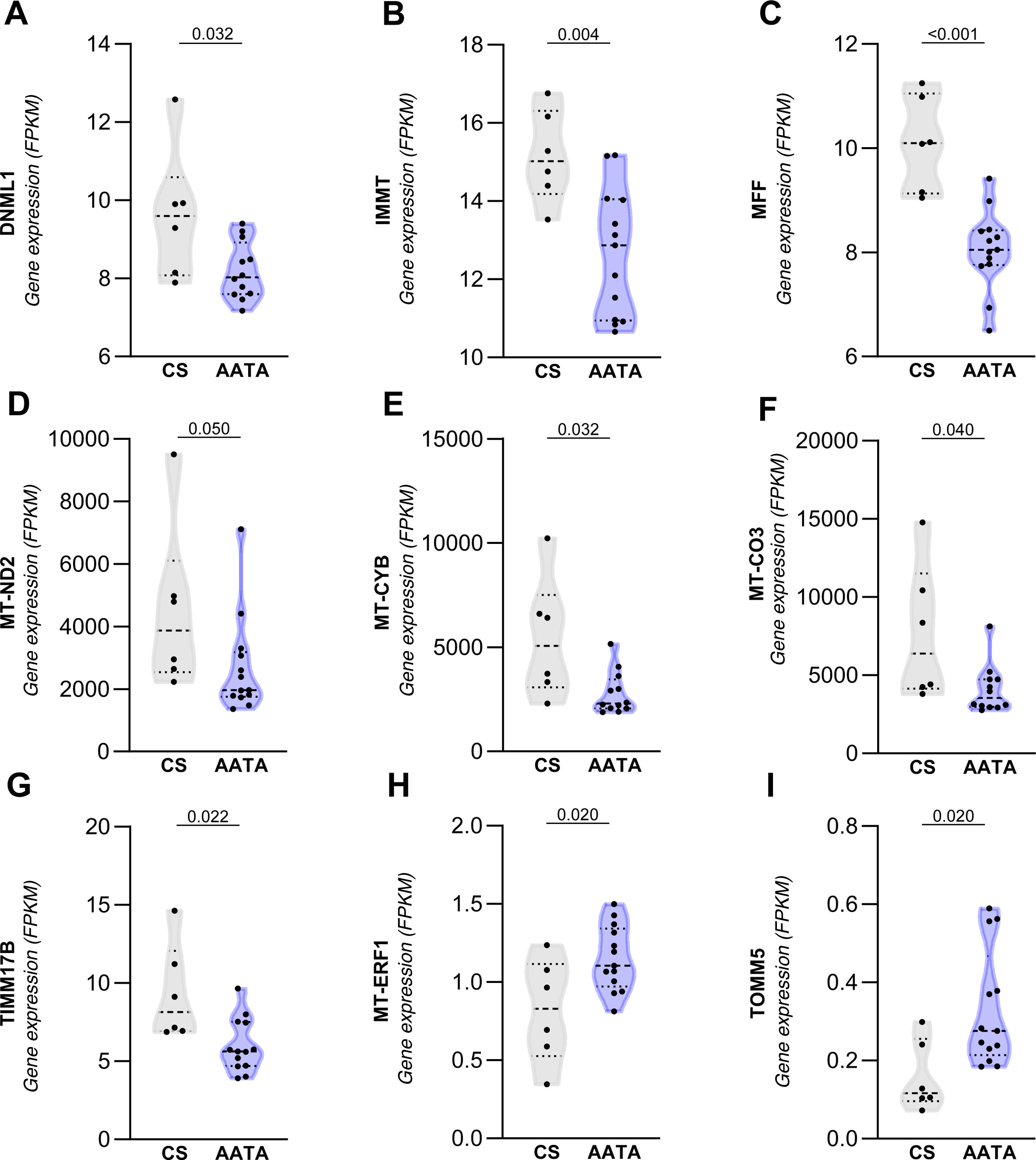
Finally, with regard to the genes involved in mitochondrial dysfunction, which are indicators of mitochondrial biogenesis and mitophagy processes, the following were found to be significantly reduced: DNML1, which encodes the DRP1 protein involved in mitophagy; IMMT, MT-CO3, MT-CYB, and MT-ND2 (which form part of complex I of the mitochondrial electron transport chain), as indicators of mitochondrial biogenesis; MFF, which encodes the mitochondrial fission factor; and the mitochondrial inner membrane translocase TIMM17B, related to mitophagy (Fig. 4). Conversely, the expression of genes related to these processes was found to be increased. These genes include the TOMM5 gene, which encodes a mitochondrial outer membrane translocase enzyme associated with mitophagy and autophagy, and MT-ERF1, which is related to mitochondrial biogenesis (Fig. 4). After evaluating the influence of age on the expression of genes related to mitochondrial dysfunction, only a negative and statistically significant association was observed for the DNM1L and IMMT genes, with a ß of −.055 and −.094, respectively (Appendix B Table 1 of the Supplementary material).

Figure 4.

Differential gene expression of markers related to mitochondrial dysfunction in ascending aortic aneurysm (ATAA) patients. A–G. The expression of the DNML1, IMMT, MFF, MT-CO3, MT-CYB, MT-ND2, and TIMM17B genes was significantly decreased in the patient group, while the expression of the MT-ERF1 and TOMM5 (H and I) genes was increased. The studies were performed on ascending aorta samples from patients with ATAA (n = 13) and healthy controls ([CS] n = 6). The Wilcoxon rank sum test was used to evaluate differences between groups. A p-value <.05 was considered statistically significant.

No significant differences were found in the expression of genes involved in cellular and vascular senescence in aortic tissue between the patient group and controls (Appendix B Fig. 1 of the Supplementary material).

Bioinformatic analysis of differentially regulated genes

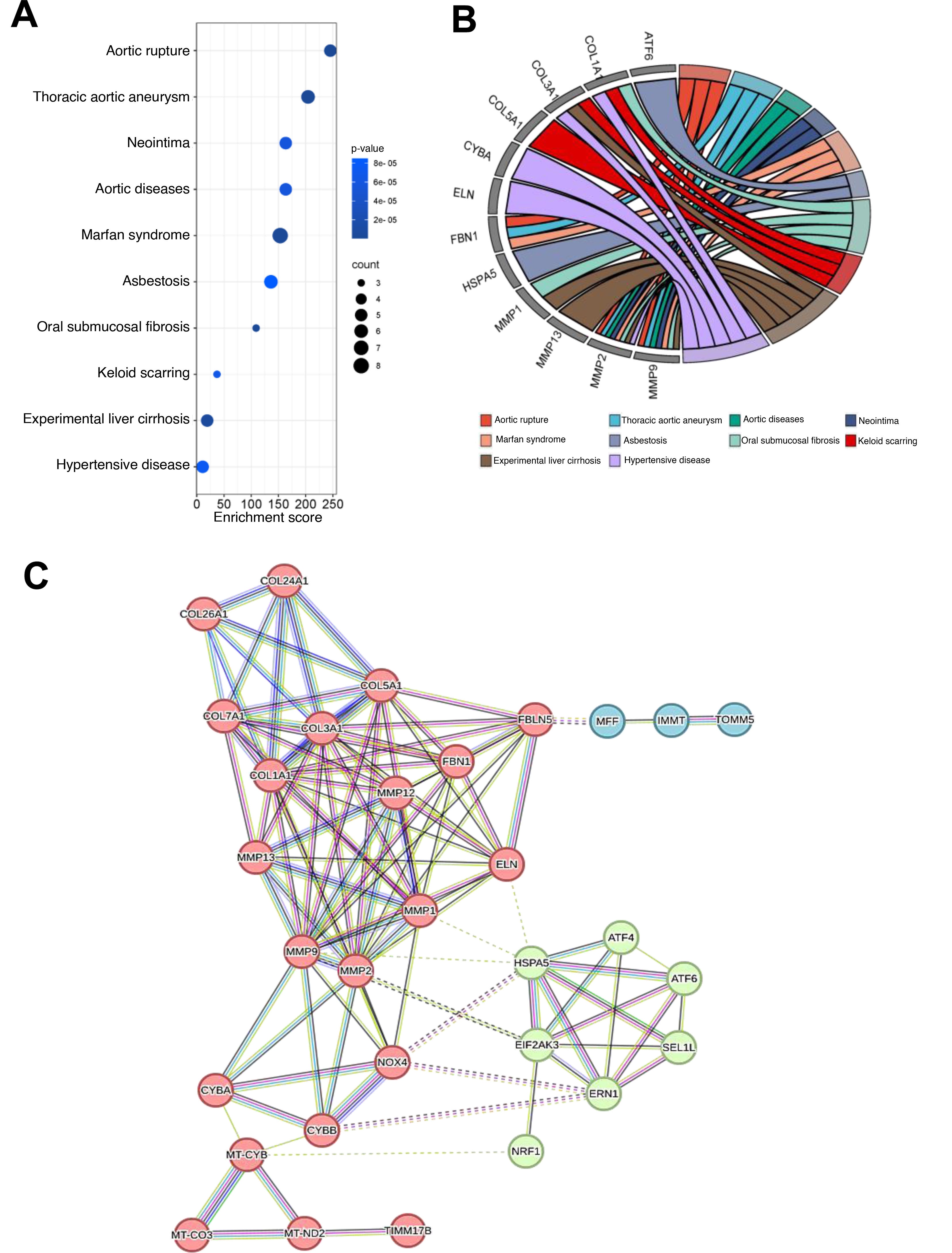
An enrichment analysis of the differentially regulated genes described in the previous section was performed using ORA and the DisGeNET and OMIM databases. Based on this ORA analysis, the 10 main pathological processes of interest were identified, including aortic rupture, aortic aneurysm, aortic diseases, Marfan syndrome, and hypertensive disease (Fig. 5A). Fig. 5B shows a string diagram of the 12 differentially expressed genes with the highest contribution to each enriched pathological process.

Figure 5.

Enrichment analysis. An enrichment analysis was performed using Over Representing Analysis (ORA) with the differentially regulated genes shown in Figs. 2–4, using the DisGeNET and OMIM databases.

A. Circle chart showing the 10 main diseases identified using this strategy, represented by ORA analysis with k-Medoid reduction in redundancy. B. String chart showing the 12 genes with the highest contribution to each disease. C. Protein-protein interaction analysis, with an average clustering coefficient of .663 and a p-value <.001, showed interactions between proteins related to ECM remodelling, ER stress, oxidative stress, and mitochondrial dysfunction. Three groups were distinguished: group number 1 (in red) is related to the ECM remodelling process, ECM integrity and mitochondrial dysfunction (average clustering coefficient: .756 and p-value <.001); group 2 (in green) is related to ER stress and the response to misfolded proteins (average clustering coefficient: .900 and p-value <.001); and group 3 (in blue) is related to mitochondrial membrane integrity with an average clustering coefficient of .667 and a p-value <.001. The dotted lines delimit each of the groups.

Next, an IPP analysis was performed based on differentially regulated genes. These proteins were grouped with an average clustering coefficient of .663 and a p-value <.001 (Fig. 5C). These proteins showed interaction in three groups related to the ECM remodelling process and mitochondrial dysfunction (1, in red), ER stress, response to the accumulation of misfolded proteins and oxidative stress (2, in green), and mitochondrial membrane integrity (3, in blue).

Discussion

Aortic dissection is the most serious complication of thoracic aortic disease. Although thoracic aortic disease is a life-threatening condition, it can be prevented if people at risk are identified early and undergo surgical repair. Current risk stratification is based solely on aortic diameter. However, recent studies have emphasised the inaccuracy of this parameter in detecting aortic complications prior to rupture.35,36 Therefore, there is an urgent need to identify new risk markers and treatment strategies for patients with ATAA. The identification of pathological mechanisms and effector pathways that lead to thoracic aortic disease and are involved in the development of aneurysms would significantly improve this stratification of individual aortic disease risk and, therefore, result in a major improvement in the field of personalised medicine.

This study aims to elucidate the pathogenic mechanisms underlying non-syndromic degenerative aortic aneurysms by a transcriptomic study of the ascending aortic wall in patients with ATAA. Several mechanisms that contribute to the formation of aortic aneurysms have been described in the literature. The most prominent of these are alterations to the ECM due to reduced synthesis, changes to its composition and maturation, and accelerated degradation due to an imbalance between proteolytic enzymes and their inhibitors.37–39 Increases in cell death rates (particularly of SMC), vascular inflammation, cell (trans)differentiation processes, atherosclerosis, and ageing phenomena in the vascular wall have also been observed, although many of these data come from studies of AAA.1,11,40,41 Some of the phenomena mentioned above have been linked, at least in part, to genetic variants and mutations.12,40 It is important to note that aneurysm formation appears to be the consequence of several independent yet interacting pathophysiological processes.41 Despite the large volume of data generated primarily through histological analysis of aneurysm samples, the pathophysiology of AAA remains poorly understood.

Several genes play a key role in the development of different aneurysmal conditions, both in the case of AAA and, as has been recognised in recent years, also in TAA, especially those genes that encode enzymes that degrade the ECM, such as MMPs and their inhibitors. Gene expression analyses performed in our transcriptomic study revealed altered expression not only of some metalloproteinases, but also of proteins that are part of the ECM or are involved in its synthesis and maintenance. Thus, we were able to detect changes in the expression of genes that encode ECM components involved in the formation and maintenance of elastin and collagen fibres, which were found to be differentially expressed in ATAA tissue. As expected, the expression of MMP1, 2, and 9 increased, accompanied by an increase in the expression of different collagens, most likely as a compensatory response, as occurs in other aneurysmal diseases. Additionally, decreased expression of genes encoding elastin and FBLN5 was observed. It should be noted that our group has previously demonstrated decreased expression of FBLN5 in human AAA and its involvement in aortic dilatation in an experimental model.42 In contrast, FBN1 expression increased in the ATAA of the patients, perhaps as a mechanism aimed at alleviating the loss and rupture of elastic fibres that occurs in this disease. In fact, histological analyses confirmed reduced cellularity in the middle layer of the aneurysmal aorta and decreased orientation and integrity of elastic fibres, while collagen deposition and disorganisation increased in the ATAA wall. Overall, medial degeneration, fragmentation, and disorganisation of the ECM, loss of vascular smooth muscle cells, and accumulation of mucoid extracellular matrix in the wall of the patients’ degenerative ATAA were observed. All histological features and differential gene expression found in the AATA tissue of our cohort appear to be independent of the age difference between patients and control subjects, as no differences in expression were found in various genes encoding proteins related to vascular senescence.43–47

Finding therapies that improve the management of patients with ATAA remains challenging. Previous publications by our group and others have demonstrated that ER stress and mitochondrial dysfunction play a key role in the aetiopathogenesis of aortic aneurysms. These studies have also shown that specifically reducing both mitochondrial stress and ER stress limits aneurysm progression and has cardioprotective effects.18–22 Despite remarkable advances in understanding the pathophysiological role of mitochondrial dysfunction and evidence of the involvement of the ER in aortic aneurysms, knowledge of its involvement in the development of ATAA is limited.29,48–51 This study reveals alterations in the expression of various genes that encode proteins involved in the response pathways to misfolded proteins and proteins involved in biogenesis, mitochondrial dysfunction, and mitophagy. This provides new evidence of the importance of both aetiopathogenic processes in the wall of degenerative ascending aortic aneurysms. Analysis using regression models determined that age does not influence the differential expression of most of these genes. However, their relative contribution to the development of atherosclerosis and/or ascending aortic aneurysm cannot be determined because both ER stress and mitochondrial dysfunction are involved in the pathophysiology of both diseases.18,21,29,52–55

In summary, our data suggest that chronic endoplasmic reticulum stress and mitochondrial dysfunction may be crucial contributors to atherosclerosis in the aortic wall, thereby promoting the onset and progression of ATAA. While these preliminary results require validation in a larger cohort, they enable us to propose the blockade of ER stress and the maintenance of mitochondrial homeostasis through reducing mitochondrial stress as a therapeutic intervention to slow the development of ATAA.

CRediT authorship contribution statement

Rafael Almendra Pegueros, Elvira Pérez Marlasca, and María Galán processed and isolated the genetic material from the human samples, performed the histology, and interpreted the transcriptomic results. Antonio J. Barros Membrilla selected the patients and provided their clinical data. Josep Julve contributed to the analysis and interpretation of the data and the critical review of the content. José Martínez González contributed to the critical review of the results. Cristina Rodríguez participated in the conception and design of the study and critically reviewed the content, María Galán conceived and designed the study and wrote the manuscript. All authors reviewed the manuscript and approved the final version.

Funding

This work was funded by a grant from the Fundación Española de Arteriosclerosis (Beca FEA 2023-Investigación Básica III). In addition, the study received funding from the Instituto de Salud Carlos III (ISCIII; PI20/01004; PI21/01048) and “ERDF, a way to make Europe”.

Rafael Almendra Pegueros is funded through a PFIS contract (ISCIII).

Declaration of competing interest

The authors have no conflict of interests to declare.

Acknowledgement

This work was conducted within the framework of the Doctoral Programme in Pharmacology at the Universidad Autónoma de Barcelona.

Appendix A
Supplementary data

The following is Supplementary data to this article:

Icono mmc1.doc

References
[1]
D.C. Guo, C.L. Papke, R. He, D.M. Milewicz.
Pathogenesis of thoracic and abdominal aortic aneurysms.
Ann N Y Acad Sci, 1085 (2006),
[2]
G. Filardo, J.T. Powell, M.A. Martinez, D.J. Ballard.
Cochrane database.
Syst Rev, 2 (2015),
[3]
P.E. Norman, J.T. Powell.
Site specificity of aneurysmal disease.
Circulation, 121 (2010), pp. 560-568
[4]
V. Mohan, D. Ameta, B. Gaurav, P. Haranahalli.
Aortic aneurysm.
J Transl Int Med, 4 (2016), pp. 35-41
[5]
C. Olsson, S. Thelin, E. Ståhle, A. Ekbom, F. Granath.
Thoracic aortic aneurysm and dissection: increasing prevalence and improved outcomes reported in a nationwide population-based study of more than 14,000 cases from 1987 to 2002.
Circulation, 114 (2006), pp. 2611-2618
[6]
E.M.R. Gouveia, G. Silva Duarte, A. Lopes, M. Alves, D. Caldeira, E.F.R. Fernandes, et al.
Incidence and prevalence of thoracic aortic aneurysms: a systematic review and meta-analysis of population-based studies.
Semin Thorac Cardiovasc Surg, 34 (2022), pp. 1-16
[7]
J.A. Elefteriades.
Thoracic aortic aneurysm: reading the enemy’s playbook.
Yale J Biol Med, 81 (2008), pp. 175-186
[8]
F. D’Abate, D. Oladokun, A. La Leggia, R. Hinchliffe, M. Thompson, P. Holt, et al.
Transthoracic ultrasound evaluation of arch and descending thoracic aortic pathology.
Eur J Vasc Endovasc Surg, 55 (2018),
[9]
W.S. Aronow.
Treatment of thoracic aortic aneurysm.
Ann Transl Med, 6 (2018), pp. 66
[10]
E. Saliba, Y. Ying Sia.
The ascending aortic aneurysm: when to intervene?.
Int J Cardiol Heart Vasc, 6 (2015), pp. 91-100
[11]
I. El-Hamamsy, M.H. Yacoub.
Cellular and molecular mechanisms of thoracic aortic aneurysms.
Nat Rev Cardiol, 6 (2009), pp. 771-786
[12]
E.M. Isselbacher, C.L. Lino Cardenas, M.E. Lindsay.
Hereditary influence in thoracic aortic aneurysm and dissection.
Circulation, 133 (2016), pp. 2516-2528
[13]
N. Grewal, O. Dolmaci, E. Jansen, R. Klautz, A. Driessen, J. Lindeman, et al.
Are acute type A aortic dissections atherosclerotic?.
Front Cardiovasc Med, 9 (2023),
[14]
J.A. Elefteriades, E.A. Farkas.
Thoracic aortic aneurysm: clinically pertinent controversies and uncertainties.
J Am Coll Cardiol, 55 (2010), pp. 841-857
[15]
H. Achneck, B. Modi, C. Shaw, J. Rizzo, G. Albornoz, D. Fusco, et al.
Ascending thoracic aneurysms are associated with decreased systemic atherosclerosis.
Chest, 128 (2005), pp. 1580-1586
[16]
J.A. Elefteriades, J.W. Olin, J.L. Halperin, B.A. Ziganshin.
Diseases of the aorta.
Hurst’s the heart, 14th ed.,
[17]
J.S. Vapnik, J.B. Kim, E.M. Isselbacher, B.B. Ghoshhajra, Y. Cheng, T.M. Sundt 3rd, et al.
Characteristics and outcomes of ascending versus descending thoracic aortic aneurysms.
Am J Card, 117 (2016), pp. 1683-1690
[18]
M. Navas-Madroñal, C. Rodriguez, M. Kassan, J. Fite, J.R. Escudero, L. Canes, et al.
Enhanced endoplasmic reticulum and mitochondrial stress in abdominal aortic aneurysm.
Clin Sci (Lond), 133 (2019), pp. 1421-1438
[19]
A.M. Siegert, G. Garcia Diaz-Barriga, A. Esteve-Codina, M. Navas-Madroñal, D. Gorbenko Del Blanco, J. Alberch, et al.
A FBN1 3’UTR mutation variant is associated with endoplasmic reticulum stress in aortic aneurysm in Marfan syndrome.
Biochim Biophys Acta Mol Basis Dis, 1865 (2019), pp. 107-114
[20]
Y. Qin, Y. Wang, O. Liu, L. Jia, W. Fang, J. Du, et al.
Tauroursodeoxycholic acid attenuates angiotensin II induced abdominal aortic aneurysm formation in apolipoprotein e-deficient mice by inhibiting endoplasmic reticulum stress.
Eur J Vasc Endovasc Surg, 53 (2017), pp. 337-345
[21]
M. Navas-Madroñal, R. Almendra-Pegueros, L. Puertas-Umbert, F. Jiménez-Altayó, J. Julve, B. Pérez, et al.
Targeting mitochondrial stress with Szeto-Schiller 31 prevents experimental abdominal aortic aneurysm: crosstalk with endoplasmic reticulum stress.
Br J Pharmacol, 180 (2023), pp. 2230-2249
[22]
H. He, B. Zeng, X. Wu, J. Hou, Y. Wang, Y. Wang, et al.
Higher matrix stiffness promotes VSMC senescence by affecting mitochondria-ER contact sites and mitochondria/ER dysfunction.
FASEB J, 37 (2023),
[23]
C. Giorgi, D. De Stefani, A. Bononi, R. Rizzuto, P. Pinton.
Structural and functional link between the mitochondrial network and the endoplasmic reticulum.
Int J Biochem Cell Biol, 41 (2009), pp. 1817-1827
[24]
J.M. Timmins, L. Ozcan, T.A. Seimon, G. Li, C. Malagelada, J. Backs, et al.
Calcium/calmodulin-dependent protein kinase II links ER stress with Fas and mitochondrial apoptosis pathways.
J Clin Invest, 119 (2009), pp. 2925-2941
[25]
C.X.C. Santos, L.Y. Tanaka, J. Wosniak, F.R.M. Laurindo.
Mechanisms and implications of reactive oxygen species generation during the unfolded protein response: roles of endoplasmic reticulum oxidoreductases, mitochondrial electron transport, and NADPH oxidase.
Antioxid Redox Signal, 11 (2009),
[26]
K. Zhang, R.J. Kaufman.
From endoplasmic-reticulum stress to the inflammatory response.
Nature, 454 (2008), pp. 455-462
[27]
C. Zhang, T.W. Syed, R. Liu, J. Yu.
Role of endoplasmic reticulum stress, autophagy, and inflammation in cardiovascular disease.
Front Cardiovasc Med, 4 (2017), pp. 29
[28]
I. Van der Pluijm, J. Burger, P.M. van Heijningen, A. IJpma, N. van Vliet, C. Milanese, et al.
Decreased mitochondrial respiration in aneurysmal aortas of Fibulin-4 mutant mice is linked to PGC1A regulation.
Cardiovasc Res, 114 (2018), pp. 1776-1793
[29]
J. Oller, E. Gabandé-Rodríguez, M.J. Ruiz-Rodríguez, G. Desdín-Micó, J.F. Aranda, R. Rodrigues-Diez, et al.
Extracellular tuning of mitochondrial respiration leads to aortic aneurysm.
Circulation, 143 (2021), pp. 2091-2109
[30]
L. Puertas-Umbert, J. Alonso, E. Roselló-Díez, A. Santamaría-Orleans, J. Martínez-González, C. Rodríguez.
Rolipram impacts on redox homeostasis and cellular signaling in an experimental model of abdominal aortic aneurysm.
Clin Investig Arterioscler, 36 (2024), pp. 108-117
[31]
D. Sánchez-Infantes, M. Nus, M. Navas-Madroñal, J. Fité, B. Pérez, A.J. Barros-Membrilla, et al.
Oxidative stress and inflammatory markers in abdominal aortic aneurysm.
Antioxidants (Basel), 10 (2021), pp. 602
[32]
N. Grewal, B.J.J. Velders, A.C. Gittenberger-De Groot, R. Poelmann, R.J.M. Klautz, T.J. Van Brakel, et al.
A systematic Histopathologic evaluation of type-a aortic dissections implies a uniform multiple-hit causation.
J Cardiovasc Dev Dis, 8 (2021), pp. 12
[33]
M.K. Halushka, A. Angelini, G. Bartoloni, C. Basso, L. Batoroeva, P. Bruneval, et al.
Consensus statement on surgical pathology of the aorta from the Society for Cardiovascular Pathology and the association for European cardiovascular pathology: II. Noninflammatory degenerative diseases - nomenclature and diagnostic criteria.
Cardiovasc Pathol, 25 (2016), pp. 247-257
[34]
O. Leone, A. Corsini, D. Pacini, B. Corti, M. Lorenzini, V. Laus, et al.
The complex interplay among atherosclerosis, inflammation, and degeneration in ascending thoracic aortic aneurysms.
J Thorac Cardiovasc Surg, 160 (2020), pp. 1434-1443.e6
[35]
E. Neri, L. Barabesi, D. Buklas, L.A. Vricella, A. Benvenuti, E. Tucci, et al.
Limited role of aortic size in the genesis of acute type a aortic dissection.
Eur J Cardiothorac Surg, 28 (2005), pp. 857-863
[36]
L.A. Pape, T.T. Tsai, E.M. Isselbacher, J.K. Oh, P.T. Ogara, A. Evangelista, et al.
Aortic diameter &or = 5.5 cm is not a good predictor of type a aortic dissection: observations from the international registry of acute aortic dissection (IRAD).
Circulation, 116 (2007), pp. 1120-1127
[37]
G.M. Longo, W. Xiong, T.C. Greiner, Y. Zhao, N. Fiotti, B.T. Baxter.
Matrix metalloproteinases 2 and 9 work in concert to produce aortic aneurysms.
J Clin Invest, 110 (2002), pp. 625-632
[38]
S. Goodall, M. Crowther, D.M. Hemingway, P.R. Bell, M.M. Thompson.
Ubiquitous elevation of matrix metalloproteinase-2 expression in the vasculature of patients with abdominal aneurysms.
Circulation, 104 (2001), pp. 304-309
[39]
J.S. Ikonomidis, J.A. Jones, J.R. Barbour, R.E. Stroud, L.L. Clark, B.S. Kaplan, et al.
Expression of matrix metalloproteinases and endogenous inhibitors within ascending aortic aneurysms of patients with bicuspid or tricuspid aortic valves.
J Thorac Cardiovasc Surg, 133 (2007), pp. 1028-1036
[40]
R. He, D.C. Guo, W. Sun, C.L. Papke, S. Duraisamy, A.L. Estrera, et al.
Characterization of the inflammatory cells in ascending thoracic aortic aneurysms in patients with Marfan syndrome, familial thoracic aortic aneurysms, and sporadic aneurysms.
J Thorac Cardiovasc Surg, 136 (2008), pp. 922-929
[41]
K. Shimizu, R.N. Mitchell, P. Libby.
Inflammation and cellular immune responses in abdominal aortic aneurysms.
Arterioscler Thromb Vasc Biol, 26 (2006), pp. 987-994
[42]
M. Orriols, S. Varona, I. Martí-Pàmies, M. Galán, A. Guadall, J.R. Escudero, et al.
Down-regulation of Fibulin-5 is associated with aortic dilation: role of inflammation and epigenetics.
Cardiovasc Res, 110 (2016), pp. 431-442
[43]
Y. Zhu, T. Tchkonia, T. Pirtskhalava, A.C. Gower, H. Ding, et al.
The Achilles’ heel of senescent cells: from transcriptome to senolytic drugs.
Aging Cell, 14 (2015), pp. 644-658
[44]
B. Bai, P.M. Vanhoutte, Y. Wang.
Loss-of-SIRT1 function during vascular ageing: hyperphosphorylation mediated by cyclin-dependent kinase 5.
Trends Cardiovasc Med, 24 (2014), pp. 81-84
[45]
H. Wan, D. Liu, B. Liu, M. Sha, W. Xia, C. Liu.
Bioinformatics analysis of aging-related genes in thoracic aortic aneurysm and dissection.
Front Cardiovasc Med, 10 (2023),
[46]
Y.B. Somani, J.A. Pawelczyk, M.J. De Souza, P.M. Kris-Etherton, D.N. Proctor.
Aging women and their endothelium: probing the relative role of estrogen on vasodilator function.
Am J Physiol Heart Circ Physiol, 317 (2019), pp. H395-H404
[47]
A. Valentini, M.A. Cianfarani, U. Tarantino, N. Di Daniele, A. Bertoli.
Osteoprotegerin as a biomarker of geriatric frailty syndrome.
Aging (Albany NY), 11 (2019), pp. 4900-4909
[48]
Y. Li, P. Ren, A. Dawson, H.G. Vasquez, W. Ageedi, C. Zhang, et al.
Single-cell transcriptome analysis reveals dynamic cell populations and differential gene expression patterns in control and aneurysmal human aortic tissue.
Circulation, 142 (2020), pp. 1374-1388
[49]
L. Wang, Z. Yang, S. Wang, Y. Que, X. Shu, F. Wu, et al.
Substitution of SERCA2 Cys(674) accelerates aortic aneurysm by inducing endoplasmic reticulum stress and promoting cell apoptosis.
Br J Pharmacol, 179 (2022), pp. 4423-4439
[50]
B.S. Tavris, A.S. Peters, D. Böckler, S. Dihlmann.
Mitochondrial dysfunction and increased DNA damage in vascular smooth muscle cells of abdominal aortic aneurysm (AAA-SMC).
Oxid Med Cell Longev, 2023 (2023),
[51]
S. Vats, K. Sundquist, Y. Li, X. Wang, M.G. Hong, J. Sundquist, et al.
Characterization of the mitochondrial genetic landscape in abdominal aortic aneurysm.
J Am Heart Assoc, 12 (2023),
[52]
E. Thorp, G. Li, T.A. Seimon, G. Kuriakose, D. Ron, I. Tabas.
Reduced apoptosis and plaque necrosis in advanced atherosclerotic lesions of Apoe-/- and Ldlr-/- mice lacking CHOP.
Cell Metab, 9 (2009), pp. 474-481
[53]
M. Civelek, E. Manduchi, R.J. Riley, C.J. Stoeckert Jr., P.F. Davies.
Chronic endoplasmic reticulum stress activates unfolded protein response in arterial endothelium in regions of susceptibility to atherosclerosis.
Circ Res, 105 (2009), pp. 453-461
[54]
J.I. Abe, K.A. Ko, S. Kotla, Y. Wang, J. Paez-Mayorga, I.J. Shin, et al.
MAGI1 as a link between endothelial activation and ER stress drives atherosclerosis.
JCI Insight, 4 (2019),
[55]
H.A. Cooper, S. Cicalese, K.J. Preston, T. Kawai, K. Okuno, E.T. Choi, et al.
Targeting mitochondrial fission as a potential therapeutic for abdominal aortic aneurysm.
Cardiovasc Res, 117 (2021), pp. 971-982
Copyright © 2024. Sociedad Española de Arteriosclerosis
asdasdasd
Article options
Tools
Supplemental materials