To evaluate whether a radioguided approach allows a higher intraoperative detection rate of adenoma and a minimally invasive parathyroidectomy (MIP), with the same or better cure rate of hyperparathyroidism secondary to parathyroid adenoma.
MethodsThis was an observational, prospective, single-center study involving 254 consecutive patients with primary hyperparathyroidism, between 2017 and 2022. A total of 258 procedures were performed: 129 non radioguided (NRS) and 129 radioguided (RS) (112 with intravenous 99mTc- MIBI and 17 with ultrasound-guided intralesional 99mTc-MAA injection) with an intraoperative gamma probe and gamma camera. Follow-up was performed for at least one year.
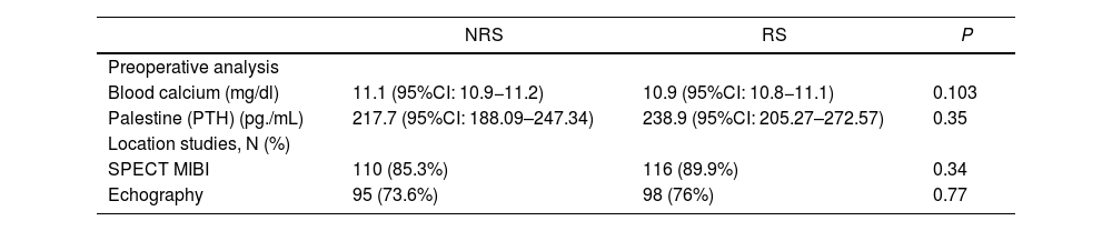
ResultsThere were no differences between the groups in terms of age, sex, preoperative calcium or parathyroid hormone levels, adenoma localization with 99mTc-MIBI-gammagraphy and ultrasound, and surgical morbidity.
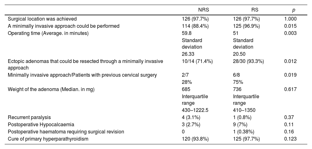
Intraoperative surgical localization was 97.7% in both groups. Statistically significant differences supported RS: It allowed to perform MIP (RS: 96.9%, NRS 88.4%; p = 0.015), also in patients with previous neck surgery (RS: 75%, NRS: 28%; p = 0.019) and with ectopic adenomas (RS: 93.3%, NRS: 71.4%; p = 0.012). The operative time was significantly shorter (RS: 51 min, NRS: 59.79 min; p = 0.005). There were no significant differences in the postoperative complications between the groups.
Biochemical cure at six months was achieved in RS: 97.7% and NRS: 93.8% (p = 0.12).
ConclusionsRadioguided MIP is useful in ectopic adenomas and in patients with previous cervical surgery and allows a minimally invasive approach more frequently. It is a safe surgery, easily reproducible by an endocrine surgeon and requires usual equipment found in operating rooms.
Evaluar si un abordaje radioguiado permite una mayor tasa de detección intraoperatoria del adenoma y una paratiroidectomía mínimamente invasiva (PMI), con la misma o mejor tasa de curación del hiperparatiroidismo primario por adenoma paratiroideo.
MetodologíaEstudio observacional, prospectivo y unicéntrico con 254 pacientes consecutivos afectos de hiperparatiroidismo primario, entre 2017 y 2022. Se realizaron un total de 258 procedimientos: 129 no radioguiados (CNR) y 129 radioguiados (CR) (112 con 99mTc- MIBI intravenoso y 17 con inyección intralesional de 99mTc-MAA) con gammasonda intraoperatoria y gammacámara. Seguimiento postoperatorio durante al menos un año.
ResultadosSin diferencias entre ambos grupos en cuanto a edad, sexo, niveles preoperatorios de calcio u hormona paratiroidea, localización del adenoma con gammagrafía con 99mTc-MIBI y ecografía, y morbilidad quirúrgica.
La localización quirúrgica intraoperatoria fue del 97,7% en ambos grupos. Diferencias estadísticamente significativas a favor de CR: Permitió realizar PMI (CR: 96,9%, CNR 88,4%; p = 0,015), también en pacientes con cirugía previa de cuello (CR: 75%, CNR: 28%; p = 0,019) o adenomas ectópicos (CR: 93,3%, CNR: 71,4%; p = 0,012). El tiempo operatorio fue significativamente menor (CR: 51 min., CNR: 59,79 min.; p = 0,005). No hubo diferencias significativas en las complicaciones postoperatorias entre los grupos.
La curación bioquímica se alcanzó en CR: 97,7% y CNR: 93,8% (p = 0,12).
ConclusionesLa cirugía radioguiada es útil en adenomas ectópicos y en pacientes con cirugía cervical previa y permite un abordaje mínimamente invasivo con mayor frecuencia. Es una cirugía segura, fácilmente realizable por un cirujano endocrino y utiliza equipamiento habitual en los quirófanos de cirugía general.
Parathyroid adenoma is the most common cause of primary hyperparathyroidism (PHPT) and surgery is the only curative treatment.1
The classic surgical approach to PHPT, defined as hypercalcemia due to chronic autonomous elevated secretion of PTH, or at times, inappropriately normal levels of this hormone in relation to serum calcium levels, has been bilateral cervical exploration, with evaluation of the four glands and excision of the macroscopically pathological one(s). However, this has changed radically since the development of more precise diagnostic procedures (high-resolution cervical ultrasound and especially SPECT-CT in MIBI Scintigraphy)2 that enable more accurate anatomical localisation of the diseased gland3 and, consequently, its excision by means of a minimally invasive parathyroidectomy (MIP).4 This is currently the procedure of choice in patients with a single adenoma5 with two positive localisation tests, while classic cervicotomy continues to be indicated in cases of multiglandular involvement, a concomitant indication for thyroid surgery, or parathyroid carcinoma.
Despite the high success rates of MIP excision (95–96.9%),6 some patients in whom the procedure is not successful (often due to involvement of more than one gland or its ectopic location) could benefit from preoperative marking of the affected gland(s), increasing the effectiveness of the selective surgical technique. In this context, radioguided minimally invasive parathyroidectomy (RMIP), described for the first time in 1997 by Norman and Chheda,7 has become widespread in recent years through the preoperative administration of 99mTc-MIBI.
This procedure reduces surgery time,8–10 with a high cure rate11 and few resulting complications8,9 and postoperative pain, while achieving optimal cosmetic results, but also enables immediate confirmation of the success of the resection, which in turn reduces the risk of persistent/recurrent PHPT.12 In this context, it may obviate the need for intraoperative frozen section studies or parathyroid hormone (PTH) analysis.13,14 This, in turn would reduce surgical time and costs.11
RMIP may be especially indicated in more compromised clinical scenarios, such as patients with a history of cervical surgery8,15,16 or with ectopic lesions,8,10,17–19 enabling a targeted surgical approach in both circumstances.
In our centre we began to implement the MIP with a protocol established in 2017, and later the RMIP was incorporated in March 2020.20
The objective of this study was to evaluate whether, in patients with primary hyperparathyroidism with surgical indication in our hospital, the performance of a radioguided surgical approach, compared to patients operated on without this technique, enabled a higher rate of intraoperative localisation of the adenoma, and made it possible more frequently to perform a minimally invasive approach, with the same or better cure rate for hyperparathyroidism and with similar surgical times and morbidity rates.
Material and methodsWe conducted an observational, prospective, single-centred, two-cohort study on a consecutive series of patients diagnosed with PHPT due to parathyroid adenoma with surgical indication,21 to perform minimally invasive surgery. Only if the adenoma was technically impossible or not located, the incision would be extended to a classic cervicotomy. All patients were included. The study was approved by the hospital's Research Ethics Committee (CEIC in its Spanish acronym).
Between March 2017 and March 2020, 129 non-radioguided surgeries (NRS) were performed and between March 2020 and December 2022, 129 radioguided surgeries (RS) were done. The surgical team was made up of four surgeons, who performed both types of approach. The study was completed in December 2022, and we chose a similar number of surgeries in the two groups as a cut-off point, so that they were comparable with each other, and a minimum follow-up time of 1 year to pinpoint cure and definitive morbidity. No patients were lost to follow-up.
Table 1 shows the characteristics of the two groups under study. These were two homogeneous groups, with a mean age of 61 years (14–88 years) and a median age of 63.
As a requirement to perform minimally invasive selective surgery, patients had to present two positive, consistent preoperative localisation imaging methods in the location of the diseased gland, with MIBI (always with SPECT-CT) and cervical ultrasound being the first-line imaging techniques. In cases where one of these was negative or the results were discordant, this was completed with other studies (methionine or choline PET and/or 4D CT). In patients with concomitant nodular thyroid pathology with no surgical indication for the thyroid, thyroid scintigraphy with 99mTc was also performed to avoid the confounding factor between nodular parathyroid and thyroid pathology without excluding these from minimally invasive surgery (48 patients). Ectopy of parathyroid adenoma was defined as the non-localisation of the lesion in an orthotopic situation.
The study included 258 procedures performed on 254 patients, as four were operated on twice, all four being due to persistence of PHPT. In 15 cases, the patient had a history of cervical surgery.
The radioguided surgery (RS) group included 112 procedures performed by intravenous administration of 99mTc-MIBI (185 MBq, 1 h before the intervention) and 17 (6.58%) by intralesional ultrasound-guided administration of 99 mTc-MAA protocols (37 MBq on the day of the intervention), previously implemented in our unit20 in patients with negative 99Tc-MIBI scintigraphy. In both procedures, a scintigraphic planar image was acquired that had to be compatible with the preoperative one, and if it was not, a three-dimensional image and SPECT-CT was required, as well as marking skin on the lesion preoperatively in the Department of Nuclear Medicine. Between administration of the radioisotope and the start of surgery, the interval was always less than an hour.
Surgery was performed under general anaesthesia in all patients.
Before the incision, in patients in the RS group the neck was assessed with the help of a gamma probe detector (Gamma probe, Neo ® 2000, Neoprobe Corporation, Dublin, Ohio, USA) and intraoperative gamma camera (Sentinella ®, Oncovision, Valencia, Spain) (Figs. 1, 2). Surgery began with a transverse cervical incision, 1.5–2.5 cm in length, approximately 1.5 cm above the sternal manubrium. The incision was placed laterally, while in the three cases with perioperative findings of bilateral location of double adenoma, the incision was a central transverse cervicotomy shorter than 3 cm. Access to the adenoma was always by lateral approach.
All surgical specimens were studied anatomopathologically by frozen section analysis for intraoperative confirmation of a pathological parathyroid gland (intraoperative PTH value was not measured). In the radioguided surgery group the surgical incision was closed if it was confirmed by gamma camera and gamma probe that the excised specimen had captured the radioisotope and corresponded to the pathological gland in its macroscopic image (Fig. 3).
The time from the patient’s admission to theatre up to their exit (anaesthetic and surgical time, which we refer to in the study as operating time), was recorded by independent members of the operating theatre staff.
Patient cure was defined as the presence of normal serum calcium 6 months after surgery. Oral supplementation of calcium in our unit was performed according to the analytical values of serum calcium, this being necessary if it was <7.5−8 mg/dL with calcitriol 0.25 mcg/12 h and calcium carbonate 1 g/8 h.22 If a progressive hematoma was detected, the patient was sent back to the operation room to achieve haemostasis.
Pre- and postoperative laryngoscopy was performed on all patients.
Data for the study was collected using IBM SPSS ® Statistics.25 Continuous quantitative variables that followed normal distribution were analysed using Student’s t-test for independent samples, while those that did not follow normal distribution were analysed using Mann-Whitney U. Qualitative variables were compared using Chi-square. Written consent for the study was obtained from all patients.
ResultsThe total number of patients was 254, with 258 procedures, since four patients were operated on twice, all four due to persistence of PHPT. Three of them had a second adenoma diagnosed at least one year after the first intervention, and the fourth had doubtful indication for the second surgery. No patients were excluded from surgery.
There were no significant differences between the two groups (Tables 2) in terms of preoperative serum calcium levels (NRS 11.1 mg/dL and RS 10.9 mg/dL, p: 0.103) nor in the mean PTH value (NRS 217.71 pg/mL and RS 238.92 pg/mL, p: 0.35). There was not any statistical significance between the two groups in terms of preoperative location in the imaging studies previously performed for the identification of the parathyroid lesion. The SPECT-CT MIBI scintigraphy was diagnostic in 89.9% of the RS group and 85.3% of the NRS group (p: 0.34). Cervical ultrasound was performed in 76% of the RS group and 73.6% in the NRS group (p: 0.77).
Preoperative results.
| NRS | RS | P | |
|---|---|---|---|
| Preoperative analysis | |||
| Blood calcium (mg/dl) | 11.1 (95%CI: 10.9−11.2) | 10.9 (95%CI: 10.8−11.1) | 0.103 |
| Palestine (PTH) (pg./mL) | 217.7 (95%CI: 188.09–247.34) | 238.9 (95%CI: 205.27–272.57) | 0.35 |
| Location studies, N (%) | |||
| SPECT MIBI | 110 (85.3%) | 116 (89.9%) | 0.34 |
| Echography | 95 (73.6%) | 98 (76%) | 0.77 |
In the event that ultrasound and scintigraphy produced discordant results, PET-Methionine was performed (58 patients up to March 2019 that located 91.5% of the adenomas); PET-Choline in 34 patients, started in March 2019, located 94.3% of the adenomas, and/or 4D cervical CT scans on 17 patients that located 80% of the adenomas. In cases of coexistence of thyroid nodularity, thyroid scintigraphy with 99-Tc was undertaken in 48 cases (18.6%) to avoid diagnostic confounding factors.
The intraoperative surgical location of the lesions was 97.7% in both groups (Table 3), however, excision by a minimally invasive approach was achieved in 96.9% of the patients in the RS group (in 4 patients the initial incision had to be widened), compared to 88.4% (15 patients) of the cases in the NRS group. In cases where a minimally invasive approach could not be performed, a classic cervicotomy had to be performed during the surgical procedure to remove the lesion. This difference was statistically significant (p: 0.015).
Study results.
| NRS | RS | p | |
|---|---|---|---|
| Surgical location was achieved | 126 (97.7%) | 126 (97.7%) | 1.000 |
| A minimally invasive approach could be performed | 114 (88.4%) | 125 (96.9%) | 0.015 |
| Operating time (Average. in minutes) | 59.8 | 51 | 0.003 |
| Standard deviation | Standard deviation | ||
| 26.33 | 20.50 | ||
| Ectopic adenomas that could be resected through a minimally invasive approach | 10/14 (71.4%) | 28/30 (93.3%) | 0.012 |
| Minimally invasive approach/Patients with previous cervical surgery | 2/7 | 6/8 | 0.019 |
| 28% | 75% | ||
| Weight of the adenoma (Median. in mg) | 685 | 736 | 0.617 |
| Interquartile range | Interquartile range | ||
| 430–1222.5 | 410–1350 | ||
| Recurrent paralysis | 4 (3.1%) | 1 (0.8%) | 0.37 |
| Postoperative Hypocalcaemia | 3 (2.7%) | 9 (7%) | 0.11 |
| Postoperative haematoma requiring surgical revision | 0 | 1 (0.38%) | 0.16 |
| Cure of primary hyperparathyroidism | 120 (93.8%) | 125 (97.7%) | 0.123 |
Data is specified in N (%) unless otherwise noted.
Regarding the presence of previous cervical surgery, a minimally invasive approach was possible in 75% of patients with a history of cervical surgery in the RS group (6/8), and only in 28% in the NRS group (2/7), the difference being significant (p: 0.019).
In turn, 93.3% of the ectopic lesions in the RS group (28/30) and 71.4% in the NRS group (10/14) were successfully removed by a minimally invasive access, which also represented a statistically significant difference (p: 0.012).
The operating time for RS was 51 min compared to 59.79 min for NRS, significantly shorter (p: 0.003).
The mean hospital stay was less than 20 h in both groups (due to the administration timetable of a public hospital).
There were no significant differences between the two groups in terms of definitive surgical complications. Recurrent paralysis was recorded in 5 cases (1.93%), 4 (3.1%) in the NRS group, and 1 (0.77%) in the RS group (p: 0.37). Transient postoperative hypocalcaemia with calcium and vitamin D requirements occurred in 12 procedures (4.65%), 9 in the RS group and 3 in the NRS group (p: 0.108). One single patient (0.38%) in the RS group required re-intervention due to a progressive haematoma.
The 12-month follow-up showed that the biochemical cure rate was higher in the radioguided surgery group, with 125 cases with normocalcemia (97.7%) compared to 120 in the non-radioguided group (93.8%), although this difference was not significant.
DiscussionThe surgical treatment of PHPT has become increasingly selective, thanks to the technological advances of imaging studies on the localisation of the diseased gland, which have enabled excision by selective, minimally invasive surgery.
Correct preoperative assessment of patients is of extreme importance in order to establish the indication for minimally invasive parathyroidectomy; to identify patients who have a single lesion; and to establish their anatomical location. In this direction, we consider that the routine acquisition of SPECT-CT in parathyroid scintigraphy with 99mTc-MIBI9,23,24 in the cases of our series, enabled us to specify the location of the parathyroid adenoma and its size and relationship with nearby structures, favouring its excision through a minimally invasive access.
In recent times, preoperative marking of the lesion with different doses of radiotracers in RS has been introduced, in line with different protocols.25–27 The radioguided surgical procedure maintained a high rate of detection in our case, as well as resection of parathyroid adenoma,20 similar to other series.9,11 The cure rate was even higher in the RS group, among other reasons due to the successful resection of a synchronous lesion. In three RS patients, two synchronous adenomas were removed, thus preventing the persistence of the disease. We consider that this improvement was due to re-evaluation with imaging on the day of the intervention (due to the protocol applied) as well as the in vivo image using the intraoperative portable gamma camera.
In recent years, several techniques have been incorporated for intraoperative localisation of the parathyroid glands (indocyanine green, and near infrared autofluorescence, etc.).28–30 These techniques have been used mainly as a support in thyroid surgery, to reduce the rate of inadvertent resection of parathyroid glands and to check the viability of the glands after surgery. However, there have been few studies regarding its usefulness in cases of surgery for parathyroid adenomas.29 The advantage of using the MIBI with a gamma camera and probe is that, on the one hand, it has already been proven preoperatively that they are capable of locating the adenoma(s), which is especially important in ectopic lesions. In addition, it can facilitate localisation in situ, from localisation with closed skin in the position of the anaesthetised patient to enable deep dissection, since radiation detection is not interfered with by the rest of the tissues or the depth of the adenoma. All this facilitates a minimally invasive approach.
In our study, no correlation was found between the PTH value and glandular weight. We think this can be explained by the adenoma weight ranging from 200 mg to 17,000 mg, and the fact that there was no agreement between some large, heavy adenomas of a cystic nature and their metabolic activity, which was low.
In the follow-up, persistent or recurrent PHPT was recorded in a higher percentage of patients in the non-radioguided group, which suggests that in some of these patients resection of the adenoma(s) may have been incomplete.
Radioguided surgery makes it possible to perform more cases with a minimally invasive access. In 11.6% of the patients included in the NRS group, surgical access had to be extended during the intervention itself to remove the diseased gland and this only occurred in 3.1% of the RS group.
RS has significantly reduced operating time. This can be partly explained by the greater experience acquired by the team (our surgical team performs more than 40 parathyroidectomies per year), however we consider that this is also closely related to intraoperative detection devices. On the one hand, the acoustic and counting signal from the gamma probe serves as a guide for the surgeon in plane dissection. On the other hand, the intraoperative gamma camera image enables immediate (“ex-vivo”) confirmation of the excision of the diseased parathyroid gland, which reduces the operating time by not requiring other intraoperative studies for its confirmation (frozen section analysis of the lesion or intraoperative determination of PTH), and in turn guarantees the success of the intervention. Team also contributes to the reduction of operating time. Although the statistically significant difference in time between the two groups is just under 9 min, any reduction in operating time is helpful for the patient and the healthcare system. Efficiency in operating theatre usage times can enable more procedures to be performed in the same time frame and demonstrates that coordinated teams achieve these goals.
Radioguided surgery offers advantages over conventional NRS,31 especially in more compromised clinical settings, such as patients with a history of cervical surgery16 or with ectopic lesions.8,9,15,17–19 In both circumstances this enables a selective approach and also reduces the risk of persistent or recurrent PHPT by ensuring complete excision of the lesion causing PHPT.12
In this context, detection rates of 68–96% have been described in reoperated patients.8–10,15 without increasing the complications inherent in cervical reoperation.15 In the series we present, the radioguided procedure facilitated minimally invasive resection of the parathyroid adenoma in most patients with a history of cervical surgery (75%), while in the NRS group, most patients finally required a classic cervicotomy to locate and remove the gland and in only 28% of cases this could be performed with a minimally invasive access.
Regarding the selective approach to the ectopic glands, 8,17–19,32 their excision has been described in up to 90% of cases, and even 60% with minimally invasive access.33 In our case, radioguided surgery enabled the removal of the ectopic gland with a minimally invasive approach in a significantly higher percentage of cases (93.3%.RS vs. 71.4% NRS). In addition, we believe that the incorporation of the radioguided procedure has meant that a minimally invasive approach has been achieved in a greater number of patients with ectopic lesions than in the previous period. As radioguided surgery allowed for improved detection and increased surgical success, more patients with ectopic adenomas were indicated for a minimally invasive approach beforehand. Previously, many of them were operated on from the beginning through classic cervicotomy.
In principle, the existence of multiglandular involvement was an exclusion criterion for minimally invasive surgery.9 However, in our series, we were able to remove more than one lesion using RMIP (three double adenomas) diagnosed in perioperative studies.
There were no significant differences between the two groups regarding postoperative complications. RS is therefore effective and safe,8–10 with few complications, similar to other series,22 as observed in our results, and enables smaller incisions to be made more frequently, which allows for better cosmetic results.
One of the theoretical limitations of radioguided parathyroid surgery is the difficulty that can be posed by the frequent coexistence of non-surgical nodular thyroid pathology, especially in endemic areas, with the risk of falsely positive findings that actually correspond to thyroid nodules that keenly capture and retain MIBI, with an intensity similar to or greater than parathyroid adenomas. In fact, in some published series10 these patients are ruled out for RMIP. Both the performance of 99Tc-thyroid scintigraphy (48 patients) among the previous diagnostic studies, as well as re-evaluation of the MIBI on the day of the surgery and intraoperative gamma camera imaging enables differentiation between thyroid nodules and parathyroid adenomas.
One of the drawbacks is that the procedure must be well protocolised and requires strict coordination of the services involved, since the radiotracer gradually becomes less active and disappears from the parathyroid tissue. In our case, we achieved good synchronisation of the human teams, the surgical team and the nuclear medicine team, without this having involved an extraordinarily large investment of time. However, each group must establish its own methodology.
The limitations of our study are that it was an observational study, however, the number of cases was large; it included all cases with an indication for surgery; and no patient was lost to follow-up, which provided an absence of bias or error.
In conclusion, RMIP is of great interest in ectopic adenomas and in patients with previous cervical surgery and makes it possible to perform minimally invasive approaches more frequently. It is a safe surgery, easily reproducible for an endocrine surgeon, using equipment that is common in our operating theatres.
CRediT authorship contribution statement- -
Pilar Salvador Egea: Conceptualisation, formal analysis, research, methodology, project management, supervision, validation, writing of the initial draft and editing of the final manuscript.
- -
Isabel Blanco Saiz: Conceptualisation, formal analysis, research, methodology, supervision, validation and editing of the final manuscript.
- -
Emma Anda Apiñániz: Research, methodology, supervision, validation and editing of the final manuscript.
- -
Aitor Redondo Expósito: Data collection, formal analysis, methodology, validation, writing of the initial draft and editing of the final manuscript.
- -
Cristina Erce García: Data collection, formal analysis, methodology, validation, writing of the initial draft and editing of the final manuscript.
- -
Irati Pérez Otermin: Data collection.
- -
Naomi Cruz Vásquez: Data collection.
This research has not received any specific grants from public sector agencies, the commercial sector or non-profit entities.


