Buscar en
Cirugía Española (English Edition)
Toda la web
Inicio Cirugía Española (English Edition) Portal Venous System Aneurysm
Journal Information
Vol. 91. Issue 1.
Pages e1 (January 2013)
Image of the Month
Full text access
Portal Venous System Aneurysm
Aneurisma del sistema venoso portal
Visits
2435
E.M. Guerra del Barrio
Corresponding author
domingoelena@telecable.es

Corresponding author.
, L. Pastor Hernández, M. Pérez-Peña, C. González Huerta
Servicio de Radiodiagnóstico, Hospital Álvarez Buylla, Mieres, Asturias, Spain
This item has received
Article information
Full Text
Download PDF
Statistics
Figures (3)
Show moreShow less
Full Text

The patient was studied for macrocytosis and high ferritin levels and tests were showing normal liver function.

The ultrasound showed a “cystic” image at the head of the pancreas joining with the splenic vein (Fig. 1), and colour Doppler was recorded. An abdominal CT scan showed an aneurysm of the splenic vein and the superior mesenteric vein (Fig. 2), more obvious in MIP reconstructions (Fig. 3).

Fig. 1
(0.19MB).
Fig. 2
(0.11MB).
Fig. 3
(0.06MB).

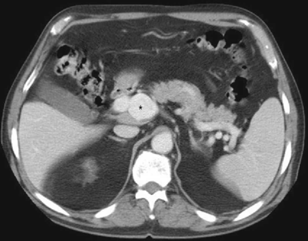
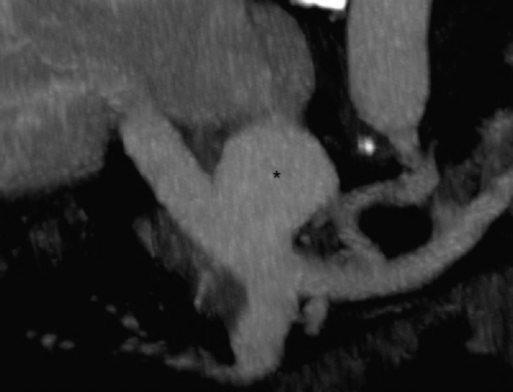
These findings are typical of portal venous system aneurysm.

Although venous aneurysms are rare, it is important not to confuse them with cystic lesions in the ultrasound. In this sense, the colour Doppler is very useful.

The majority are asymptomatic; vigilance is recommended, with monitoring by Doppler ultrasound. If there are complications or symptoms, they will require surgery.

Diagnosis of a case: portal venous system aneurysm

Please cite this article as: Guerra del Barrio EM, et al. Aneurisma del sistema venoso portal. Cir Esp. 2013;91:e1.

Copyright © 2011. AEC
Article options
Tools
es en pt

¿Es usted profesional sanitario apto para prescribir o dispensar medicamentos?

Are you a health professional able to prescribe or dispense drugs?

Você é um profissional de saúde habilitado a prescrever ou dispensar medicamentos