


To describe the ultrasound biomicroscopic (UBM) features and complications associated with iris cysts.
DesignA retrospective case series.
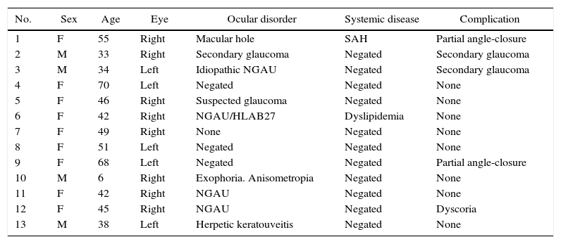
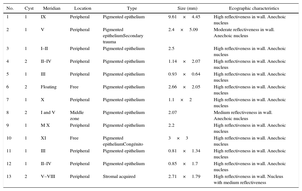
MethodsThirteen patients with iris cysts were identified in a 10 year period study at a ophthalmologic reference center in Mexico City. The variables included demographic data, ocular and medical history, clinical course, and complications. All patients were examined by UBM, and type, number, location, and acoustic characteristics of cysts were evaluated. Descriptive statistics were performed.
ResultsThirteen patients were included (8 men and 5 women). The mean age was 44.5±15.5 years (range 6–70 years). The origin most prevalent was neuroepithelial (92.3%), and 7.7% had stromal cysts. Regarding to location 76.9% were found in the periphery, and 69.2% between meridians II and VI. All cysts showed a moderate to high reflectivity in the wall. Complications were present in 38.5% of cases (15.4% partial angle closure, 15.4% secondary angle closure glaucoma and 7.7% dyscoria).
ConclusionsMost cysts are derived from iris pigmented epithelium, with a benign course and a minor rate of complications. The UBM is an indispensable tool that allows us to plan more specific and conservative treatments, with less damage to ocular structures and, therefore, better visual prognosis.
Describir los hallazgos ultrabiomicroscópicos y complicaciones de pacientes con quistes iridianos.
DiseñoSerie de casos, restrospectivo.
MétodoSe incluyó a 13 pacientes con diagnóstico de quistes de iris, confirmado mediante ultrabiomicroscopia (UBM) en un periodo de 10 años (2002–2012) en un centro oftalmológico de la ciudad de México. Se incluyeron datos demográficos, historia clínica médica y ocular, características clínicas y ultrabiomicroscópicas (tipo, número, localización y hallazgos acústicos), así como complicaciones asociadas. Se realizó un análisis descriptivo, incluyendo medias y desviación estándar.
ResultadosLa distribución por sexo fue 8 mujeres y 5 hombres, con edad promedio de 44,5 años±15,5 (rango de 6 a 70 años). El 92,3% fueron quistes del epitelio pigmentado y 7,7% del estroma; el 76,9% se encontraron en la periferia y 69,2% entre los meridianos de las II y las VI horas del reloj. Todos los quistes mostraron una pared con reflectividad moderada a alta. El 38,5% presentó complicaciones (el 15,4% cierre parcial del ángulo camerular; el 15,4% glaucoma secundario de ángulo cerrado y el 7,7% discoria).
ConclusionesLa mayoría de los quistes de iris son derivados del epitelio pigmentado, de curso benigno y con una baja tasa de complicaciones. La UBM es una herramienta indispensable que nos permite planear tratamientos localizados, específicos, más conservadores y menos destructivos, con un daño potencial menor de las estructuras oculares y, por lo tanto, mejor pronóstico visual.
