


To determine and compare the diagnostic precision in glaucoma of two deep learning models using infrared images of the optic nerve, eye fundus, and the ganglion cell layer (GCL).
MethodsWe have selected a sample of normal and glaucoma patients. Three infrared images were registered with a spectral-domain optical coherence tomography (SD-OCT). The first corresponds to the confocal scan image of the fundus, the second is a cut-out of the first centered on the optic nerve, and the third was the SD-OCT image of the GCL. Our deep learning models are developed on the MatLab platform with the ResNet50 and VGG19 pre-trained neural networks.
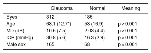
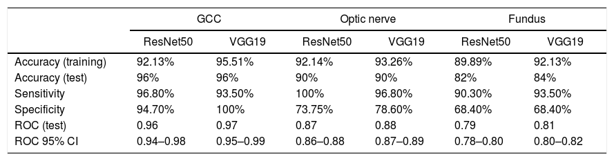
Results498 eyes of 298 patients were collected. Of the 498 eyes, 312 are glaucoma and 186 are normal. In the test, the precision of the models was 96% (ResNet50) and 96% (VGG19) for the GCL images, 90% (ResNet50) and 90% (VGG19) for the optic nerve images and 82% (ResNet50) and 84% (VGG19) for the fundus images. The ROC area in the test was 0.96 (ResNet50) and 0.97 (VGG19) for the GCL images, 0.87 (ResNet50) and 0.88 (VGG19) for the optic nerve images, and 0.79 (ResNet50) and 0.81 (VGG19) for the fundus images.
ConclusionsBoth deep learning models, applied to the GCL images, achieve high diagnostic precision, sensitivity and specificity in the diagnosis of glaucoma.
Determinar y comparar la precisión diagnóstica en glaucoma de dos modelos de aprendizaje profundo, usando imágenes en infrarrojo del nervio óptico, del fondo de ojo y de la capa de células ganglionares (CCG).
MétodosHemos seleccionado una muestra de pacientes normales y con glaucoma. Se recogieron tres imágenes en infrarrojo con un tomógrafo de coherencia óptica de tipo spectral-domain (SD-OCT). La primera corresponde a la imagen de barrido confocal del fondo de ojo, la segunda es un recorte de la primera centrada en el nervio óptico, y la tercera fue la imagen del corte SD-OCT de la CCG. Nuestros modelos de aprendizaje profundo se desarrollaron en la plataforma MATLAB con las redes neuronales preentrenadas ResNet50 y VGG19.
ResultadosSe recogieron 498 ojos de 298 pacientes. De los 498 ojos, 312 son glaucomatosos y 186 son normales. En la prueba, la precisión de los modelos fue de 96% (ResNet50) y 96% (VGG19) para las imágenes de la CCG, de 90% (ResNet50) y 90% (VGG19) para las imágenes del nervio óptico y de 82% (ResNet50) y 84% (VGG19) para las de fondo de ojo. El área ROC en la prueba fue de 0,96 (ResNet50) y 0,97 (VGG19) para las imágenes de la CCG, de 0,87 (ResNet50) y 0,88 (VGG19) para las imágenes del nervio óptico, y de 0,79 (ResNet50) y 0,81 (VGG19) para las imágenes de fondo de ojo.
ConclusionesLos dos modelos de aprendizaje profundo analizados, aplicados sobre las imágenes de la CCG, ofrecen una alta precisión diagnóstica, sensibilidad y especificidad en el diagnóstico de glaucoma.
