


Several factors must be taken into account when cementing an endodontic post reinforced with fiberglass, among them we can mention selection of the cementing agent. Market available cements differ with respect to application mode, working time, polymerization and chemical composition. It is therefore necessary to count with sufficient knowledge of all their characteristics and behavior, not only from the clinical approach, but also in the laboratory. The evolution of resin cements is nowadays geared to technique simplification so as to decrease time and margin of error during clinical process. Moreover, previous studies have demonstrated that these changes have decreased adhesion force to dentin.
ObjectiveThe purpose of the present study was to observe the behavior of two resin cementing agents, assessing their adhesion strength in intra-root dentin: the BisCem®, Bisco Inc. system, formed by a dual self-adhesive cement, and the ParaCore® Automix (Coltene/Whaledent) system which is a dual cement system requiring a chemical curing self-conditioning agent and dentin adhesives (ParaBond® Coltene Whaledent).
Material and methodsThirty six single rooted teeth we encapsulated in acrylic and worn down until reaching intra-root dentin. Following manufacturer's instructions, 18 samples were executed for each cement, and then in a universal testing device they were subjected to shearing tests (guillotine test) at a speed of 1mm per minute.
ResultsIt was observed that BisCem® exhibited lesser adhesion force than ParaCore® Automix. After statistically analyzing outcome by means of a «T» Student test, results revealed significant difference between both cements.
ConclusionParaCore® Automix, requiring previous dentin conditioning (ParaBond®), exhibited greater adhesion force.
Para cementar un endoposte reforzado con fibra de vidrio se deben de tomar en cuenta varios factores, entre ellos, la selección del agente cementante. Los cementos disponibles en el mercado difieren por la modalidad de aplicación, tiempo de trabajo, polimerización y composición química, por ello es necesario contar con el conocimiento de todas sus características y su comportamiento no sólo clínico sino también en el laboratorio. Hoy en día la evolución de los cementos de resina va encaminada a la simplificación de la técnica con el fin de reducir tiempo y margen de error durante el proceso clínico, sin embargo, previos estudios han demostrado que estos cambios han reducido la fuerza de adhesión a la dentina.
ObjetivoEl propósito de este estudio es observar el comportamiento de dos agentes cementantes de resina, evaluando su fuerza de adhesión en dentina intrarradicular, el sistema BisCem® de Bisco Inc., el cual es un cemento autoadhesivo dual y el sistema ParaCore® Automix de Colténe/Whaledent; cemento dual que requiere de un agente autoacondicionador y un adhesivo dentinarios de curado químico (ParaBond® de Colténe Whaledent).
Material y métodosSe encapsularon 36 dientes unirradiculares en acrílico y se desgastaron hasta descubrir la dentina intrarradicular, siguiendo las especificaciones del fabricante, se realizaron 18 muestras para cada cemento y después se sometieron a pruebas de cizalla a una velocidad de 1mm por minuto en una máquina de ensayo universal.
ResultadosSe observó que BisCem® presenta una menor fuerza de adhesión que ParaCore® Automix. Después de analizar estadísticamente los resultados a través de la prueba «T» de Student, los resultados mostrando una diferencia significativa entre ambos cementos.
ConclusiónParaCore® Automix que requiere previo acondicionado de dentina (ParaBond®) presentan una mayor fuerza de adhesión.
Presently, use of fiberglass endodontic posts is a common part of the daily routine of a dental practice. Posts made of composite resin reinforced with fiberglass must be cemented with resin cements in order to form a functional monoblock, since both possess an elastic model which is similar to that of dentin, generating lesser stress and risk of root fracture as well as protecting tooth remnants and restoration.
Success of any restoration largely depends on the cementing agent, which is defined as the means of fixation of two solid surfaces; according to Machi R., cement is a liquid which flows, humidifies surfaces, penetrates into its irregularities and fills spaces between both surfaces, to later harden ensuring contact between those surfaces.1
Constant research has favored the development of new cements offering greater adhesion force combined with a simple and effective placement technique. These cements are biocompatible, insoluble to the oral environment, esthetic and possess mechanical properties which outperform the rest of the cements.1
Resin cements possess a composition similar to that of resin used as reconstruction material. Nevertheless, they contain lesser amounts of inorganic filling material so as to render it more fluid. In general, they are composed of an inorganic matrix, monomers, diluting agents and inorganic filling material composed of silanized micro-filling (silica or zirconium).2
Conventional resin cements require previous application of an adhesive system able to penetrate into the dentin to polymerize within it; adhesion can be understood as the state in which both surfaces are kept together by means of interfacial forces or energies, based on chemical and mechanical mechanisms, or both, with the mediation of an adhesive agent (ISO/TR 11405;1994 [E]).3
A suitable adhesive agent must be able to humidify or impregnate the surface, possess low surface tension so as to be able to flow into the irregularities of the solid matter, as well as be able to change from liquid phase to solid without experiencing major dimensional changes.4
This concept, applied to dentistry since 1955 by Michael Buoncore, presently refers to a process of resinous monomers’ demineralization and infiltration, with the aim of creating a mechanical lock between adhesive and tooth structure, seal dentine tubules and thus recover and preserve homeostasis of the internal milieu of the dentin-pulp complex.5
Evolution of adhesive systems requires dentin conditioning prior to their placement. This dentin conditioning implies all chemical alterations of the dentin surface by means of acid or chelating agents with the purpose of removing or modifying the structure of dentin debris and simultaneously de-mineralizing dentin surface.6,7
Development of adhesives is geared to technique simplification, nevertheless, all of them contain conditioning agents in varied amounts, a primer and an adhesive,8 therefore, four categories can be established:
- 1.
Conditioner and rinse, primer and adhesive (3 steps).8
The primer counts with an hydrophilic monomer which bonds with collagen fibers and polymerizes, forming a hybrid or inter-diffusion layer9,10 as well as adhesive resin which, when co-polymerizing with the previous component, forms resin prolongations and anastomosis (Tags and micro-Tags).11,12
- 2.
Conditioning and rinsing, primer-adhesive (2 steps).
- 3.
Self-conditioning primer and adhesive (2 steps).
This technique does not require use of acids. Dentin conditioning is achieved by incorporating an acid resin into the primer which, when applied to the dental sub-stratum, modifies dentin debris and creates a small demineralization front; after acting for a few seconds, acid radicals neutralize with the de-mineralized hydroxyapatite crystals, resulting in a de-mineralized and infiltrated tissue to which liquid resin is later applied.13
- 4.
Conditioner-primer-adhesive (1 single step).
This is a combination of one solution conducted in a single step from a single container.
During the last decade self-adhesive cements have been introduced to clinical dental practice. They are portrayed as an ideal alternative since they exhibit in a single product all advantages of conventional cements, the ability of self-adhesion, fluoride release like glass ionomers as well as mechanical properties of dimensional stability and micro-mechanic retention provided by resinous cements.1
Application technique is one of the main reasons for using this type of cements, where application is solved in one single clinical step; after mixing base paste and catalyst paste, or activating capsules, it is directly applied into the surfaces to be adhered, therefore, manipulation errors are curtailed.14
Even though dentin morphology, especially intraradicular dentin, bears influence on adhesion force, it is of the utmost importance to achieve excellent tissue preparation and material handling when preparing the space which will lodge the post and its cementing material.
During cementation process the following factors are of the foremost importance: removal of dentin debris composed of materials resulting from the de-obturation process, dentin, plasticisized guttapercha caused by drill frictions, sealing elements, etc,15 in addition to irrigation materials and cements like eugenol16,17 which are used during root canal treatments;18 the time elapsed from the moment after endodontic treatment and post manufacturing, access difficulty to the canal in order to achieve adequate adhesive and cementing agent application without causing bubbles, without forgetting that single step adhesives exhibit chemical incompatibility when used in combination with dual resin cements or chemical polymerized cements.19
MATERIAL AND METHODSFor the present study, 36 single rooted human teeth were used. Teeth were free of caries and had been extracted due to orthodontic reasons or periodontal problems. They were cleansed and kept hydrated in water for 7 days; after this time the crown portion was cut with a low speed diamond disc (Brasseler®). Root portions were then randomly encapsulated in two different colors of acrylic (Nic Tone®) (Figure 1).

Specimens were worn down in the metallographic polisher with number 600 emery until the point when dentin was uncovered and a smooth surface was obtained. One of the groups was selected, and with a teflon shaper and a press, BisCem® Bisco Inc. cement samples were manufactured according to manufacturer's instructions (Figure 2).

The other group of specimens had previously been treated with Parabond® (Coltene, Whaledent) according to manufacturer's instructions. ParaCore® Automix, (Coltene/Whaledent) samples were built upon them (Figure 3).

Both materials were photo-polymerized with a Bluephase® light-curing light (Ivoclar Vivadent). This lamp possesses 830 millivolts power according to the Bluephase® Ivoclar Vivadent radiometer. Both groups were subjected to light exposure for 30seconds according to manufacturer's instructions. Specimens were stored in 100% humidity for 24hours in a chamber at 37°C. Each sample was measured with a digital vernier, from north to south and from east to west; both measurements were added up and divided into two to obtain diameter. With this measurement in hand, the formula Pi x r2 was applied to thus obtain the area of all the samples. Mechanical guillotine test at a 1mm per minute speed to observe adhesion strength was used in an universal testing machine Instron® 5567 USA (Figure 4).
 test in Instron® 5567 USA universal testing machine.")
Once the results were obtained, they were subjected to the t student test for independent testing, using IBM SPSS 18.0 software package, with 5% statistical significance level in all tests.
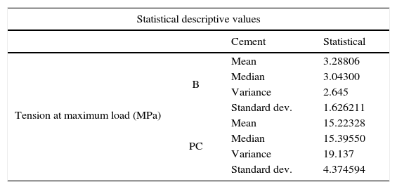
RESULTSAccording to central tendency measurements, ParaCore® required greater load tension for sample displacement (Table I).
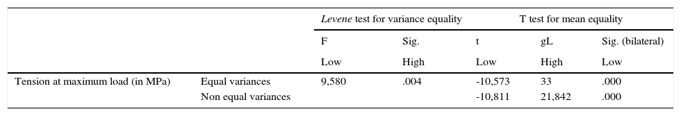
Using Levene's F test the hypothesis of significant differences among variants (p = 0.004 < 0.05) was verified, and the hypothesis of data normality was verified through the Kolmogorov-Smirnov test with 0.200 values for both groups (Figure 5).

According to t student test (bilateral sig = 0.000 < 0.05) it is possible to refute the null hypothesis and conclude that within the mentioned confidence level, when comparing BisCem® and ParaCore® there is significant difference in the resistance to displacement in intra-radicular dentin (Table II).
DISCUSSIONThe present research shows differences in resistance to displacement of two cementation systems. Important differences were observed in the adhesion forces of both cements at the time of applying shearing force at a 1mm per minute speed.
Previous research endeavors revealed that cements which had undergone previous dentin treatment using self-conditioning adhesives presented thin demineralization areas, but did not exhibit penetration, according to microscopic evidence observed by Al-Assaf et al,20 Yang et al21 and Monticelli et al.22 It has been observed that resins of self-conditioning adhesives exhibit lower acidity than 37% phosphoric acid, and do not achieve such a deep conditioning either in enamel or dentin according to Tay and Pashley;23,24 nevertheless, Pashley and Carvalho25 showed that creation of larger or lesser inter-diffusion does not bear influence on the final result, a minimum existing interaction with the substrate is enough. Moreover, research conducted by Hanning et al,26 Hayakawa et al,27 Santini et al28 have revealed that bonding strength and penetration of these adhesives exhibited similar values to those of conventional adhesives.
Beher et al29 observed that self-adhesive cements presented an adhesion degree comparable to conventional cementing agents such as silicate cement and zinc phosphate. There is no evidence of de-mineralization or penetration into the dentin;20–22 this might be due to the high viscosity of these cements and the rapid de-activation of acid monomers caused by the acid-base reaction (Munck et al).30 The present research purports to show the differences of resistance to displacement of two cementing agents.
CONCLUSIONSIn the present work, the cement requiring previous dentin conditioning (ParaCore® Automix, Coltene Whaledent with ParaBond® Coltene Whaledent) exhibited greater adhesion when compared to self-adhesive cement (BisCem®, Bisco Inc.).
It is important to consider that technique simplifications might cause adhesion strength detriment, compromising thus formation of post-resindentin monoblock.
Most of these studies were performed in vitro, it would be advisable to further the aforementioned with long term in vivo studies.
Graduate, Fixed Oral Prosthesis Specialty.
This article can be read in its full version in the following page: http://www.medigraphic.com/facultadodontologiaunam