


Odontomas are the most prevalent benign tumors in the mouth, they are normally diagnosed after routine X-ray examinations. Between 2010-2015, a total of 1,261 oral surgical interventions were practiced at the School of Dentistry, University of Cartagena. In them, 12 cases of odontoma were identified after reviewing respective clinical histories. In some cases, when odontomas are accompanied by other characteristics, they can be associated to syndromes, such as Gardner's syndrome. The present report informs of a case with certain characteristics such as possible presence of colorectal polyposis, supernumerary teeth and bone excrescences present in a 12 year old male, therefore, studies were undertaken to determine association to the aforementioned syndrome. Location of odontomas was an inherent characteristic and found to be altered in the previously mentioned case.
Los odontomas son los tumores benignos más prevalentes en la cavidad oral, los cuales, por lo general, son diagnosticados mediante exámenes radiográficos de rutina. En la Facultad de Odontología de la Universidad de Cartagena se realizaron un total de 1,261 cirugías orales entre los años 2010 y 2015, donde se presentaron 12 casos con diagnóstico de odontomas, los cuales fueron identificados al inspeccionar sus respectivas historias clínicas. En algunos casos, los odontomas acompañados de otras características se asocian a síndromes, tales como el síndrome de Gardner, este reporte menciona un caso con ciertas características, como la posible presencia de poliposis colorrectal, órganos dentales supernumerarios y excrecencias óseas presentes en un niño de 12 años de edad, por lo cual se realizaron estudios para determinar la asociación a mencionado síndrome. La localización de los odontomas es una característica particular de ellos, que se encuentra alterada en el caso anteriormente resaltado.
Odontomas are benign, non-aggressive tumors, composed of enamel, dentin and pulp tissue.1 They are the product of growth of differentiated mesenchymal and epithelial cells, where ameloblasts and odontoblasts form enamel and dentin and are anomalously or defectively deposited.2,3 During the first decades of last century, the term odontoma meant not only odontogenic tumors, but rather included odontogenic and non odontegenic cysts as well as varied ossifying fibroid lesions of the jaws. Odontoma etiology is unknown, in it, several factors are implicated such as trauma, infections, genetic mutations, odontoblastic hyperactivity or alterations of the dental development control gene. Most of these tumors are discovered during the patient’ s second to third decade of life, and do not exhibit clear gender predilection.4–7
Odontomas are classified into composite (all tooth tissues are found, with a distribution pattern sorted out in multiple structures called denticles; mainly located in the anterior section or the upper jaw) and complex (dental structures with a disordered distribution pattern, no individualized denticles are observed since they are fused, mainly located in the posterior region of the lower jaw).8,9 This condition can be visualized as an amorphous, mineralized, asymptomatic mass, X-rays reveal a radio-opaque mass, surrounded by a radio-lucid halo with regular borders.7 In some cases, odontomas are associated to certain syndromes, such as Gardner's syndrome.9,10
Odontomas are considered hamartomas rather than true neoplasia, due to the fact that they originate from odontogenic epithelium in the oral cavity. Odontomas have been widely reported in literature as being the odontogenic tumor most frequently found in the jaws,6 nevertheless, there are very few reports on complex odontomas in the anterior region of the upper jaw, as is the case of the clinical case here presented.
The main purpose of this series of cases is to analyze the apparition frequency of odontomas, their location, prevalence according to gender and age of diagnosis in patients seeking treatment at the School of Dentistry of the University of Cartagena in the last five years, as well as reporting a case of complex odontoma in the anterior region of the upper jaw.
RESULTSAll clinical cases involving surgical intervention and treated during 2010-2015 at the School of Dentistry of the University of Cartagena were reviewed. Total number of cases was 1,261 out of which 12 had been diagnosed as odontoma; assessment was conducted on prevalence according to type of odontoma, location, gender and age of patient.
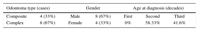
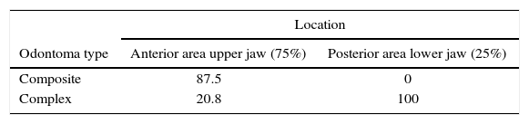
Out of 12 total cases, 4 were found in females (33%) and 8 (67%) in males. Diagnosis age for odontomas was found in the second (58.55%) and third (41.67%) decades of life (Table I). Anatomical- pathological-clinical study confirmed presence of 8 complex odontomas (66.66%) and 4 composite (33.33%) odontomas; 75% of all odontomas were located in the anterior area of the upper jaw (in 87.5% diagnosis was composite odontoma and 20.8% was complex odontoma) 25% were located at the posterior areas of the lower jaw, all of them were diagnosed as complex odontoma (Table II).
A 12 year old male patient sought treatment at the School of Dentistry of the University of Cartagena, due to the absence of upper incisors. Personal pathological history information was non-contributory. When the patient was seven years old, he attended stomatological consultation, he was referred to orthopedics treatment to receive bi-maxillary expansion by reason of exhibiting transverse micrognathism and dental crowding in the lower jaw (present up to the present age) at that time, no condition was associated to the delay in dental eruption.
In order to assess the patient, panoramic X-rays were taken as well as an imaging study by means of computerized axial tomography. Intraoral examination revealed healthy mucosae and expansion of the cortical bone in the anterior section of the upper jaw, ankylosis, presence of palatal tori and mandibular bilateral tori (Figure 1). General physical examination revealed bilateral bone excrescences in the upper section of the shoulder blades. At a later date a colonoscopy was conducted in order to assess presence of intestinal polyps as well as association to Gardner's syndrome (which proved to be negative).

From the radiographic perspective, two radio-opaque areas surrounding the crowns were observed at teeth 11, 12, 21, 22. At both lower premolar areas, a radiolucid halo was observed (Figure 2). Presumptive diagnosis was proposed to which effect the following was effected: local anesthesia with 2% lidocaine carpulae (1:80,000) as well as surgical approach with number 3 Bard-Parker scalpel, total enucleation of tumor lesions with gubia type pliers, straight apical elevators, extraction of supernumerary teeth with angulated elevators, synthesis with non-resorbable suture and needle. Anti-inflammatory medication was prescribed for the postoperative period and the patient was remitted to the Orthodontics and Maxillary Orthopedics section. The patient's mother received recommendations. After the procedure, previous diagnosis was replaced for one of complex odontoma due to confirmation provided by morphological characteristics and later histological examination, in addition to supernumerary teeth confirmation (Figures 3 and 4). Microscopically, the lesion was composed of a disorganized mixture of dental tissues, among which dentin, enamel, pulp tissue and cement could be found, as well as some foci of odontoblastic cells. No malignancy characteristics were found in analyzed samples (odontomas).
DISCUSSION


Odontomas are benign tumors frequently found in the mouth. In most cases, they do not exhibit symptomatology, the most common sign is delay in the eruption of permanent dentition,11 they are normally diagnosed through routine radiographic examination. Panoramic and periapical X-rays are diagnostic aids essential in first instances.12
Several authors refer that in the search of odontomas, age is a non -determinant factor, nevertheless, there are reports in scientific literature pointing out that the most common age for odontoma diagnosis is the second decade of life,5 concurring thus with the present study.
Hissatomi et al, in 2002, analyzed 107 odontoma cases; their results revealed greater incidence in females,10 which differs with results obtained in the present series of cases, which revealed greater percentages of the condition in males.
Literature reviews and epidemiological studies indicate that the preferred site for composite odontomas location is the anterior-superior (upperanterior) section of the upper jaw; for complex odontomas, preferred location is the posterior region of the lower jaw. Nevertheless, in the clinical case presented in this study, location of complex odontoma was in the anterior-superior region of the upper jaw.
CONCLUSIONIn the present study we described an unusual case of complex odontoma location at the upper jaw's anterior-superior region. This odontoma was preventing eruption of permanent teeth in this area. Additionally, supernumerary teeth were located in the area of lower premolars, and bilateral bone excrescences in the region located above the shoulders; these clinical signs could be related to Gardner's syndrome; nevertheless, this hypothesis was discarded after conducting an endoscopy procedure and observing absence of intestinal polyps. It is of the utmost importance to conduct suitable stomatological and radiographic examinations in order to observe oral clinical findings and abstain from isolating them from other types of lesions that might be perceived during the course of a general physical examination, this is of the utmost importance so as not to omit presence of other syndromes. Moreover, it is important to offer the best possible multi-disciplinary handling, bearing in mind at all timesg anomalies that might be generated by odontomas.
It is relevant to mention the low index of odontoma appearance during the years 2010-2015; 1,261 oral surgical procedures were performed during this period at the University of Cartagena, out of those, only 12 cases were odontomas.