


When compared to general population, subjects afflicted with cleft lip and palate present alterations in craniofacial growth and development as well as high incidence of dental anomalies which vary according to studied population; agenesis, presence of supernumerary teeth, abnormal crown morphology and taurodontism can be counted amongst them.
ObjectiveTo assess prevalence of dental anomalies found in Colombian children with non syndromic cleft lip and palate sequels, being treated at health providing institutions.
MethodologyA cross-sectioned descriptive, observational study was conducted on a sample of 258 medical histories and panoramic X-rays of Colombian children treated at different health providing institutions in the city of Bogota, Colombia. The sample was composed of 156/258 males (60.55%) and 102/258 (39.5%) females. Average age was 9.8 years (± 3.3 years).
ResultsBased on studied X-rays, it was determined that 38.4% (99/258) children presented full left unilateral cleft lip and palate sequels, 31.0% (80/258) exhibited bilateral cleft and 30.6% (79/258) suffered right unilateral cleft lip and palate. Most frequent dental anomalies found were; dental agenesis, supernumerary teeth and size anomalies. Prevalence for said anomalies were: dental agenesis, over 90%; supernumerary teeth: 40% and size anomalies: 30%.
ConclusionHigh prevalence of dental anomalies was found in children with cleft lip and palate in Bogota in concordance with information reported in scientific literature.
Comparados con la población general, los sujetos con labio y paladar hendido presentan alteraciones en su crecimiento y desarrollo craneofacial y una alta prevalencia de anomalías dentales, que varía según la población estudiada, entre ellas: agenesias, presencia de dientes supernumerarios, morfología coronal anormal y taurodontismo.
ObjetivoEvaluar la prevalencia de anomalías dentales encontradas en niños colombianos con secuelas de hendiduras labio palatinas no sindrómicas, atendidos en instituciones prestadoras de salud.
MetodologíaSe realizó un estudio observacional descriptivo transversal en una muestra de 258 historias clínicas y radiografías panorámicas de niños colombianos de diferentes instituciones prestadoras de salud de la ciudad de Bogotá-Colombia. De los cuales 60.5% (156/258) eran hombres y 39.5% (102/258) mujeres. El promedio de edad fue 9.8 (± 3.3) años.
ResultadosDe las radiografías evaluadas se determinó que 38.4% (99/258) de los niños presentaban secuelas de labio y paladar hendido unilateral izquierdo completo, 31.0% (80/258) bilateral y 30.6% (79/258) con unilateral derecho. Las principales anomalías dentales encontradas fueron: agenesias dentales, dientes supernumerarios, anomalías de tamaño. La prevalencia encontrada para cada una de ellas fue: agenesias dentales: mayor del 90%. Dientes supernumerarios: 40% y en anomalías de tamaño estuvo alrededor del 30%.
ConclusiónSe encontraron altas prevalencias en anomalías dentales en los niños con labio y paladar hendido en Bogotá, similar a lo reportado en la literatura científica.
Cleft lip and palate cases (CLP) are the most common craniofacial malformations; they constitute congenital structural deficiencies caused by defects in the fusion of cranio-facial processes which form primary and secondary palate.1,2 They possess multifactorial etiology and varied frequency, according to environmental and socio-cultural factors. Thus, prevalence has been reported as 1 out of 800 live births in South America, 1,8 in 1,000 live births in Europe, 1 in 750 in Asia and 1 in 1200 in Africa. In Colombia, according to the III National Study in Oral Health (1988) ENSAB III, prevalence close to 0.2% has been reported, (13-17). Reports in ENSAB IV for 2014 revealed 0.07%, although margins of error were high due to the sample's characteristics.3–6
CLP sequels generate esthetic, psychological and functional alterations. Esthetic alterations are mostly related to lack of continuity in the upper lip as well as scars from surgical interventions. Psychological alterations are caused by disorders in the feelings of subjects with CLP sequels as well as frame of mind disturbances and lack ability to relate to other people. Lastly, functional alterations are within the framework of phonation, deglutition and mastication disorders.
When compared to general population, CLP subjects exhibit alterations in craniofacial development and growth1,2,7 as well as high prevalence of dental anomalies (agenesis, presence of supernumerary teeth, abnormal crown morphology and taurodontism,5,8 which varies according to studied population. In general, in this type of population a 90% increase of dental anomalies is observed when compared to non-affected subjects.9
Within reported frequencies of dental anomalies for this population, we can find the following; microdontia: 37%, dental agenesis: over 20%.8,10,11 Küchler12 reported a 15.2% taurodontism frequency. Likewise, higher presence of supernumerary teeth has been reported for CLP population when compared to general population.
Within this context, the target of the present research project was to assess prevalence of dental anomalies found in Bogota in children with sequels of non syndromic palate and lip clefts, who were treated in Health Care providing institutions in that city.
MATERIAL AND METHODSA cross-sectioned, descriptive and observational study was conducted in children with sequels of cleft lip and palate treated in public and private health care institutions. Children's ages ranged from 5-15 years, children had not received definitive oral rehabilitation or restorative care, and were equally lacking pre-existent orthodontics and/ or maxillary orthopedic treatment. Sample size was 258 children and was calculated by way of approximation to expected frequencies (CLP) prevalence and dental anomalies.
All patients attending dental services in selected institutions and within aforementioned age ranges were accepted to form the sample. HPI statistical records were assessed in order to obtain the names of all these patients which met inclusion criteria; they had at least been subjected to first care clinical examination at the dental service; panoramic x-rays were examined at the time of attending the service as well as patients’ full medical history taken at each institution
In the study, independent variables were: sociodemographic characteristics of the child and type of cleft, result variables were dental characteristics of number, size and root development.
Based on state-of-the art circumstances, a data collecting instrument was designed in the study. Before application, the instrument was subjected to a pilot test; according to test results, adjustments were undertaken until achieving the final instrument.
X-rays were selected according to criteria of suitable sharpness, density and contrast. X-rays should lack stains, double images, marks or scratches. X-rays which were deteriorated to the point of preventing analysis of number and shape of teeth to be assessed were discarded.
Dental development was assessed according to Nolla methodology,13 referenced by Infante.14 Congenital tooth absence and supernumerary teeth presence were determined according to Na-Youngkim15 study, which examined congenital absence of upper lateral incisors or presence of supernumerary teeth.
In 27 patients the size of teeth adjacent to the cleft was assessed. This procedure was conducted in two ways: one with study models, assessing integrity of each model; considering as integrity the fact that represented dental tissues did not exhibit fissures or lack of continuity. A fine-point digital gauge with 0.01mm precision was used. This gauge was employed to measure the greatest mesio-distal and buccal-palatal dimension of each tooth in the arch, especially teeth which were adjacent to the cleft.
In order to collect data for the study, researchers were trained and qualified, radiographic and clinical criteria tests were undertaken, an intra-class correlation coefficient was obtained, with kappa superior to 0.8 (it was conducted with an N which corresponded to 10% of the total sample (± 25 subjects) where X-rays of subjects which were different to the final sample were assessed.
Research teams recorded information of gathering instruments. During information collection typing, quality control was assessed by means of filters, checkers and program checking lists. Gathered information was included in a database designed in Access format. It was then exported to Stata version 11.0 for analysis and procedure. Before analysis, data were checked and cleansed. Whenever inconsistencies were found the instrument was selected and compared with the appropriate diagnostic aid in order to minimize errors.
With respect to information analysis, an exploratory analysis was initially conducted, using descriptive statistics techniques, in order to determine variable distribution; a bivariate analysis was conducted using contingency tables, square chi (χ2) and Fisher tests according to the case and distribution in number and normalcy. Paired t test was conducted for root development.
Statistical significance lesser than 0.05 had to be presented in the evaluation bivariate analysis.
During development of the present study ethical considerations of the Helsinki Declaration as well as Resolution Number 00084301616 of the Colombian Ministry of Health were observed. This research project was considered risk-free and received the endorsement of the Ethics Committee of the School of Dentistry, National University of Colombia.
RESULTSInformation was gathered on 258 children with cleft lip and palate sequels; 60% were male (156/258) and 39.5% were female (102/256). Average age was 9.8 years (± 3.3), mean was 10, minimum age 5 and maximum age 15 years. Over 70% of patients counted with some sort of affiliation to some type of Social Security Regime (30.2% contributory, 44.6% subsidized) 8.5% attended the service as private patients; the rest did not inform of any affiliation (16.7%). With respect to origin it can be said that 96.5% (249) of subjects participating in the study were from the Capital and 9 (3.5%) came from another Colombian city.
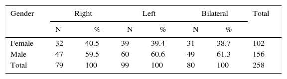
In assessed children, 38.4% (99/258) exhibited full left unilateral CLP sequels, 31.0% (80/258) presented bilateral CLP and 30.6% (79/258) full right unilateral CLP. With respect to distribution amongst gender and CLP type, no significant differences were observed between both genders (p=0.9) males exhibited greater numbers. Left Unilateral CLP was the most frequently found circumstance in both male and female patients (Table I).
Dental agenesis: Prevalence of agenesis in studied population was 93.0% (240/258) regardless of missing tooth, with an average of 1.8 (± 1.0) missing teeth. With respect to agenesis and gender distribution, greater number of dental agenesis in males was observed (58.7%) compared with females (44.2%) with significant differences (p=0.04)
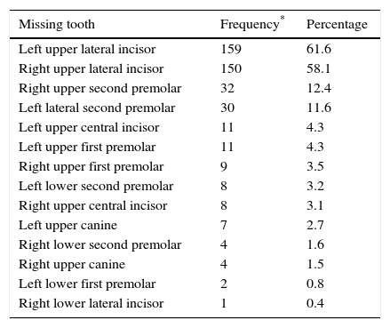
Left upper lateral incisors were the most frequently absent teeth (61.6%) followed by their contra-lateral counterparts (58.1%) (Table II). The most frequent combination of missing teeth were those found between lateral and premolar in the affected site (8.5%) followed by central and premolar. (7.7%). No significant differences were found with respect to distribution of agenesis and type of CLP (p=0.23 square chi (χ2), Fisher)
Distribution of dental agenesis according to tooth type.
| Missing tooth | Frequency* | Percentage |
|---|---|---|
| Left upper lateral incisor | 159 | 61.6 |
| Right upper lateral incisor | 150 | 58.1 |
| Right upper second premolar | 32 | 12.4 |
| Left lateral second premolar | 30 | 11.6 |
| Left upper central incisor | 11 | 4.3 |
| Left upper first premolar | 11 | 4.3 |
| Right upper first premolar | 9 | 3.5 |
| Left lower second premolar | 8 | 3.2 |
| Right upper central incisor | 8 | 3.1 |
| Left upper canine | 7 | 2.7 |
| Right lower second premolar | 4 | 1.6 |
| Right upper canine | 4 | 1.5 |
| Left lower first premolar | 2 | 0.8 |
| Right lower lateral incisor | 1 | 0.4 |
With respect to dental agenesis type and their relationship with CLP types, significant differences were found for right unilateral CLP at the right lateral incisor (p=0.00); with respect to left unilateral CLP differences were found for left lateral incisor (p=0.00), left central incisor (p=0.02), left upper canine (p=0.05) and left first premolar (p=0.05).
Supernumerary teeth: Prevalence of supernumerary teeth in studied population was 42.6% with an average of 1.4 teeth (± 0.7) Most children presented only one supernumerary tooth, whereas 13.2% exhibited more than one, there even was a patient with six supernumerary teeth. No significant differences were found with respect to gender and supernumerary teeth (p=0.36).
Significant differences were found with respect to CLP type and amount of supernumerary teeth (p=0.02); the greatest number of supernumerary teeth were found in left unilateral CLP. With respect to supernumerary tooth type, significant differences (p < 0.05) were found only in upper lateral supernumerary teeth.
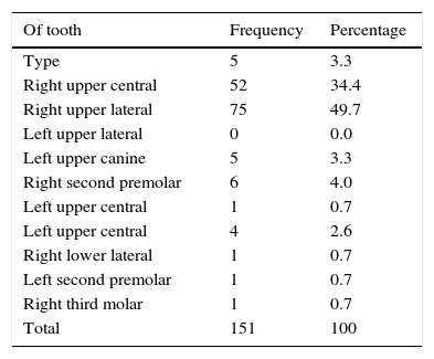
Upper lateral central Incisors were the most frequent supernumerary teeth found (its presence was simultaneously found with the normal-nomenclature tooth) (Table III). It is important to note that in the present case, the object of observation were teeth of CLP subjects.
Frequency of supernumeraries according to tooth type.*
| Of tooth | Frequency | Percentage |
|---|---|---|
| Type | 5 | 3.3 |
| Right upper central | 52 | 34.4 |
| Right upper lateral | 75 | 49.7 |
| Left upper lateral | 0 | 0.0 |
| Left upper canine | 5 | 3.3 |
| Right second premolar | 6 | 4.0 |
| Left upper central | 1 | 0.7 |
| Left upper central | 4 | 2.6 |
| Right lower lateral | 1 | 0.7 |
| Left second premolar | 1 | 0.7 |
| Right third molar | 1 | 0.7 |
| Total | 151 | 100 |
Tooth size anomalies: Anomalies which were clearly different from normal tooth size (not associated to radiographic distorsion) were radiographically assessed and determined. Each tooth was compared with its contra-lateral opponent, Thus, microdontia and macrodontia were observed in 75 subjects (29.1%).
Proportion of dental size anomalies in studied subjects were 4% macrodontia and 96% microdontia. Microdontia cases were mainly found in the left upper lateral incisor adjacent to the cleft (supernumerary teeth smaller than expected size were excluded from the analysis).
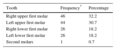
A sub-sample of 27 study models of this population was equally taken. Upper incisor size was measured with a digital gauge, it was observed that 82.5% exhibited tooth-size differences. Of these, 69% presented a difference lesser than 0.6mm, the rest of the percentage exhibited differences greater than 0.6mm taurodontism was included within the categories of size anomalies; it was observed in 19.4% (50) of cases, its prevalence was higher in upper permanent molars (Table IV).
Taurodontism distribution according to tooth presenting it.
| Tooth | Frequency* | Percentage |
|---|---|---|
| Right upper first molar | 46 | 32.2 |
| Left upper first molar | 44 | 30.7 |
| Right lower first molar | 26 | 18.2 |
| Left lower first molar | 26 | 18.2 |
| Second molars | 1 | 0.7 |
No significant differences were found (p=0.37) with respect to CLP type and dental size anomalies (microdontia, macrodontais and taurodontism) (Figure 1). Differences according to gender did not exhibit statistical significance (p=022).

Anomalies in eruption sequence: Anomalies in eruption sequence were observed in 12% (31/258) of all patients. No statistical differences were found according to gender (0.32), but they were encountered with respect to CLP type (p=0.02). Greater frequency of sequence anomalies were found in children with left unilateral CLP sequels (58.1%) especially in upper canines.
With respect to Nolla stage, it was observed that there were canine-to-canine differences among homologous teeth, but no significant differences were found. In canines, differences of up to two Nolla root development stages were found, but this did not exhibit statistical significance.
Position anomalies: Presence of position anomaly was observed in 22.1% cases (57/258) of studied population; rotations were the most common position anomalies (72%) followed by mesial and distal versions (17%) and (10.5%).
Transposition anomaly was observed in 4.3% of children; the most frequent (2.7%) was seen in cases when the upper canine assumed the position of the upper first premolar. Tooth impaction was observed in 3.8% of cases (10/258); the most frequent impaction was observed at the left upper first molar (1.2%). Likewise, impaction of upper and lower molars was observed, but in percentages lower than 1%.
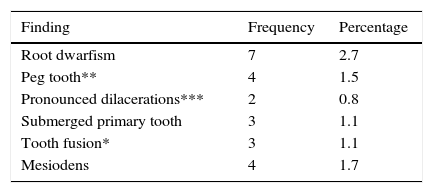
Other findings: In this group of studied children presence of peg teeth was detected (especially lateral incisors), as shown in their clinical history. Likewise, radiographic findings also exhibited marked root laceration and root dwarfism among others (Table V).
Distinct tooth characteristics in cleft lip and palate patients.
| Finding | Frequency | Percentage |
|---|---|---|
| Root dwarfism | 7 | 2.7 |
| Peg tooth** | 4 | 1.5 |
| Pronounced dilacerations*** | 2 | 0.8 |
| Submerged primary tooth | 3 | 1.1 |
| Tooth fusion* | 3 | 1.1 |
| Mesiodens | 4 | 1.7 |
* Fusion took place between lower lateral incisors and lower incisors of the same dental quadrant. ** Dilacerations were observed in upper lateral incisors as well as *** peg-shaped crown.
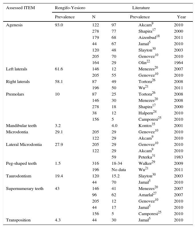
High values of dental agenesis were found (93.0%). These values were similar to those reported by Akcam9 in 2010, who indicated 97.1% frequencies at that time. Values of the present study were above those reported by Shapira17 in 2000 (77%), Aizenbud18 in 2011 (68%), Jamal5 in 2010 (67%) and Slayton30 in 2003 (47.5%). Table VI depicts a comparison of our findings with other findings reported in literature.
Comparison of present study's findings with other findings in literature.
| Assessed ITEM | Rengifo-Yesioro | Literature | |||
|---|---|---|---|---|---|
| Prevalence | N | Prevalence | Year | ||
| Agenesis | 93.0 | 122 | 97 | Akcam9 | 2010 |
| 278 | 77 | Shapira17 | 2000 | ||
| 179 | 68 | Aizenbud18 | 2011 | ||
| 44 | 67 | Jamal5 | 2010 | ||
| 120 | 48 | Slayton30 | 2003 | ||
| 205 | 70 | Genovez10 | 2010 | ||
| 164 | 29 | Olin22 | 1964 | ||
| Left laterals | 61.6 | 146 | 12 | Menezes20 | 2007 |
| 205 | 55 | Genovez10 | 2010 | ||
| Right laterals | 58.1 | 87 | 49 | Tortora26 | 2008 |
| 196 | 50 | Wu21 | 2011 | ||
| Premolars | 10 | 87 | 25 | Tortora26 | 2008 |
| 146 | 30 | Menezes20 | 2008 | ||
| 278 | 18 | Shapira17 | 2000 | ||
| 38 | 12 | Halpern24 | 2010 | ||
| 156 | 5 | Camporesi25 | 2010 | ||
| Mandibular teeth | 3.2 | 4.0 | Kontos35 | 2001 | |
| Microdontia | 29.1 | 205 | 29 | Genovez10 | 2010 |
| 122 | 29 | Akcam9 | 2010 | ||
| Lateral Microdontia | 27.9 | 205 | 29 | Genovez10 | 2010 |
| 122 | 29 | Akcam9 | 2010 | ||
| 59 | Peterka31 | 1983 | |||
| Peg-shaped teeth | 1.5 | 316 | 18-34 | Walker29 | 2009 |
| 196 | No data | Wu21 | 2011 | ||
| Taurodontism | 19.4 | 120 | 15.2 | Slayton30 | 2003 |
| 44 | 70 | Jamal5 | 2010 | ||
| Supernumerary teeth | 43 | 146 | 41 | Menezes20 | 2007 |
| 96 | 62 | Amarlal27 | 2007 | ||
| 205 | 12 | Genovez10 | 2010 | ||
| 44 | 17 | Jamal5 | 2010 | ||
| 156 | 5 | Camporesi25 | 2010 | ||
| Transposition | 4.3 | 44 | 30 | Jamal5 | 2010 |
Lateral incisors were the most frequent absent teeth; this was similar to Baek's19 reports in Korea, Menezes, 20 Genovez 10 in Brazil and Jamal.5 Percentages of dental agenesis (over 50%) of lateral incisors were similar to those reported by Wu21 in 2011.
In scientific literature, different authors18,22 mention that the upper second premolar (USP) was the most commonly found absent tooth, with percentages above 10%; these percentages were similar to those reported by Shapira23 in 1999, Halpern24 in 2010 and Camporesi25 in 2010. Frequencies found in the present study were well below the percentage reported by Menezes,20 who indicated a percentage above 30%, and by Tortora26 (25%). With respect to central incisor absence, results of our study concurred with those found in literature:21 they were close to 3%.
With respect to mandibular teeth agenesis, findings of the present study were similar to those reported in 2010 with percentages close to 4% for premolars, but they were below reported frequency for incisors (0.4% in this study, whereas literature reports frequencies above 2%).
With respect to supernumerary teeth, in the present study, prevalence found was in the 43% range: they were similar to those reported by Menezes20 for Brazil and Amarlal27 in India; they differ from reports by Genovez10 who indicated 12% prevalence of supernumerary teeth, Jamal5 reported 17% frequency and Camporesi and Baccetti25 reported 5.2% frequency in 2010. Ramos28 found significant differences with respect to supernumerary teeth and gender; he established greater number in males than in females, this fact was not observed in the present study.
Literature21 reports frequency of microdontia-type dental anomalies, where, like in the present study, greater frequency in teeth adjacent to the cleft29 as well as in lateral incisors are observed.12 In the present study, microdontia percentages were similar to those reported by Jamal,5 Genovez10 and Akcam9 in studies published in 2010, where they reported prevalences above 30%.
With respect to microdontia in lateral incisors a 28% frequency was observed, this was similar to reports of Genovez10 and Akcam9 who indicated presence of 29% frequency. Additionally, it can be said that frequency found in the present study was below that reported by Peteka in 199331 who informed of a prevalence close to 60% in a population of Portuguese children.
With respect to Taurodontism prevalence, results of the present study (20%) were very different from those reported in literature for this type of population: literature shows ambivalent percentages, there are studies12 reporting 15.2% frequencies and others which inform of an over 70% frequency.5,30 Literature reports did nor individualize teeth which presented taurodontism as was the case in this project.
When describing patients with oral and facial clefts, literature32 informs of delays in development and eruption patterns of the permanent dentition, and alterations in different degrees; similar findings were observed in the present study. Nevertheless, no evidence was found in differences by gender, in contraposition to other findings in literature which show greater female prevalence.
Rodrigues, 33 in 2005, when studying root development, found differences in lateral incisors, whereas in the present study these differences were found in canines. With respect to CLP type, no differences were found in root development, this concurs with studies executed by Solis32 since 1998.
Jamal5 reported transposition prevalence in the vicinity of 30%. These figures were well above those found in the present study (4.3%). Nevertheless, concurring with literature,34 greater transposition frequency between canine and upper first premolar was found.
With respect to tooth impaction, the present study is one of the first to evidence percentages of 3.8% since literature21 only mentions impaction frequency but does not report values for these impactions, underlining statistical differences (p=0.01) where higher impaction frequency was observed in patients with Left Unilateral CLP.
Several findings of the present study show similarities with literature reports when studying peg-shaped26,35 laterals, but with different frequencies. Concurrence with literature reports were also found with anomalies such as dilacerations and root dwarfism.5
CONCLUSIONSIn order to conclude the present article it is important to highlight that high prevalence of dental anomalies were found in Bogota in children with left clip and palate, in a similar proportion as that reported in scientific literature. Within the limited scope of the present study, use of panoramic X-rays from a single radiological center was established, nevertheless, due to Colombian Health System and characteristics of care provided for Colombian population, standardization was impossible, nevertheless, minimization of this variability was attempted with the valuator's standardization (kappa over 80%) as well as use of radiographs before initiating any type of treatment. It is important to continue characterizing population with sequels of cleft lip and palate and care, targeting daytoday improvement of care offered to this population.
This article can be read in its full version in the following page: http://www.medigraphic.com/facultadodontologiaunam
Graduate in Maxillary Orthopedics and Orthodontics, National University of Colombia Hospital de la Misericordia Foundation (HOMI).