Diagnosis and treatment of retained teeth becomes necessary in order to avoid later complications jeopardizing the integrity of the dental arch. To this end a multidisciplinary work is instituted comprising from the early evaluation of the tooth until it is perfectly aligned in the arch, using the Orthodontics and Surgery services. According to each patient’s needs, orthodontic traction after surgical exposure may be the only treatment or it may be the first part of a more complex treatment.
ObjectiveTo apply traction to the upper right canine and to appropriately position it in the arch.
Case reportFemale patient, mesofacial, retained upper right canine, upper arch with a triangular shape and the lower one with a squared shape; severe crowding in both arches, the patient shows lateral upper incisors in crossbite, lower dental midline deviated to the left, molar class I on both sides and canine class not assessable on either side.
ResultsSuccessful traction of the upper right canine was achieved, taking the tooth to its proper position into the maxillary arch; molar and canine class I was achieved on both sides-arch shape was improved, appropriate overjet and overbite was achieved, and the profile and the incisors’ long axis improved.
ConclusionFull fixed appliances offer an option frequently used with traction applied to center of the alveolar process, using wire ligature from the buttons to the rigid archwire; this technique assures a good control system.
El diagnóstico y tratamiento de las piezas retenidas se hace necesario con el fin de evitar complicaciones posteriores que comprometan la integridad del arco dental. Para tal fin se plantea un trabajo multidisciplinario que abarque desde la evaluación temprana de la pieza hasta que ésta se encuentre en perfecta alineación en el arco, utilizando los servicios de ortodoncia y cirugía. La tracción ortodóntica posterior a la exposición quirúrgica puede ser única o proponerse como la primera parte de un tratamiento más complejo de acuerdo a las necesidades de cada paciente.
ObjetivoTraccionar el canino superior derecho y posicionarlo adecuadamente en la arcada.
Presentación del casoPaciente femenino de 13 años de edad, mesofacial, presenta ausencia de canino superior derecho, la arcada superior de forma triangular y la inferior de forma cuadrada; con apiñamiento severo en ambas arcadas, presenta incisivos laterales superiores en mordida cruzada, línea media dental inferior desviada a la izquierda, clase I molar ambos lados y clase canina no valorable en ambos lados.
ResultadosSe logró traccionar exitosamente el canino superior derecho y llevarlo a su posición adecuada dentro de la arcada maxilar; con ello se logró clase I molar y canina de ambos lados, mejorando la forma de arcadas, sobremordida horizontal y vertical adecuada, se mejoró el perfil y eje axial de incisivos.
ConclusiónLa aparatología fija completa ofrece una alternativa comúnmente utilizada con la tracción aplicada al centro del proceso alveolar utilizando ligadura metálica del botón hacia el arco rígido, esta técnica asegura un buen sistema de control.
Included dental organs may cause lesions to neighboring teeth, infection or cysts and represent a difficultproblemdue to its esthetic and functional implications. The orthodontist has several therapeutic options but to achieve success it is essential to diagnose the dental impaction early.1,2
A retained canine is defined as the canine that, having reached its normal time for eruption (11 to 13 years old for the upper and 10 to 11 years old for the lower) and its full development (formed tooth) remains included or locked inside the maxilla or mandible, keeping its pericoronary sac intact.1,3
This impaction can be intraosseous (covered by bone) or submucosal (covered by gingiva). It is more common in the upper canine than in the lower. The more frequently impacted teeth are the upper and lower third molars followed by the lower second premolars, the upper canines and the upper central incisors.4,5
Its incidence varies from 0.9-2% up to a 7% in older than 11 years old individuals. Therefore ectopic canines represent the third most frequently included and retained teeth. In 60% of the cases they are located in the palate, 30% in the labial and 10 in between. It occurs more frequently in women (1.17%) than in men (0.51%).6 When surgery is necessary, the crown of the canine is exposed with an apical reposition of the graft if the canine is on the buccal or just releasing the crown from the bone and mucosa if the canine is located palatally, always respecting the amelocemental junction.7
The options for therapeutic management vary depending on the type of retention (buccal or palatal), its severity and the patient’s age. Most patients require surgical intervention, removal, surgical exposure or transplant; with or without orthodontic traction to achieve a correct alignment when the early extraction of the deciduous canine had no success. The best option is surgical exposure of the teeth and orthodontic tradition for its best positioning. This treatment must be performed early to prevent damage to the adjacent teeth, asides from being able to upright the canine when it is still high in the vestibule in the case of labial retentions.8
The prognosis for moving retained teeth depends on a variety of factorssuch as the position of the retained tooth according to the adjacent teeth, its angulation, distance to be moved, root dilacerations and possible ankylosis or root resorption.9,10
In general, horizontally retained canines, ankylosed canines or canines close to incisors (in the horizontal plane) or located more apically are the most difficult to manage or the ones with the poorest prognosis and therefore may need to be extracted; likewise, the chances for success are reduced with age. Complete fixed appliances offer a commonly used alternative with the traction applied by means of an elastomeric chain or elastic thread or with a rigid arch wire. This technique offers a good control system.11,12
Case report13-year-old patient female patient from Mexico City comes to the Orthodontic Clinic of the Postgraduate Studies and Investigation Division of the National University of Mexico, with the chief complaint «I do not like my teeth because they are crooked».
Clinical examinationMesofacial patient with a straight profile, slightly retrusive chin and slight protrusion of the lower lip. Her nasolabial angle is 90 degrees. A slight hyperactivity of the mentalis muscle is observed (Figure 1).
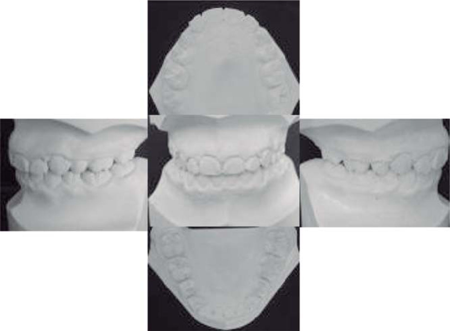
In the intraoral examination, the patient presents absence of the upper right canine, triangular upper arch and squared lower arch. Severe crowding is observed in both arches and the upper lateral incisors present an anterior crossbite. She has a 4mm. overjet and a 2mm overbite. The lower dental midline is deviated to the left. She is a molar class I on both sides and the canine class is non-assessableon both sides as well (Figure 2).
Radiographic examinationOn the panoramic radiograph we observed the retention of the upper right canine, eruptingupper and lower permanent second molars and included lower third molars (Figure 3).
The cephalometric analysis revealeda skeletal class ipatient due to retrognathia with vertical growth, lower dental protrusion and proclination and narrow airways.
Diagnosis- •
13-year-old female patient.
- •
Skeletal class II due to retrusive mandible.
- •
Straight profile with hyperactivity of the mentalis muscle.
- •
Crossbite of the upper lateral incisors (Figure 4)
- •
Retained upper right canine.
- •
Molar class I.
- •
Non-assessable canine class.
- •
4mm overjet and 2mm overbite.
- •
- •
Severe crowding on both arches, triangular upper arch and squared lower arch.
- •
Vertical growth.
- •
Lower dental protrusion and proclination.
- •
Improve profile
- •
Maintain molar class I
- •
Obtain canine class I
- •
Correct overjet and overbite
- •
Improve arch form
- •
Improve the inclination of the lower incisors
- •
Placement of a Hyrax-type expansion screw.
- •
Extraction of upper and lowerfirst premolars.
- •
Edgewise fixed appliances.
Phase 1: Aligning and leveling
0.0175 multistrand archwire
0.014 Niti archwire
0.016 Niti archwire
0.016 SS archwire
Phase 2: Space closure
0.016×0.016 SS upper and lower archwires wit closing loops.
Ideal 0.016×0.022 SS upper and lower archwires.
Phase 3: Finishing
¼ heavy boxelastics for two weeks.
Retention
Lower canine to canine fixed retention and circumferential retainer on the upper.
TreatmentA Hyrax-type expansion screw was placed to correct the upper arch form. It was activated V turn by day and V turn by night for 12 days (Figure 5). Once the expansion was accomplished, the screw was fixated with wire ligature and left for retention for a period of 3 months (Figure 6).
Afterwards, the patient was referred to surgery for the surgical exposure of the upper right canine and placement of a lingual button to initiate orthodontic traction (Figure 7). The Hyrax was removed and the patient was referred to the surgeon for the extraction of the upper and lower premolars.
Edgewise appliances, bands on upper and lower first molars with upper double tubes and lower single tubes were placed. We placed a 0.0175 multistrand archwire and began the orthodontic traction of the canine with ligature wire. The upper left lateral incisor was ligated proximally with an elastic thread. Canine distalization was initiated with passive lacebacks (Figure 8).
Afterwards, the upper left and the lower canines were moved distally with an elastomeric chain, the lingual button was removed from the upper right canine and replaced with a bracket. Leveling was continued with 0.014, 0.016 Niti and 0.016 SS archwires (Figure 9). Subsequently, 0.016×0.016 SS archwires with closing loops were placed (Figure 10). After space closure, an 0.016×0.022 SS cinched ideal archwire was used with an elastic chain from secondpremolarto second premolar upper and lower (Figure 11). Fixed appliances were removed and we placeda fixed lower retainer from canine to canine and a circumferential retainer on the upper arch (Figure 12and13).
ResultsWith this treatment we managed to performa successful orthodontic traction of a retained canine and position it correctly in the maxillary dental arch (Figures 14 to 16) while obtaining:
- •
A straight profile
- •
Molar and canine class I on both sides
- •
Periodontal health
- •
Centered dental midline
- •
Adequate overjet and overbite
- •
Paraboloid arch form
The management of a retained maxillary canine is not complete with just its alignment; final periodontal health is a fundamental key to assess treatment success for the maxillary retained canine.
Several strategies for the interceptive treatment of the displaced canine have been proposed but in animpaction case a surgical-orthodontic approach is required.
In previous papers it has been proposed acombined surgical (flap) and orthodontic (direct traction towards the center of the alveolar bone) approach with the purpose of simulating the canine’s physiological eruption pattern.3,11
On this matter, it needs to be said that the present clinical case was treated with the same standard surgical-orthodontic approach with the purpose of guiding the retained canine towards the center of the alveolar bone in the maxillary arch.
This technique allows the repositioned canine to be surrounded by a physiological amount of gingiva at the end of orthodontic treatment. This result is similar to the findings of the longitudinal research done by Quirynen et al.4
ConclusionsIt is fundamental to know the location of retained and included canines before their surgical exposure.
When the treatment was finished, positive changes were achieved by performing the orthodontic traction of the upper right canine and positioning it correctly in the dental arch. In doing so, canine class I was achieved and we improved arch form, the overjet and the overbite as well as the profile and the incisor’s inclination.
The radiographic characteristics prior to treatment assessed in the panoramic radiographs are useful indicators for the duration of the orthodontic traction but they are not valid predictors for the final periodontal status of the orthodontically repositioned impacted canine.
Complete fixed appliances are a commonly used alternative in combination with traction applied to the center of the alveolar process and the use ofa lingual button and ligature wire tied to the rigid arch wire. This technique ensures a good control system.


