


Impacted teeth are present in 1 to 5% of orthodontic patients. The most common causes are: lack of space for the eruption, prolonged bone retention barrier or early loss of deciduous teeth, abnormal position of the tooth to erupt, presence of alveolar cleft, anchylosis, neoplasias, dental or alveolar trauma and root dilaceration. The central incisor is the most frequently impacted incisor; the frequency varies between 0.06 and 0.2%. Surgical-orthodontic treatment is an option for the correction of dental impactation. This article shows the case of a patient of nine years of age who presented the clinical absence of the upper right central incisor. By using radiographic and tomographic examination the impaction was confirmed.
MethodSurgical-orthodontic management, surgical exposure and orthodontic traction of the upper right central incisor.
ConclusionIt is important to perform comprehensive treatments, taking into consideration the risks and benefits for the patient thus minimizing treatment complications of impacted teeth.
Los dientes impactados se presentan en 1 a 5% de los pacientes ortodóncicos. Las causas más comunes son locales: falta de espacio para la erupción, barrera ósea prolongada retención o pérdida temprana de dientes deciduos, posición anormal del diente a erupcionar, presencia de hendidura alveolar, anquilosis, formación neoplásica, trauma dental o alveolar y dilaceración radicular. El incisivo central es el incisivo que más frecuentemente se impacta, esta frecuencia varía entre 0.06 a un 0.2%. El tratamiento ortodónticoquirúrgico es una opción para la corrección de las impactaciones dentales. El presente artículo muestra el caso de una paciente de nueve años de edad que presenta la ausencia clínica del incisivo central superior derecho, mediante examen radiográfico y tomográfico se confirma su impactación.
MétodoManejo ortodónticoquirúrgico, exposición quirúrgica y tracción ortodóncica del órgano dentario 11.
ConclusiónEs importante realizar tratamientos integrales, tomando en cuenta las consideraciones riesgo-beneficio para el paciente y así minimizar las complicaciones del tratamiento de los dientes impactados.
Definition. The term dental impaction refers to a tooth located within the bone and/or soft tissue, and that probably will not erupt spontaneously.
Impacted teeth are present in 1 to 5% of orthodontic patients.1 According to the review performed by Bishara,2 the causes of tooth impaction are divided into general and local factors. The common causes are usually local: lack of space, prolonged retention or early loss of deciduous teeth, abnormal position of the tooth to erupt, presence of alveolar cleft, anchylosis, neoplasias, dental or alveolar trauma, and root dilaceration.3 Among the general causes there are: febrile diseases, endocrine disorders, hypovitaminosis and inheritance. Impacted teeth may cause injury to the neighboring teeth, infections, or cysts and represent a problem due to their functional and aesthetic implications.
The orthodontist offers a variety of therapeutic options, but in order to achieve success it is essential to diagnose dental impaction in a timely manner.4
Surgical-orthodontic treatment is an option for the correction of dental impaction. It is important to make an adequate diagnosis and treatment planning of impacted teeth thus avoiding further functional and aesthetic complications that may compromise the integrity of the arches as well as the psychosocial development of the patient. A multidisciplinary work must be performed when evaluating the impacted organ and routine follow-ups should be installed until it is incorporated into the arch.5
Clinical diagnosis- •
Check sequence of dental eruption.
- •
Significant eruptiondelay.
- •
Comparison of quadrants.
- •
Check the presence and position of the impacted tooth.
- •
Assess morphology and structure of the impacted tooth.
- •
Show possible obstacles for orthodontic traction.
- •
Surgical intervention, removal, or simple exposure with the placement of an orthodontic device during or after surgery.
- •
Orthodontic traction.
The prognosis of this type of treatment will depend on several factors such as: position of the impacted tooth in relation to the adjacent teeth, angulation, distance that the tooth must travel up to the correct position, root canal dilaceration and possible presence of anchylosis and root resorption.
MÉTODONine-year-old female patient with no pathological history, skeletal class 1, straight profile, with mixed dentition, bilateral class 1, insufficient space in maxillary and mandibular arch, moderate osseous-dental discrepancy, deviated midline; clinically the absence of the right upper central incisor was observed, in the radiographic examination the right upper central incisor is observed near the anterior nasal spine, located perpendicularly to the occlusal plane (Figures 1 to 3).


The present case describes the use of the Edgewise 0.018″ technique for the traction of the right upper central incisor.
Objectives- •
Maintain the facial profile.
- •
Correct the maxillary and mandibular tooth-bone discrepancy.
- •
Maintain space for the correct position of canines and premolars.
- •
Bilateral class I molar relationship.
- •
Bilateral canine class I.
- •
Achieve normal overjet and overbite.
- •
Correct dental midline.
- •
Surgical approach of the permanent upper right central incisor and placement of button for orthodontic traction.
- •
Traction of the permanent upper right central incisor and incorporation into the dental arch.
0.018″ slot Edgewise appliances; moderate upper and lower anchorage: Nance button appliance and lingual arch.
Treatment began with 0.016″ braided arch wires (Figure 4).
Phase I
Alignment and leveling.
Space consolidation for traction of upper right central incisor.
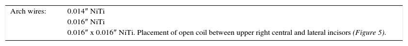
| Arch wires: | 0.014″ NiTi |
| 0.016″ NiTi | |
| 0.016″ x 0.016″ NiTi. Placement of open coil between upper right central and lateral incisors (Figure 5). |

Surgical phase: tooth exposure, placement of button and ligature for the traction of the tooth # 11.
0.016″ x 0.022″ stainless steel arch wire (Figure 6).
Phase III
Traction and incorporation of the right central incisor to the arch (Figure 7).

Withdrawal of button, placement of bracket in tooth # 11.
Phase IVDetailing and occlusal consolidation.
0.017″ x 0.025″ SS arch wire with ideal bends.
Midline correction elastics.
Retention
RESULTSAt the end of treatment, the patient showed a suitable facial profile, coordinated arches, good dental alignment, permanent dentition with partial eruption of maxillary and mandibular second molars, bilateral canine class I, class I molar on both sides, normal overjet and overbite, coincident dental midlines. Clinically it may be observed that the upper right central incisor was fully incorporated to the dental arch and radiographically, root parallelism was achieved (Figures 8 to 11).
DISCUSSION


Combined orthodontic-surgical procedures represent a good option to treat dental impaction with good aesthetic and functional results.
Johnston makes reference to the fact that in clinical practice, the following factors are used to determine if an impacted tooth may be aligned successfully in its correct position: the position and direction of the impacted tooth, the length of radicular formation and the degree of root dilaceration.1
Depending on the case, it is advisable to perform early treatment of impacted teeth as delay can lead to secondary problems such as root dilacerations or possible anchylosis of the affected teeth. During initial clinical examination the presence of retained deciduous teeth after 12 years of age, absent permanent teeth and the use of a routine X-rays should be considered for a correct diagnosis.6
To obtain favorable results it is necessary to perform controlled traction forces combined with radiographic assessments on a regular basis.7 Likewise, the intervention of other specialties such as periodontics and endodontics may increase treatment success.
CONCLUSIONSIt is advisable to perform early treatment of impacted teeth since possible side effects may arise such as anchylosis of the affected teeth. It is also important to assess the position of the impacted tooth as well as its root length for prognosis and treatment.
Surgical-orthodontic treatment of impacted teeth is usually successful, but relatively long so it is important to mention this to both the patient and their parents so they can be warned of this situation.