


This study aimed to biochemically and histopathologically investigate the effect of sunitinib on oxidative testicular damage induced by ischemia/reperfusion in rats.
Material-methodExperimental animals were divided into three groups of six rats each: testicular torsion–detorsion (TTD), sunitinib+testicular torsion–detorsion (STD), and sham control (SC). Sunitinib (25mg/kg) was administered orally to the STD group by gavage. Normal saline (0.9% NaCl) was administered orally to the TTD and control groups as the solvent. One hour after administration of sunitinib and 0.9% NaCl, all animal groups were done torsion–detorsion. Then, all the rats were killed by high-dose anesthesia, and their testicles were removed. Biochemical and histopathological examinations were performed on the removed testicular tissues.
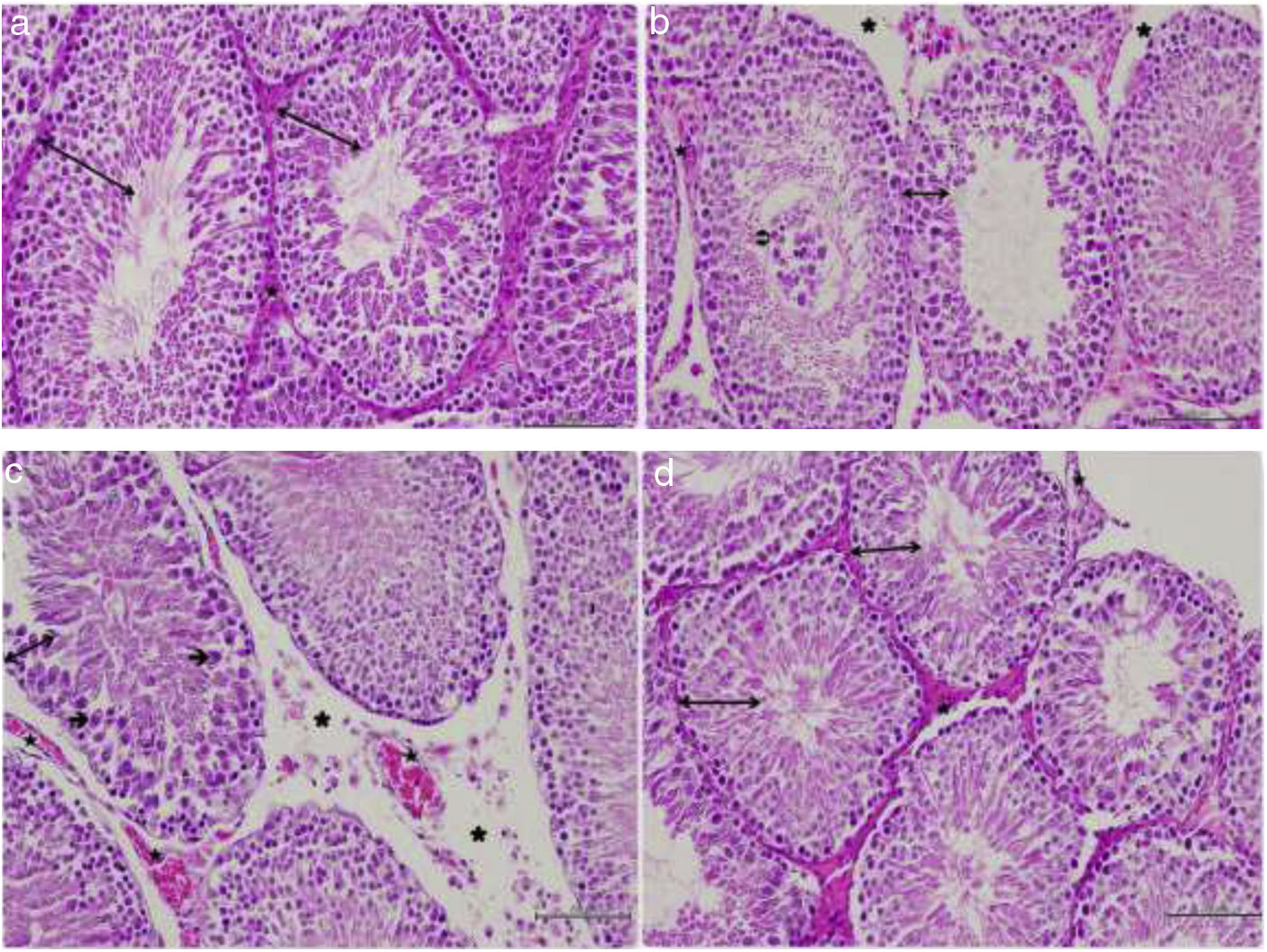
ResultsMalondialdehyde; it was observed that the results in the STD group were close to those of the SC group and statistically significant lower compared to the TTD group (p=0.001). The glutathione values were statistically significantly higher in the STD group compared to the TTD group (p<0.001). Nuclear factor kappa B values, revealing a statistically significant difference between the TTD and STD groups (p<0.001). The TNF-α levels were measured and indicating that the results of the STD group were statistically significantly lower than those of the TTD group (p<0.001). Histopathologically, animal tissues given sunitinib were observed to resemble normal tissues.
ConclusionSunitinib was shown to prevent histopathological changes in testicular tissue against ischemia/reperfusion damage.
Este estudio tuvo como objetivo investigar bioquímica e histopatológicamente el efecto del sunitinib en el daño testicular oxidativo inducido por isquemia/reperfusión en ratas.
Material-métodoLos animales experimentales se dividieron en tres grupos de seis ratas cada uno: torsión-detorsión testicular (TTD), sunitinib + torsión-detorsión testicular (STD) y control simulado (SC). El sunitinib (25 mg/kg) se administró por vía oral al grupo STD por sonda. Se proporcionó por vía oral solución salina normal (NaCl al 0,9%) a los grupos TTD y control como disolvente. Una hora después de la administración de sunitinib y NaCI al 0,9%, se realizó una torsión-detorsión a todos los grupos de animales. A continuación, todas las ratas fueron sacrificadas con anestesia de alta dosis y se les extrajeron los testículos. Se realizaron exámenes bioquímicos e histopatológicos de los tejidos testiculares extraídos.
ResultadosMalondialdehído; se observó que los resultados en el grupo STD eran cercanos a los del grupo SC y estadísticamente significativos más bajos en comparación con el grupo TTD (p = 0,001). Los valores de glutatión fueron estadísticamente significativos más altos en el grupo STD en comparación con el de TTD (p < 0,001). Los valores del factor nuclear kappa B, revelaron una diferencia estadísticamente significativa entre los grupos TTD y STD (p < 0,001). Se midieron los niveles de TNF-α e indicaron que los resultados del grupo de ETS fueron estadísticamente significativos más bajos que los del ETV (p < 0,001). Desde el punto de vista histopatológico, se observó que los tejidos de los animales a los que se les administró sunitinib se parecían a los tejidos normales.
ConclusiónSe demostró que el sunitinib previene los cambios histopatológicos en el tejido testicular frente al daño por isquemia/reperfusión.
Artículo
Comprando el artículo el PDF del mismo podrá ser descargado
Precio 19,34 €
Comprar ahora