


The first metatarsophalangeal joint arthrodesis is indicated for the treatment of various pathologies as a technique to reduce pain and improve the support of the first radius. Numerous surgical techniques and fixation methods have been described, with the combination of a dorsal plate and an interfragmentary screw being the one that has shown to be the most stable construct in biomechanical studies. Our aim is to analyze the radiological results after metatarsophalangeal arthrodesis of the hallux using a dorsal plate associated or not with an interfragmentary screw. The differences in terms of consolidation rates and complications in patients diagnosed with hallux rigidus, hallux valgus, hallux varus and failure of previous surgeries were evaluated.
Materials and methodsA retrospective cohort study of 55 patients with a mean age of 65.10 years in whom a dorsal plate was used was performed. Patients were divided into two groups depending on whether or not an interfragmentary screw was used. The minimum follow-up was 6 months after surgery. The assessment of the pre and postoperative radiological results was based on the variation of the hallux angle, the intermetatarsal angle and the dorsal metatarsophalangeal angle of the hallux, as well as the cases of nonunion identified in each study group.
ResultsThe radiological results, statistically significant differences (p<0.05) were only found in the dorsal metatarsophalangeal angle between both study groups. No statistically significant differences were found regarding the radiological evaluation of the pre and postoperative hallux angle and intermetatarsal angle. An equal decrease of each angles was observed in both study groups. Regarding the consolidation rate, statistically significant differences (p<0.05) were found between group A, which associated an interfragmentary screw, presenting a consolidation rate of 92%, and group B, which did not associate an interfragmentary screw, and that presented a union rate of 63%.
ConclusionHallux metatarsophalangeal arthrodesis of the hallux with a dorsal plate and interfragmentary screw show best results regarding consolidation rate and complications compared to those cases in which an interfragmentary screw was not used.
La artrodesis de la articulación metatarsofalángica (MTF) del hallux es una intervención quirúrgica indicada para el tratamiento de diversas afecciones, cuyo objetivo es aliviar el dolor y mejorar el apoyo del primer radio. Existen diversas técnicas quirúrgicas y métodos de fijación para llevar a cabo la artrodesis de dicha articulación, siendo la combinación de placa dorsal y tornillo interfragmentario la que ha demostrado en diversos estudios biomecánicos mayor estabilidad. Nuestro objetivo es analizar los resultados radiológicos tras artrodesis metatarsofalángica del hallux utilizando placa dorsal asociada o no a tornillo interfragmentario, valorando las diferencias en relación a la consolidación y complicaciones en pacientes diagnosticados de hallux rígidus, hallux valgus, hallux varus y fracaso de cirugías previas.
Material y métodosRealizamos un estudio de cohortes retrospectivo de 55 casos con una edad media de 65,10 años, intervenidos de una artrodesis de la articulación metatarsofalángica del hallux con placa dorsal, divididos en 2 grupos de estudio, según asocien o no a tornillo de compresión sin cabeza, con un seguimiento de al menos 6 meses postoperatorios. Valoramos los resultados radiológicos pre y postoperatorios basándose en la variación de los ángulos estudiados (ángulo del hallux, ángulo intermetatarsal y ángulo metatarsofalángico dorsal del primer dedo), así como los casos de seudoartrosis encontrados en cada grupo de estudio.
ResultadosEn cuanto a los resultados radiológicos, únicamente se encuentran diferencias estadísticamente significativas (p<0,05) en relación con el ángulo de dorsiflexión posquirúrgico entre ambos grupos de estudio. No se encontraron diferencias estadísticamente significativas en relación con el análisis radiológico del ángulo del hallux e intermetatarsal pre y posquirúrgico, ya que se observa que disminuyen igual en ambos grupos de estudio. El grupo A, que asocia tornillo interfragmentario, presentó una tasa de consolidación del 92%, frente al grupo B, sin tornillo a compresión, que fue del 63%, siendo estas diferencias estadísticamente significativas (p<0,05).
ConclusionesLa artrodesis metatarsofalángica del hallux con placa dorsal asociada a tornillo interfragmentario obtiene mejores resultados en lo que se refiere a consolidación y complicaciones respecto a aquellos casos en los que no se utiliza el tornillo interfragmentario.
Arthrodesis of the metatarsophalangeal joint (MTP) of the hallux is a surgical intervention indicated for the treatment of various ailments, such as advanced hallux rigidus and hallux valgus, arthritis derived from inflammatory diseases such as rheumatoid arthritis, and as rescue surgery after performing other procedures.1–4
The goal of first MTP joint arthrodesis is to relieve pain and improve support of the first radius.
There are various surgical techniques and fixation methods to carry out arthrodesis of said joint, such as parallel or crossed screws, cerclages, Kirschner wires (KW), intramedullary devices, staples or dorsal plates with or without interfragmentary screws.1,2,5,6 Currently, there is no consensus on which is the best fusion method, although various biomechanical studies have shown greater stability with the combination of dorsal plate and interfragmentary screw.6
Pseudarthrosis, symptomatic or not, is one of the most relevant complications in arthrodesis of this joint. The consolidation rate varies in studies between 71% and 100%7–10 of cases depending on the procedure used, with no significant difference currently being demonstrated between the techniques used. Other factors to take into account when studying the result of arthrodesis are the condition for which the procedure was indicated, comorbidity, the age and sex of the patient, and the postoperative position of the joint.
The optimal postoperative position of the first MTP joint is neutral rotation, between 5° and 15° of valgus and between 10° and 15° of dorsiflexion with respect to the ground (20–25° with respect to the first metatarsal).11,12 In our practice, this degree of dorsiflexion is determined intraoperatively with a simulation loading test with a flat board.
A lower dorsiflexion angle than indicated would increase pressure on the interphalangeal joint, favouring the development of osteoarthritis at that level, while a greater angle would increase pressure at the head of the first metatarsal, overloading the sesamoids and causing a claw deformity of the interphalangeal joint and difficulties with footwear.13
The objective of this study was to analyze the radiological results after MTP arthrodesis of the hallux using a dorsal plate associated or not with an interfragmentary screw, assessing the differences in relation to consolidation and complications in patients diagnosed with hallux rigidus, hallux valgus, hallux varus and failure of previous surgeries.
Material and methodsPatient selectionWe carried out a retrospective cohort study between January 2016 and January 2021, with a mean follow-up of 22.7 months (SD 12.5 months) of all patients operated on by the Foot and Ankle Unit of our centre, through the digital medical record (HCIS, Dedalus, Milan, Italy).
Regarding the inclusion criteria in the study, those patients who underwent an arthrodesis of the MTP joint of the hallux with a dorsal plate associated or not with an interfragmentary headless compression screw, with a preoperative diagnosis of hallux valgus, hallux rigidus, hallux varus or post-surgical recurrence of hallux valgus. Patients who did not complete a regulated clinical and radiological follow-up of at least 6 months postoperatively were excluded from the analysis.
Demographic data, history (BMI, tobacco habit), previous forefoot surgeries, data on the surgical technique (approach, type of synthesis), postoperative management (follow-up time and use of post-surgical shoe) and complications (superficial or deep infection, pseudoarthrosis, discomfort in relation to the implanted material, osteoarthritis in neighbouring joints and reintervention) were collected.
The patients were grouped into two study groups according to the type of implant used: group A, cases treated with a cannulated interfragmentary screw associated with a neutralizing locked dorsal plate, and group B, cases treated without an interfragmentary screw, in which only the blocked dorsal plate was implanted, using the oval hole to provide compression.
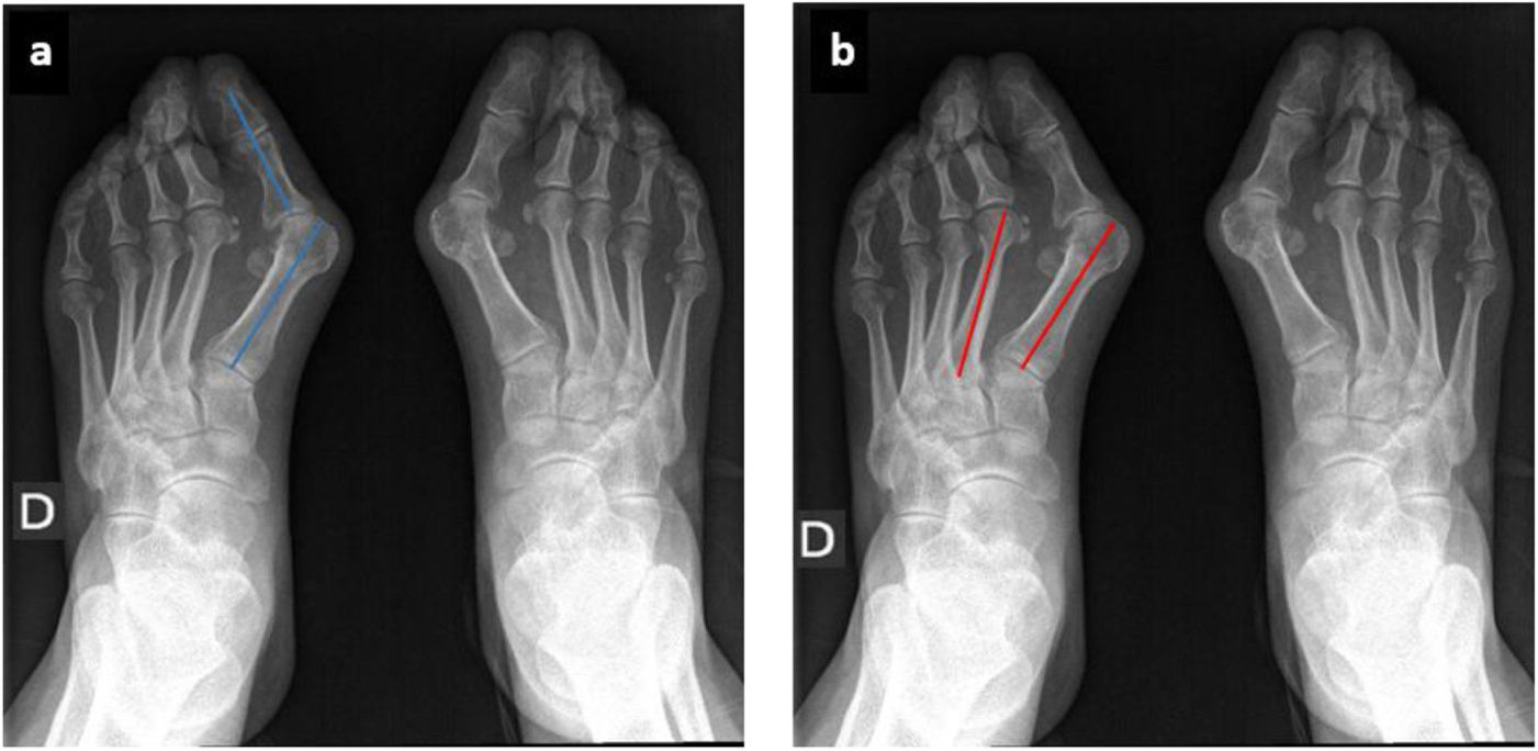
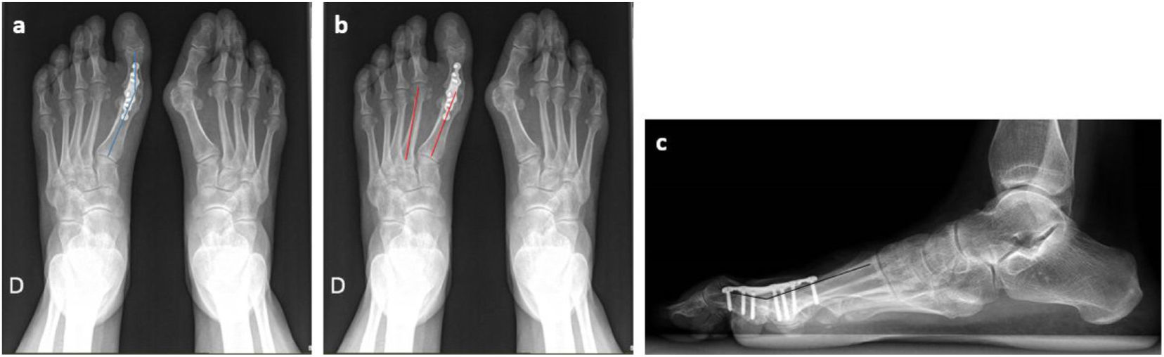
Study variablesA radiological study was performed based on pre- and post-surgical weight-bearing dorsoplantar and lateral radiographs (Figs. 1 and 2). The parameters studied in the dorsoplantar radiograph in all cases were the hallux angle (angle resulting from the diaphyseal axis of the first metatarsal and that of the first phalanx) and the intermetatarsal angle (angle between the diaphyseal axis of the first and second metatarsal). In the post-surgical lateral radiograph, the dorsal metatarsophalangeal angle of the first toe obtained with the implant was analyzed. Vue PACS software (Carestream Health, Inc., Rochester, NY, USA) was used to perform all measurements.
 Dorsoplantar weight-bearing radiograph. Hallux angle (blue) and intermetatarsal angle (red).")
 Dorsoplantar radiograph in postoperative weight bearing. Hallux angle (blue) and intermetatarsal angle (red). (c) Postoperative weight-bearing lateral radiograph. Dorsal metatarsophalangeal angle of the first toe (black).")
A consolidated arthrodesis was defined as one with signs of union in at least 3 of the 4 cortices (medial and lateral in the dorsoplantar projection, and dorsal and plantar in the lateral projection).12,14
The state of consolidation and the angles were analyzed independently by two researchers. In case of discrepancy or determination of nonunion, the result was verified by an attending physician from the Foot and Ankle Unit.
Surgical techniqueThe same treatment sequence was performed in all patients, with the exception of whether or not an interfragmentary screw was used, a decision made according to the surgeon's preferences.
In the supine position, with ischaemia at the level of the corresponding ankle and peripheral block associated with local anaesthetic, a medial approach was performed in 53 cases (96%), at the level of the intersection between the plantar and dorsal skin, and dorsal in two cases (4%). Next, capsulotomy and dorsal cheilectomy were performed to adapt the dorsal surface of the first metatarsal to the configuration of the plate and joint dislocation. The articular surfaces were prepared by motorized concave–convex drilling guided by an KW previously aligned with the diaphyseal axis of the first metatarsal and the first phalanx of the hallux. The articular surfaces were also bloody with successive perforations carried out with the help of a 1.6mm KW. Under radioscopic control, the final position of the arthrodesis was checked, keeping the finger in a neutral position in the coronal plane and a dorsiflexion angle of 5–15°. This position was temporarily fixed with an KW through the axis of the first spoke, confirming this position with the help of a flat board simulating the load.
In group A, a 2.5–3.00mm cannulated headless compression screw (Zimmer Biomet, Warsaw, IN, USA) was placed in a medial-proximal to lateral-distal direction across the joint. A dorsal plate (ALPS® Total Foot System, Zimmer Biomet, Warsaw, IN, USA) was subsequently implanted in both treatment groups, available in two sizes: small (2.5mm) or large (3.5mm), with cortical screws or locked according to the patient's needs, which has a preconformation of 14° or 7° of dorsiflexion depending on the size, and 5° of valgus to allow adequate anatomical alignment. In those patients where the interfragmentary screw was not placed, the oval hole present in the plate was used to perform compression. After verifying the correct position of the arthrodesis and the material under radioscopic control, the skin was closed with 4.0 monofilament and a compressive bandage of the foot and ankle was applied.
All patients were allowed immediate weight bearing with the help of a rigid-soled post-surgical shoe for 6–8 weeks, allowing weight bearing with normal footwear after this period. They were scheduled for weekly consultations to monitor the surgical wound, and at 6- and 12 weeks post-surgery with weight-bearing radiographs, extending the follow-up if consolidation of the arthrodesis was not observed at that time.
Statistical analysisThe data were analyzed with the statistical program Stata version 16 (StataCorp LLC, College Station, TX, USA), studying the entire sample together and comparing the two groups with each other based on the type of treatment received. For quantitative variables we used the Student's t test, and for qualitative variables the Chi square test, considering results with a value of p<.05 significant.
ResultsFifty-five cases were studied in a total of 52 patients. Group A included 39 cases (71%): 6 men (15%) and 33 women (85%), and group B 16 cases (29%): 6 men (38%) and 10 women (63%). The age at the time of surgery in group A was 67.91 years (SD 8.34 years), compared to group B, in which it was 58.27 years (SD 10.81 years) (p<.001). The mean BMI in group A was 28.54 (SD 4.53), and in group B was 29.68 (SD 5.68). In group A, 8 cases (21%) were smokers and in group B, 3 cases (19%) were smokers. No statistically significant differences were observed in relation to sex, BMI or tobacco use.
There were also no statistically significant differences in relation to preoperative diagnosis between both groups. Table 1 shows the pre-surgical diagnoses of each group.
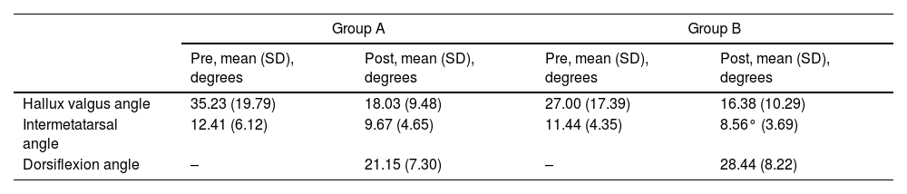
In relation to the radiological results, the pre-surgical hallux valgus angle in group A was 35.23° (SD 19.79°) and 27.00° (SD 17.39°) in group B. The post-surgical in group A it was 18.03° (SD 9.48°) and 16.38° (SD 10.29°) in group B.
The pre-surgical intermetatarsal angle was 12.41° (SD 6.12°) in group A and 11.44° (SD 4.35°) in group B, and the post-surgical angle was 9.67° (SD 4.65°) in group A and 8.56° (SD 3.69°) in group B (Table 2).
Radiological results of hallux valgus, intermetatarsal and dorsiflexion angles in both study groups.
| Group A | Group B | |||
|---|---|---|---|---|
| Pre, mean (SD), degrees | Post, mean (SD), degrees | Pre, mean (SD), degrees | Post, mean (SD), degrees | |
| Hallux valgus angle | 35.23 (19.79) | 18.03 (9.48) | 27.00 (17.39) | 16.38 (10.29) |
| Intermetatarsal angle | 12.41 (6.12) | 9.67 (4.65) | 11.44 (4.35) | 8.56° (3.69) |
| Dorsiflexion angle | – | 21.15 (7.30) | – | 28.44 (8.22) |
SD: standard deviation.
No statistically significant differences were found in relation to the radiological analysis of the pre- and post-surgical hallux valgus and intermetatarsal angles, since it is observed that both decrease equally in both study groups.
The post-surgical dorsiflexion angle was 21.15° (SD 7.30°) in group A, and 28.44° (SD 8.22°) in group B, these differences being statistically significant (p<.05).
Complications were observed in 15% of the cases in group A (6 cases) compared to 50% of the cases in group B (8 cases), this difference being statistically significant (p<.05). The most frequent complications were pseudoarthrosis, followed by two superficial infections (one in each study group), discomfort in relation to the implanted material in three cases (two in group A and one in group B), two interphalangeal osteoarthritis (one in each study group) and one case of interfragmentary screw breakage.
Eighty-four percent of the total procedures were consolidated at the end of follow-up (46 of the 55 cases). Group A presented a consolidation rate of 92% (36 cases), and group B 63% (10 cases), this difference being statistically significant (p<.05).
The multivariate study identifies that the most important risk factor for the development of pseudarthrosis is the degree of dorsiflexion (p<.05), seeing that an increase in one unit in the dorsiflexion variable has a 10.5% less probability of union of the arthrodesis occurring. No statistically significant differences were found in relation to the patient's age (p>.05) with respect to the consolidation rate.
Four patients had to undergo further surgery due to complications derived from the initial surgery, two from each study group (5% of group A and 13% of group B). In three cases this entailed the removal of material due to discomfort, and in another an arthrodesis with autograft support because of symptomatic pseudarthrosis. The rest of the patients with nonunion had not required intervention at the time of this study.
DiscussionNumerous techniques have been described for performing MTP arthrodesis, including one or two interfragmentary screws, locked or nonlocked dorsal plates, and staples. When performing a hallux arthrodesis, it is essential to adequately prepare the joint, as well as reduce and fix it. Multiple studies have reported good results with various techniques. However, one of the complications that we can face is nonunion of the arthrodesis, these rates being different depending on the stability of the implant used in each surgical technique.
Politi et al.15 carried out a biomechanical study, comparing the stability of four types of fixation, and determined that the dorsal plate associated with an interfragmentary screw was the strongest construct, being up to 3 times more stable than the isolated screw, and up to 10 times more stable than the KW or the dorsal plate.
Buranosky et al.,16 in a cadaveric study, compared the stability of arthrodesis using two crossed cannulated screws versus a 6-hole dorsal plate with interfragmentary compression screw, after preparation of the articular surface with concave–convex reaming. The screws were tested to failure. They determined that the plate with a dorsal screw provides greater resistance to loading compared to screws.
The present study has shown that the use of interfragmentary compression screw associated with the dorsal plate for MTP arthrodesis of the hallux has a significantly higher consolidation rate compared to the use of the dorsal plate alone (92% vs. 63%) (p<.05).
In a retrospective study, Cone et al.14 reviewed 99 arthrodeses with dorsal titanium compression plate or interfragmentary compression screw with locked dorsal plate. The overall nonunion rate was 4%, with no statistically significant differences between both groups. However, the group with the compression screw showed less change in the postoperative dorsiflexion angle (.6° vs. 6.7°, p<.01), showing that the addition of the interfragmentary screw provides a more robust construct than the locked plate.
Hyer et al.17 conducted a retrospective study comparing several fixation models: static plate, locked plate, static plate with compression screw and locked plate with compression screw. The overall nonunion rate was 7% (9 of 130), with the lowest nonunion rate in the interfragmentary screw-locked dorsal plate group (4%, 2 of 45). However, these differences were not statistically significant (p>.05).
Gould et al.18 prospectively studied the fusion rate of 30 hallux MTP arthrodeses fixed with a dorsal plate with a compression screw through the plate. They reported a 100% consolidation rate.
Weigelt et al.19 carried out a retrospective study of 178 cases, 97 treated with a dorsal locking plate with integrated interfragmentary screw and 81 with an interfragmentary compression screw independent of the plate. They determined a nonunion rate of 6.2% (11 of 178 cases).
In our study, the overall nonunion rate is 16%, being 8% in group A and 37% in group B (p<.05). This discrepancy in the nonunion rate between both groups may be due to the difference in the number of patients included in each group (39 in A versus 16 in B) or the reason for the indication for arthrodesis. Also because the compression screw adds stability to the assembly. The difference in the dorsiflexion angle obtained between both groups may explain the difference in nonunion rate seen in our study, since as we have explained previously, this angle directly influences consolidation. The difference may be due to the moment in which the compression with the plate occurs: if it has an interfragmentary screw, it maintains the initially desired position, not modifying the dorsiflexion angle as much.
In addition to the construct used to fix the arthrodesis, some authors describe diabetes mellitus,19 together with a residual hallux valgus angle greater than 20°3,19 as risk factors for not achieving adequate union. Therefore, adequate correction of this hallux valgus deformity could reduce the risk of nonunion.
With the present study we have shown that the use of an interfragmentary screw together with a dorsal plate in the MTP arthrodesis of the hallux is associated with a higher rate of union and a lower number of complications during the follow-up period compared to arthrodesis with a dorsal plate (p<.05). Furthermore, the union rate of this fixation method is similar to that described in the literature.
The first limitation of our study is that it is retrospective, in which the study cohorts are not homogeneous. To determine whether or not the arthrodesis is union, radiographs are used, and not a CT study. Another limitation is the heterogeneity in the number and characteristics of patients included in each study group. As this is a study in which radiological and non-clinical data are studied, the clinical impact of pseudarthroses obtained radiologically has not been determined.
Despite the limitations listed, the same protocolised treatment was applied to all patients depending on the treatment group to which they belonged.
ConclusionMTP arthrodesis of the hallux with a dorsal plate associated with an interfragmentary screw obtains better results in terms of complications and consolidation compared to those cases in which the interfragmentary screw is not used, with the degree of dorsiflexion being a risk factor for developing pseudarthrosis once the arthrodesis has been performed.
Level of evidenceLevel of evidence IV.
FundingThis research has not received specific support from public sector agencies, the commercial sector or non-profit entities.
Conflict of interestsThere are no conflict of interests.
