


Objective. The objective of this study was to compare these methods Fourier (STFT) and Wavelet (WT) transforms to assess muscle fatigue during supramaximal exercise.
Methods. Twenty five subjects of both genders (13 men, age = 28.2 ± 2.7 years and 12 women, age = 23.2 ± 2.7 years) performed the Wingate test, during which the electromyographic responses of the superficial muscles of the quadriceps were examined. For analysis we used the STFT and WT transforms, which provided the following variables: median frequency (MF), slope and variance.
Results. The results showed no differences (P>0.05) in MF and slope. However, there were differences for the variance between the analysis (P<0.05).
Conclusion. It seems that both analyzes provide the same physiological parameters, however, the WT transform shows less variance between the results.
Objetivo. El propósito de este estudio fue comparar los métodos transformados de Fourier (STFT) y Wavelet (WT) para evaluar la fatiga muscular durante el ejercicio supra-máximo.
Método. Veinticinco individuos de ambos sexos (13 hombres, 28,2 ± 2,7 años and 12 mujeres, 23,2 ± 2,7 años) realizaron el test de Wingate, durante el cual las respuestas electromiográficas de los músculos superficiales de los cuádriceps fueron examinadas. Para el análisis se utilizaron las transformaciones de STFT y la WT, lo cual proporcionó las siguientes variables: frecuencia media (FM), de pendiente y variación.
Resultados. Los resultados no mostraron diferencias (P>0,05) en FM y en la pendiente. Sin embargo, hubo diferencias en la variación entre el análisis (P<0,05).
Conclusiones. Parece que ambos análisis proporcionan los mismos parámetros fisiológicos, sin embargo, la transformada de WT muestra menos variación entre los resultados.
Introduction
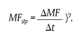
Muscle fatigue can be defined as the inability to generate force in a muscle contraction1. According to Enoka and Stuart1, muscle contraction is triggered from the sarcoplasmic membrane depolarization that modifies the electrical potential of the muscle, and this electrical potential can be captured and analyzed by surface electromyography (EMG). Numerous researchers have used EMG with different purposes2-6. For the use of EMG in order to determine muscle fatigue, it is necessary to analyze the electromyographic signal, and such analysis can be performed in time domain (RMS) or frequency domain. The latter requires the decomposition of the signal power spectrum density (PSD), to obtain the median frequency (MF), which is a frequency that divides the power spectrum in two regions of equal areas. A decrease of MF over time is related to muscle fatigue (for example, slope given by
One of the most used tools for evaluating the changes in MF during muscle fatigue is the short-time fourier transform (STFT). This transform is applied to determine the spectral content in terms of sinusoidal frequency and phase content of local portions of a signal as it changes over time8,9. This has been the main procedure used for this type of analysis, especially for isometric contractions. However, this method may not be as effective for dynamic contractions, in which EMG signals are not stationary due to variations in strength, speed and joint range of motion9, and thus not reflecting true physiological behavior of the muscle during exercise10. In this sense it becomes necessary to use alternative methods to correct this methodological problem.
A method that has been used in an attempt to minimize this problem is the Wavelet transform (WT). Wavelet analysis takes into account the dynamics of the EMG signals, which may represent a greater accuracy in the analysis. Thus, several studies have been used WT to analyze the EMG signal in isometric and dynamic maximal effort11, supramaximal constant load exercise12 and maximal exercise with constant load13. However the use of WT requires better understanding of the evaluation of muscle fatigue in exercises not as standardized in terms of control of strength and speed of movement14.
Recent studies indicate that STFT and WT would be comparable during muscle fatigue in well standardized protocols, both in a static or dynamic situation15-17, although other studies also suggest that WT has higher accuracy of the physiological information of muscle fatigue when compared to STFT18. However, further studies are needed to compare the methods of analysis of the EMG signal, especially for dynamic exercise without complete standardization of the task. Moreover, few studies have investigated the effects of supramaximal exercise on muscle responses such as fatigue. Supramaximal exercises are generally used to determine anaerobic capacity both in athletes of different modalities and non-athletes19. The literature is still scant in information on the use of either STFT or WT for an appropriate analysis of the EMG signal to assess muscle fatigue during this type of exercise.
To assess human motor performance during a supramaximal exercise, it is necessary to carry out specific tests such as the supramaximal Wingate test, often used in the literature to evaluate anaerobic performance20. Some studies have shown that the MF of EMG activity decreases over time during this type of test, which reflects the onset of muscle fatigue in evaluated muscle group21. However, this behavior was observed using the STFT analysis, where specific windows of analysis were successively standardized, as were the phases of movement analyzed, which in turn could generate data not as reliable as suggested by the studies of Karlsson et al.13. To check whether these results would be really influenced by the method of EMG analysis, a comparison of the STFT and WT methods should be done in this type of exercise.
Therefore, our hypothesis is that the WT transform enables more accurate results when compared to STFT. Thus, the purpose of this study was to compare the spectral analysis of the STFT and WT methods using the MF to assess muscle fatigue during supramaximal exercise.
Methods
Sample
The sample consisted of twenty-five (13 men, age = 28.2 ± 2.7 years and 12 women, age = 23.2 ± 2.7 years) untrained college students, who volunteered to the study. After the purpose of the study and the procedures to be performed were explained, they signed a consent form. This study was approved by the Ethics Committee of of the State University of Londrina (document 032/07; CAAE n.º 0034.0.268.000-07).
Experiment design
All participants performed the Wingate test (WT) to assess the anaerobic performance. Subjects were instructed not to ingest any substance or food containing caffeine for the duration of the experiment, as well as alcoholic beverages, and not to perform vigorous physical activity within 24 hours prior to the tests in order to avoid any interference in the results. Each subject was tested at the same time of day to minimize the effects of diurnal biological variation. The volunteers underwent a pilot study to familiarize with the testing protocols and the equipment used.
Anthropometry
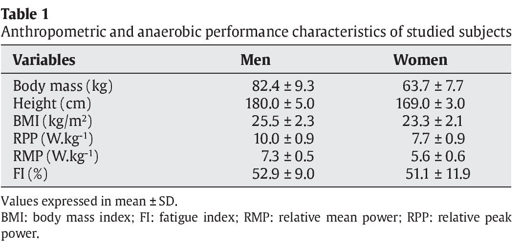
Body weight was measured on a Uranus® digital platform scale, model PS 180, with precision of 0.1 kg. Height was determined in a wooden stadiometer with accuracy of 0.1 cm. All individuals were measured and weighed barefoot, wearing only light clothes. Body mass index (BMI) was determined by the weight/height2, ratio, with body weight in kilograms (kg) and height in meters (m).
Anaerobic performance evaluation
The anaerobic performance of the subjects was assessed by WT20,22. The anaerobic performance indices were determined by a computer program (Wingate test®, Cefise, Brazil) that allowed the determination of the power generated every second during the test, the relative peak power (W.kg-1) (RPP), relative mean power (W.kg-1) (RMP) and fatigue index (FI) (%).
The protocol consisted of a warming-up of four minutes in a mechanical cycle ergometer for lower limbs (Monark® 324E, Sweden) with a load of 50 W and a pedaling cadence of 70 rpm, and at the beginning of each minute the subjects performed a six seconds sprint23.
After the warm-up, there was an interval of two minutes for the measurement of body weight, height adjustment of the bike saddle and adjustment of the effort intensity. After that, the participants began the WT, with no previous rotation, with a load corresponding to 0.075 kg. kg-1 of the body weight. At the end of the protocol, participants performed an active recovery on the same ergometer, without resistance, for a period of three minutes in an attempt to minimize possible side effects caused by stress.
A familiarization protocol was performed prior to the beginning of the study, in an attempt to reduce the learning effects and establish the reproducibility of the test. All subjects were tested in a similar situation to the experimental protocol in two separate sessions, with intervals of 48 hours. The coefficients of intra-class correlation found were 0.98, 0.95 and 0.90 for RPP, RMP and FI (%), respectively.
The bike measures corresponding to participant such as: saddle height and distance, stem height and distance and hands position were standardized for all tests, thus avoiding changes in posture and consequently possible interferences in the activation of the muscles evaluated. Temperature and relative humidity were controlled in all trials and kept between 21 and 24 ∫C and 40 and 60% respectively.
Recording and processing of electromyographic signals
The EMG signals were recorded during the entire period of the WT according to ISEK guidelines24. Before the start of each WT, participants had the bipolar active EMG electrodes model TDS 150™ (Biopac Systems®, USA), with fixed inter-electrode distance of two cm, placed on the superficial muscles of the quadriceps femoris (QF) of the right leg: vastus lateralis (VL), vastus medialis (VM) and rectus femoris (RF). After skin trichotomy and asepsis, the electrodes were positioned over each muscle following the standardization proposed by SENIAM25. The EMG activity was recorded by a 16-channel electromyograph model MP150™ (Biopac System®, USA) with sampling frequency of 2,000 Hz. The common mode rejection ratio was 95 dB, and the input limits of the signal were set at ± 5 mV. The reference electrode (ground) was placed in the right elbow (lateral epicondyle).
Signal recording and processing software was preformed with the AcqKnowledge™ 3.8.1 (Biopac Systems®, USA) software and the mathematical simulation environment Matlab 7.0 (Mathworks®, South Natick, MA, USA.) The raw EMG signals were digitally filtered with a band-pass filter of 20 Hz and 500 Hz. For spectral analysis of EMG signals, we used MF values determined using STFT and WT (Daubechies type: DB5). Using both methods we obtained the following parameters: the magnitude of changes in median frequency over time (MF [t]); slope of MF normalized by initial value, this is the EMG fatigue index (NFI)17; MF variance of the successive time windows established for the processing of the EMG signal during 30 seconds exercise. The EMG fatigue index was determined by linear regression of between the relationship of MF and the duration of exercise (30 seconds), for each muscle studied.
Statistical treatment
To compare the values found in all muscles using the techniques WT and STFT we used the Mann-Whitney U test. The significance adopted was 5%. We employed the statistical package Statistica™ 6.0® (Statsoft Inc., Tulsa, OK, USA) for data analysis.
Results
Table 1 presents the descriptive data corresponding to the anthropometrics characteristics and anaerobic performance of the participants.

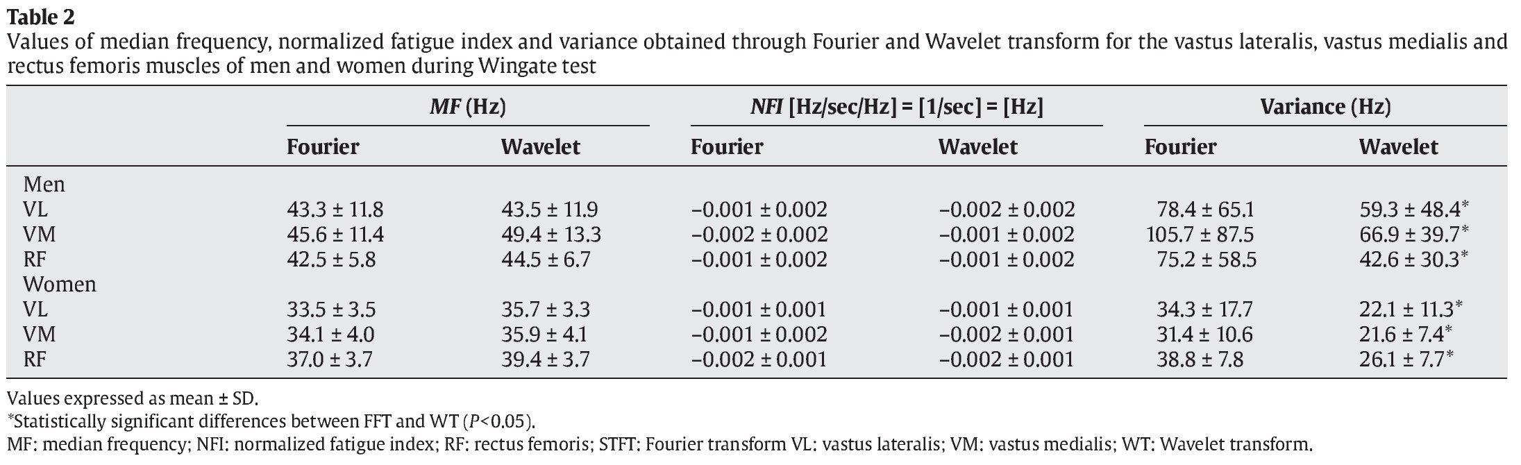
The values of median frequency (MF), normalized fatigue index (NFI) and the MF variance obtained by STFT and WT over the 30 seconds of exercise in WT for the VL, VM and RF of men and women are presented in table 2.

Regarding MF, there were no significant differences between the values obtained by both methods in any of the muscles evaluated for both genders (P>0.05). While there may be signs of muscle fatigue (negative values of the EMG index of fatigue), NFI for STFT and WT did not show any significant differences for any of the muscles evaluated in men and women (P>0.05).
Significant differences were observed in the MF variance between the values obtained by STFT and WT in all muscles analyzed in men and women (P<0.05), indicating a greater dispersion of data with the STFT analysis in relation to WT, as shown by an example for one individual (fig. 1).
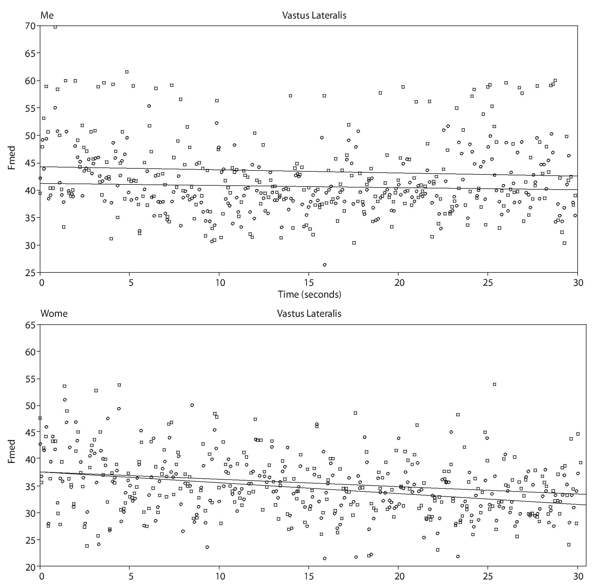
Fig. 1. Example for one individual of the electromyography fatigue index (NFI) obtained through the slope of median frequency normalized by the initial value. □ represents data corresponding to Wavelet test transform (upper line in the graphics) and ○ the Fourier transform.
Discussion
The aim of this study was to compare the EMG responses during the course of the Wingate anaerobic test, using two different methods of spectral analysis, STFT and Wavelet. From these findings we can confirm our hypothesis, since both methods provide the same physiological information about muscle fatigue, but a higher variability of data when these were analyzed by STFT.
The supramaximal effort required by the Wingate test causes the value of the median frequency of the EMG signal to decrease, which was previously verified by applying the STFT21 and corroborated by our study using both STFT and WT, thus confirming the presence of muscle fatigue. According to these authors, this behavior is associated with a high concentration of H+ ions during the course of the exercise impairing the propagation of the electrical signal, thus decreasing the frequency values.
However there is still a debate in the literature over the use of STFT analysis in dynamic muscle contractions, since this method of analysis assumes that data are stationary10. However, recent results on muscle fatigue during standardized dynamic contractions of different muscle groups showed the similarity and the effectiveness of these two methods to provide information on muscle behavior during fatiguing exercise15-17. The results of this study confirms the findings of previous ones, and also brings a new aspect of evaluation methods for nonstandardized and high intensity exercises, which was never done by other studies on the subject.
Da Silva et al.17, showed that the effect of mediation for the calculation of EMG indices of fatigue increases the association between STFT and WT. Moreover, these authors showed that WT shows less data variability and hence better accuracy of information compared to STFT. This is also supported by our results of variance, indicating less accuracy in terms of variability in data obtained from analysis by STFT compared to WT for all the muscles in both sexes. Similar results were also found by da Silva et al.17.
The use of alternative methods for analyzing the EMG signal, such as WT transform, has been proposed in an attempt to better adjust the mathematical model for dynamic tasks. In fact, Von Tscharner and Goepfert14, showed that analysis through the WT is highly effective in identifying a pattern of muscle recruitment of different muscle fiber types in a specific phase of the movement. This information would hardly be represented by the STFT analysis, which shows a large dispersion of individual MF values within the temporal window of processing and over time (successive windows), and consequently would result in larger measurement errors in dynamic situations, as suggested by other studies17,18.
Therefore, although the values of NFI were not different between methods, the values obtained by STFT showed greater data variance when compared to WT. This demonstrates that WT seems to adjust itself better to dynamic tasks, since it does not depend on the signal to be (quasi-) stationary, unlike the technical limitation imposed by conventional STFT. Thus, we suggest that the method of analysis of EMG signals via WT can provide more information and accuracy when applied to different types of muscle fibers in a specific muscle group regardless of gender during a dynamic muscle action. Also, this method could be interesting to identify the pattern of muscle recruitment during a specific phase of the movement in cyclic exercise, as suggested by Von Tscharner and Goepfert14.
In conclusion, considering the findings of this study we suggest the use of both methods of spectral analysis on supramaximal dynamic exercise, that is non-standardized in terms of speed and angle of motion and short duration (~ 30 s) when the aim is to quantify muscle fatigue with spectral EMG indices (for example, slope of MF).
Conflicts of interest
The authors declare that they have no conflicts of interest.
Acknowledgements
The authors thank the FAPESP, the CNPq and the CAPES, for graduate and post-graduate scholarships.
History of the article: Received January 7, 2012 Accepted March 5, 2012
Correspondence:
Prof. Dr. L.R. Altimari.
Departamento de Educação Física.
Universidade Estadual de Londrina.
Rodovia Celso Garcia Cid, Pr 445 km 380.
Campus Universitário, Cx Postal 6001.
CEP 86051-990, Londrina, Brazil.
E-mail:altimari@uel.br
