


Central venous access devices (CVADs) are used to deliver intravenous therapy to the bloodstream. CVAD insertion is sometimes fluoroscopically guided and thus associated with radiation dose to both the patient and the staff members within the room. The objective of this study is to assess the radiation dose to the patient through a retrospective audit and directly measure the exposure to staff members in simulated procedures. A secondary objective is to evaluate the radiation exposure to the staff and patients when utilising fluoroscopic pulse rate of 7.5pps and 4pps.
Material and methodsA retrospective audit of patients undergoing Permcath and Hickman line insertions was conducted. The patients were grouped by the pulse rate used for the duration of the study; 4 pulses per second (pps) (n=24) and 7.5 pps (n=33). A STEP OD-2 monitor and PMMA was used in a simulated environment to estimate the radiation exposure to locations that a Radiologist, Nurse and Radiographer would be standing during the procedures using the average procedure details collected in the retrospective audit. Measurements were conducted at heights to reflect a whole body estimate and an estimate to the lens of the eye.
ResultsThe results show that the median dose area product (DAP) for CVAD insertion is 0.7Gycm2 and 0.3Gycm2 for procedures done at 7.5pps and 4pps, respectively. This corresponded to an effective dose of 0.22mSv and 0.1mSv. The radiologist, nurse and radiographer were exposed to a whole-body shielded dose of 0.36μSv, 0.1μSv and 0.05μSv when 7.5pps was utilised and 0.13μSv, 0.03μSv and 0.02μSv when 4pps was used. The exposure to the head of radiologist, nurse and radiographer was 2.1μSv, 1.4μSv, and 0.6μSv in the 7.5pps studies and 0.7μSv, 0.5μSv, and 0.2μSv when 4pps was used.
ConclusionThe patient effective dose was estimated to be 0.1–0.22mSv depending on the fluoroscopic pulse rate utilised during CVAD insertions. Additionally, The radiologist, nurse and radiographer whole body and lens exposure was estimated in a simulated setting. In all cases, there was a statistically significant dose reduction when the lower fluoroscopic pulse rate was used. Thus, where possible, consideration should be given to utilising a lower pulse rate during CVAD insertions to reduce the exposure to both staff and patients.
Los dispositivos de acceso venoso central (DAVC) se utilizan para la administración de tratamiento intravenoso al torrente sanguíneo. En ocasiones, la inserción del DAVC se guía mediante fluoroscopia y, por tanto, se asocia a dosis de radiación, tanto para el paciente como para los miembros del personal presentes en la sala. El objetivo de este estudio es evaluar la dosis de radiación que recibe el paciente a través de una auditoría retrospectiva y medir directamente la exposición del personal en procedimientos simulados. Un objetivo secundario es evaluar la exposición a radiación para el personal y los pacientes cuando se utilizan frecuencias de pulsaciones fluoroscópicas de 7,5 pps y 4 pps.
Materiales y métodosSe realizó una auditoría retrospectiva de pacientes sometidos a inserciones de vías Permcath y Hickman. Los pacientes se agruparon en función de la frecuencia de pulsaciones utilizada durante el estudio; 4 pulsaciones por segundo (pps) (n=24) y 7,5 pps (n=33). Se utilizó un monitor STEP OD-2 y PMMA en un entorno simulado para calcular la exposición a la radiación en los lugares en los que un radiólogo, un enfermero y un técnico de radiología estarían de pie durante los procedimientos, utilizando los detalles promedio del procedimiento recogidos en la auditoría retrospectiva. Se realizaron mediciones por alturas para reflejar una estimación para el cuerpo completo y una estimación para el cristalino del ojo.
ResultadosLos resultados muestran que la mediana de producto dosis área (PDA) para la inserción del DAVC es de 0,7 Gy.cm2 y de 0,3 Gy.cm2 para los procedimientos a 7,5 pps y 4 pps, respectivamente. Esto corresponde a una dosis efectiva de 0,22 mSv y 0,1 mSv. El radiólogo, el enfermero y el radiógrafo estuvieron expuestos a una dosis con protección de cuerpo entero de 0,36 μSv, 0,1 μSv y 0,05 μSv cuando se utilizó la frecuencia de 7,5 pps y de 0,13 μSv, 0,03 μSv y 0,02 μSv cuando se utilizó la de 4 pps. La exposición de la cabeza del radiólogo, el enfermero y el radiógrafo fue de 2,1 μSv, 1,4 μSv, y 0,6 μSv en los estudios a 7,5 pps y de 0,7 μSv, 0,5 μSv, y 0,2 μSv cuando se utilizaron 4 pps.
ConclusiónSe calculó que la dosis efectiva del paciente era de 0,1-0,22 mSv en función de la frecuencia de pulsaciones fluoroscópicas utilizada durante las inserciones de los DAVC. Además, se calculó la exposición para el cuerpo entero y el cristalino del radiólogo, el enfermero y el técnico de radiología en un entorno simulado. En todos los casos hubo una reducción de la dosis estadísticamente significativa cuando se utilizó la frecuencia de pulsaciones fluoroscópicas más baja. Por lo tanto, siempre que sea posible, debe considerarse la posibilidad de utilizar una frecuencia de pulsaciones más baja durante las inserciones del DAVC para reducir la exposición tanto del personal como de los pacientes.
Central venous access devices (CVADs) are commonly utilised in medical practice to deliver various substances directly to the “central” circulation, typically the superior vena cava (SVC) or right atrium.1 Hickman lines and Permcaths are two such examples of these devices, with the former typically used for delivery of chemotherapy, antimicrobial agents, parenteral nutrition and performing repeated venesection, while the latter is most commonly employed for haemodialysis.2
CVADs are regularly inserted using ultrasound-guided vessel localisation, and fluoroscopic guidance to ensure the adequate position is achieved.3 While the radiation exposure associated with these procedures is often reported as being very low, the cumulative dose may be highly relevant given that this patient population – particularly oncological – is often subjected to multiple and serial radiologic examinations, while staff perform numerous procedures daily.
Many papers exist with regards to radiation exposure to paediatric and adult patient populations during insertion of CVADs. However, there is a paucity of corresponding literature for interventional staff. Furthermore, scarce published data is quantifying ‘diagnostic’ reference levels (DRL) for the insertion of these devices.4 Seminal research such as the RAD-IR papers are integral to our understanding of radiation dose during interventional radiology procedures; however, overlook insertion of CVADs using fluoroscopic guidance.5 A review by Hart et al. of radiation dose data in the United Kingdom importantly establishes a DRL for insertion of Hickman lines, recommending a dose-area product (DAP) of 3Gycm2 for such procedures.6 Subsequently, new DRLs were introduced by Cécile Etard et al. in 2017 with a recommended DAP of 0.6Gycm27 and by Greffier et al. in 2018 of 0.35Gycm2 for implantable venous access port insertion.8 Multiple researchers have reported on the benefits of using pulsed compared with continuous fluoroscopy, as well as low pulse compared with high pulse rates to achieve lower radiation dose.9–14
The objective of this study is to:
- 1.
conduct a retrospective observational audit on the radiation dose to patients utilising 7.5pps and 4pps.
- 2.
Simulate the radiation exposure to the operators at a varying distance to the body and lens when using 7.5 and 4 pulses per second (pps).
A retrospective audit of radiation records of 57 patients undergoing Permcath and Hickman line insertions was conducted between January 2016 and November 2017. Data collected were patient age, sex, site of access, and procedure type as well as radiation dose metrics including DAP, Reference Air Kerma (RAK), and fluoroscopy time (FT). This institution used 4pps as the standard fluoroscopic screening pulse rate from January 2017. Data were collected for the previous standard pulse rate setting of 7.5pps (January 2016–December 2016). The typical radiation exposure, as well as the effect of pulse rate on patient dose and staff dose, was assessed. All procedures were done on a Siemens Axiom Artis Digital Biplane system (Siemens Healthcare, Erlangen, Germany). The data was collected as part of a hospital quality project. The inclusion and exclusion criteria are listed in Table 1.
Inclusion and exclusion criteria for patients assessed in this study.
| Include (n=137) | Exclude (n=80) |
|---|---|
| • Patients undergoing Permcath and Hickmans insertion | • Paediatric patients. |
| • Between January 2016–November 2017 | • Studies were the radiation dose structured report (RDSR) was not archived or not complete. |
| • Studies where non-standard protocols were used. | |
| • Patients with unsuccessful catheterisation. |
A Monte-Carlo based software PCXMC version 2.0.1.5 (STUK-A231, Helsinki, Finland) was used to estimate the patient effective dose. The procedures were determined based on the data collected in the Radiation Dose Structured Report (RDSR) for every examination included in this study. The DAP for each patient was used as the input dose parameter and the collected information from the RDSR relating to kVp and beam filtration was used to define the beam quality in PCXMC. The patient position relative to the gantry did not change drastically between each patient case, and thus, the geometric setup in PCXMC was kept constant during the simulation of each procedure. In reality, there may have been minor changes in the geometric configuration, due to different body morphology of the patients and operator preferences. The median effective dose was reported from the PCXMC output. An effective dose conversion factor was calculated from DAP.
Staff doseCVAD insertion procedures were simulated using a 20cm PMMA phantom under automatic dose rate control and varying fluoroscopic pulse rates. This set up simulated the scattered radiation from patients which exposes the staff members. The ambient dose rate was measured using a STEP OD-02 Survey metre as a surrogate for staff exposure (PTW Freiburg GmbH, Germany). The detector is calibrated in H*(10) for whole-body exposure, and thus the directional dose equivalent H′(3, Ω) to the lens of the eye is only approximated from the ambient dose equivalent measured in this study. Only the average patient size of 20cm was considered during the simulated procedure.
The typical locations that the Radiologist, Nurse and Radiographer stood during the procedures were observed over numerous patient cases. The representation is a worst-case scenario, in which none of the people inside the room are interposed, and consequently, the exposure is maximum. The locations during an insertion via right jugular, are shown in Fig. 1, and the average distance considered from the patient are 0.6m, 1m and 1.25m for radiologist, nurses and radiographers, respectively. The exposure rate was measured at each location at the height of 1.2m and 1.7m, to simulate whole-body exposure as well as exposure to the head, respectively. Data was collected at all locations for the whole body both unshielded and shielded setups. The shielding used was a wraparound lead apron worn at this institution, which provides two layers of 0.35mm Pb equivalent attenuating material.
, Nurse (2) and Radiographer (3).")
The whole-body effective dose and the lens equivalent dose for the staff member during the insertion of Hickman lines and Permcaths are calculated based on the measured exposure rate and the median exposure time found in this study.
Statistical analysisThe patient dose is reported as the median. Data sets were tested for normality using the Shapiro Wilk test. The statistical significance of the different pulse rates and the patient dose was assessed using a Mann–Whitney analysis for non-normally distributed data. Nominal data were compared with a Chi-Squared test. A significance level of p<0.05 was used. All analyses were done in GraphPad Prism 7.04.
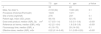
ResultsPatient doseA summary of the patient dose metrics and patient demographics are presented in Table 2.
Descriptive statistics of patient groups included in this study.
| 7.5pps | 4pps | p-Value | |
|---|---|---|---|
| n | 33 | 24 | |
| Male, No./total % | 21/33 (64) | 10/24 (42) | 0.1 |
| Procedure (Hickman/Permcath) | 10/23 | 13/11 | / |
| Site of entry (right/left) | 27/6 | 23/1 | 0.1 |
| Patient age, mean (SD), years | 56 (19) | 52 (15) | 0.5 |
| Dose area product, median (IQR), Gycm2 | 0.7 (0.5–1.4) | 0.3 (0.1–0.6) | <0.001 |
| Reference air kerma, median (IQR), mGy | 4.2 (2.1–8.4) | 1.9 (0.8–3.8) | <0.001 |
| Fluoroscopy time, median (IQR), s | 41 (18–70) | 29 (14–50) | 0.1 |
| Effective dose, median (IQR), mSv | 0.22 (0.14–0.43) | 0.1 (0.05–0.20) | <0.001 |
A decrease in fluoroscopic pulse rate from 7.5 to 4pps leads to a statistically significant reduction in DAP and RAK without a significant increase in fluoroscopy time (Table 2). This resulted in a median effective dose of 0.22 (0.14–0.43)mSv and 0.1 (0.05–0.20)mSv for the 7.5pps and 4pps groups, respectively (Fig. 2).

A linear regression analysis was calculated to predict effective dose based on DAP. A significant regression equation was found (F(1,52)=128,815, p<0.0001), with an R2 of 0.9996. The patient's predicted effective dose measured in mSv is equal to −0.0006+0.32 (DAP) when DAP is measured in Gycm2. Thus, the patient's effective dose increased approximately 0.32 for every 1Gycm2 of DAP.
Staff doseThe staff dose decreases with the decreasing of the fluoroscopic rate at the operators head and whole body. The whole-body dose is presented for both shielded and unshielded exposure to highlight the effectiveness of utilising a radioprotective apron.
The estimated shielded body dose for radiologist per procedure was 0.36μSv and 0.13μSv, respectively for 7.5 and 4pps. The dose to nurse and radiographer is gradually lower (Fig. 1); 0.1μSv and 0.03μSv for the nurse, 0.05μSv and 0.02μSv for radiographer. The study was extended to investigate occupational radiation exposure to the head, a surrogate value for ocular lens exposure. The estimated lens dose for the three staff categories is 2.11 and 0.7μSv, 1.35 and 0.47μSv, 0.63 and 0.2μSv for the radiologist, nurse and radiographer respectively for the 7.5pps and 4pps protocol.
DiscussionRadiation exposure during insertion of various CVADs varies significantly within the published literature. Our study recorded median, DAP, RAK, FT and effective doses for the insertion of Hickman lines and Permcaths of 0.7Gycm2 and 0.3Gycm2 dose area product, 4.2mGy and 1.9mGy RAK, 41s and 29s fluoroscopy time, and of 0.22mSv and 0.1mSv for fluoroscopic pulse rates of 7.5pps and 4pps respectively. Plumhans and colleagues reported much higher doses during insertion of implantable venous access devices, with means of 5.13Gycm2 for the internal jugular vein and 11.20Gycm2 for the subclavian vein. However, the latter approach involved mapping venography before device insertion.15 Jonczyk et al. investigated differences between proceduralist experience and the impact on patient dose during the insertion of the tunnelled central venous port, reporting a mean DAP of 0.573Gycm2 for ‘experienced’ radiologists and 0.682Gycm2 for ‘inexperienced’ radiologists.1 Variability also exists according to the medical specialty of the proceduralist, with a study revealing a mean Kerma-area product (KAP) of 0.832Gycm2 for tunnelled venous access catheter inserted by nephrologists.16 Similarly, dose differences have been observed between radiologist and surgeon implantation, although the former utilising venography confounded these results at the commencement of the procedure.17
The patient radiation dose from different studies has been summarised in Table 4. Additionally, our research shows that patient effective dose can be approximated using the conversion factor of 0.32mSv/Gycm2. The effective dose of 0.1–0.2mSv estimated in this study during CVAD insertion is unlikely to contribute to the development of tissue reactions, however, these procedures have the potential to contribute to the occurrence of stochastic effects.18,19 This is particularly pertinent for patients undergoing CVAD insertion which may be exposed to serial radiologic examinations over an extended period, particularly the oncological population.20
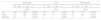
Patient radiation dose associated with central venous access device insertion.
| Reference | Year | Procedure | DAP (Gycm2) | Technique | RAK (mGy) | FT (s) | ED (mSv) | Type of study | Professional performing the procedurs | BMI |
|---|---|---|---|---|---|---|---|---|---|---|
| This study | 2017 | CVAD | 0.3 | Radiological | 1.9 | 29 | 0.1 | Retrospective observational study | Expert Radiologist | Avg |
| [Knebel] | 2011 | TIAP | 0.37 | Surgical | / | / | / | Retrospective and randomised | ExpertSurgeon | 25.1 |
| ” | ” | ” | 2.00 | Radiological | / | / | / | ” | Expert Radiologist | 25.2 |
| [Plumhans] | 2011 | TIAP | 5.13 | Jugular | / | / | / | Retrospective | Expert interventional radiologist | / |
| ” | ” | ”” | 11.20 | Subclavian | / | / | / | ” | ” | / |
| [Beathard] | 2013 | CVAD | 0.83 (KAP) | Radiological | / | / | / | Prospective | Expert Interventional Nephrologist | / |
| [Lee] | 2014 | PICC | 2.7 | C-arm Fluoroscopy | / | 52 | / | Prospective | / | / |
| [A.Rasekhi] | 2017 | CVAD | 1.51 | Radiological double lumen placement | / | 516 | / | Retrospective | Expert Radiologist | 24.6 |
| [Jonczyk] | 2018 | CVAD | 0.57 | Radiological | / | 24 | / | Retrospective | Senior Radiologist | / |
| ” | ” | ” | 0.68 | ” | / | 43 | / | ” | JuniorRadiologist | / |
DAP=dose area product; RAK=reference air kerma; FT=fluoroscopy time; ED=effective dose; CVAD=central venous access device; TIAP=totally implantable access ports; PICC=peripherally inserted central catheters.
Most of the above dose values are greater the recommended DRL of 0.3Gycm2 proposed by Greffier and colleagues in 2018, highlighting how technology has been improved in recent years.8 Moreover, our results are slightly above the median reported value of 0.6Gycm2 of Etard et al. for the central line insertion using the 7.5pps but fell below with the 4.5pps.7 It is critical to note, however, that the DRL is based on the third quartile value of median patient dose, and is thus most influential in determining unusually high dose values, rather than providing a reasonable target.4,21
Radiologist effective dose for the insertion of Hickman lines and Permcaths at our institution was estimated as 0.36μSv and 0.13μSv depending on the fluoroscopy pulse rate utilised (7.5pps and 4pps, respectively). Nurses and radiographers received less dose than radiologists, given the latter group's proximity to the primary radiation beam and scatter. Effective doses for nurses were 0.1μSv and 0.03μSv for 7.5pps and 4pps respectively, with the same pulse rates producing effective doses for radiographers of 0.05μSv and 0.02μSv. Rasekhi et al. have also measured occupational dose specific to CVAD insertion, with radiologists experiencing a mean effective dose of 0.02μSv during ‘challenging’ haemodialysis catheter placement.22
The higher dose observed at our institution may be attributable to the difference in sensitivities between the survey metre used in our study versus using personal dosimeters (TLD). Furthermore, the dose reported by Rasekhi et al. is based on real procedures where the radioprotection habits may change while our study is done in a simulated environment with fixed positions and shielding.22 Finally, the interposition between the staff members that is likely to happen in the real scenario would mean that some staff are shielded by other staff members who are standing between them and the source of scatter. Thus, our estimation of staff dose could be seen as a worst-case scenario when assuming average-sized patients and average procedure complexity. No corresponding exposure data could be found in the literature specifically for nurses and radiographers during insertion of CVADs. However, nurse effective dose levels for a variety of interventional radiology procedures have been reported between 0.018 and 1.42μSv.18,23,24
The current study was extended to investigate occupational radiation exposure to the head, a surrogate value for ocular lens exposure. The dose to the head of the radiologist, nurse and radiographer was 2.1μSv, 1.4μSv, and 0.6μSv for studies utilising 7.5pps, and 0.7μSv, 0.5μSv, and 0.2μSv for procedures using 4pps. These results represent an ‘unshielded’ measurement, which could be vastly diminished with the use of lead glasses. No data exist specifically for ocular lens exposure during CVAD placement unless results referring to more complex and more protracted procedures with much higher dose are considered.25 In practice, the use of leaded glasses would be recommended based on reported cases of lens opacities.26 However, if the staff are solely performing CVAD and not involved in complex interventional procedures, they are unlikely to exceed the occupational limit of 20mSv year for the lens of the eye.27
As demonstrated in Table 3 and Fig. 2, the pulse rate utilised during insertion of CVADs had a significant effect on patient dose and occupational whole-body and head dose. In this study, the pulse rate was reduced while all the other parameters, such as kVp, field size and filtration, remained similar in both patient groups. The results showed that a reduction in fluoroscopic pulse rate was not accompanied by an increase in fluoroscopy time which indicates that completion of the primary objectives of the procedure was achieved without the need to image more frequently to account for the reduction in temporal resolution. Similar results have been observed throughout the literature when lower pulse rates are employed for fluoroscopy studies.9–12
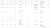
Occupational eye, body and body (shielded) dose depending on the fluoroscopy pulse rate utilised.
| Radiologist | Nurse | Radiographer | |||||||
|---|---|---|---|---|---|---|---|---|---|
| 7.5pps | 4pps | p | 7.5pps | 4pps | p | 7.5pps | 4pps | p | |
| μSv/h | μSv/h | μSv/h | |||||||
| Eye | 186 | 92.6 | <0.0001 | 119 | 59 | <0.0001 | 56 | 27 | <0.0001 |
| Body | 471 | 240 | <0.0001 | 126 | 63 | <0.0001 | 61 | 30 | <0.0001 |
| Body (S) | 32 | 16 | <0.0001 | 9 | 4 | <0.0001 | 4 | 2 | <0.0001 |
Nonetheless, these results must be interpreted with caution, and some limitations should be considered in future research studies. The use of phantoms can be regarded as an excellent approximation to evaluate equipment settings and other protocols but is limited in assessing the complexities and variance in clinical practice. Another limitation in this study is the small sample size due to the novelty of the reduced pulse rate at this centre.
ConclusionA retrospective observational audit was conducted on patients undergoing CVAD insertions revealing a median effective dose of 0.22mSv and 0.1mSv for the 7.5pps and 4pps groups respectively. The radiologist, nurse and radiographer whole body and lens exposure was estimated in a simulated setting. In all cases, there was a statistically significant dose reduction when the lower fluoroscopic pulse rate was used. Thus, where possible, consideration should be given to utilising a lower pulse rate during CVAD insertions to reduce the exposure to both staff and patients.
Authorship- 1.
Responsible for the integrity of the study: RB
- 2.
The conception of the study
- 3.
The design of the study: MKB.
- 4.
Acquisition of data: MKB, CJW, RB, DC and EY.
- 5.
Analysis and interpretation of data: MKB, CJW, RB, DC and EY.
- 6.
Statistical treatment: N.A.
- 7.
Bibliographic search
- 8.
Drafting of the work: MKB, CJW, RB.
- 9.
Critical review of the manuscript with intellectually relevant contributions: EY.
- 10.
Final approval of the version: MKB, CJW, RB , DC and EY.
Mohamed K. Badawy: Substantial contributions to the design; collection, analysis, and interpretation of data; drafting the article and final approval of the version to be published.
Chris J Witkowski: Collection, analysis, and interpretation of data; drafting the article and final approval of the version to be published.
Riccardo Baldoni (corresponding author): collection, analysis, and interpretation of data; drafting the article and final approval of the version to be published
Daniel Carrion: collection, analysis, and interpretation of data; and final approval of the version to be published.
Ertan Yildirim: Substantial contributions to the design; collection, and interpretation of data; and final approval of the version to be published.
Conflicts of interestNone.
Please cite this article as: Badawy MK, Witkowski CJ, Baldoni R, Carrion D, Yildirim E. Dosis de radiación durante la inserción guiada por fluoroscopia de un dispositivo de acceso venoso central: un estudio observacional retrospectivo. Radiología. 2021;63:5–12.

