


To use the mDIXON-Quant sequence to quantify the fat fraction of adrenal lesions discovered incidentally on CT studies. To analyze the relation between the signal loss between in-phase and out-of-phase T1-weighted sequences and the fat fraction in mDIXON-Quant. To compare the sensitivity and specificity of the two methods for characterizing adrenal lesions.
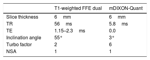
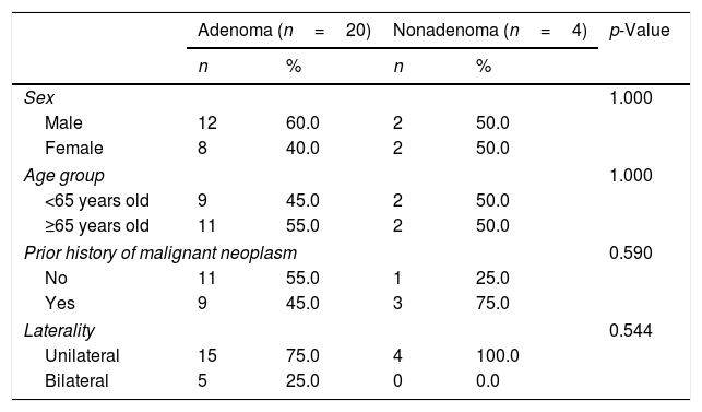
Material and methodsThis prospective descriptive study included 31 patients with incidentally discovered adrenal lesions evaluated with 3T MRI using in-phase and out-of-phase T1-weighted sequences and mDIXON-Quant; the fat fraction of the adrenal lesions was measured by mDIXON-Quant and by calculating the percentage of signal loss between in-phase and out-of-phase T1-weighted sequences.
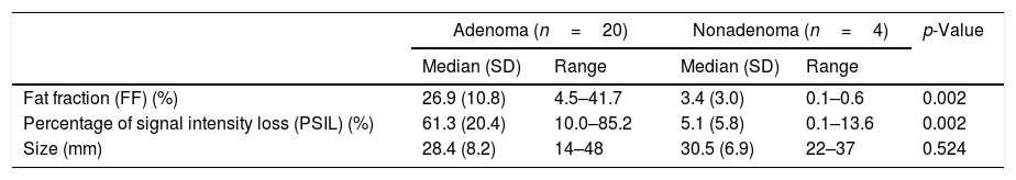
ResultsThe percentage of signal loss was significantly higher in the group of patients with adenoma (61.3±20.4% vs. 5.1±5.8% in the group without adenoma, p<0.005). The mean fat fraction measured by mDIXON-Quant was also higher for the adenomas (26.9±10.8% vs. 3.4±3.0%, p<0.005). The area under the ROC curve was 0.99 (0.96–1.00) for the percentage of signal loss and 0.98 (0.94–1.00) for the fat fraction measured by mDIXON-Quant. The cutoffs obtained were 24.42% for the percentage of signal loss and 9.2% for the fat fraction measured by mDIXON-Quant. The two techniques had the same values for diagnostic accuracy: sensitivity 96% (79.6–99.9), specificity 100% (39.8–100.0), positive predictive value 100% (85.8–100.0), and negative predictive value 80% (28.4–99.5).
ConclusionThe fat fraction measured by the modified Dixon technique can differentiate between adenomas and other adrenal lesions with the same sensitivity and specificity as the percentage of signal loss between in-phase and out-of-phase T1-weighted sequences.
Cuantificar mediante secuencia mDIXON-Quant la fracción grasa (FG) de las lesiones suprarrenales encontradas incidentalmente en estudios de TC. Analizar la relación de la caída de señal entre las secuencias potenciadas en T1 en fase y fase opuesta con la FG en mDIXON-Quant. Comparar la sensibilidad y especificidad de ambos métodos para caracterizar las lesiones suprarrenales.
Material y métodosSe realizó un estudio prospectivo descriptivo que incluyó a 31 pacientes con lesiones suprarrenales incidentales evaluados mediante RM 3T con las secuencias T1 en fase y fase opuesta y mDIXON-Quant. Se midió la FG de las lesiones suprarrenales mediante mDIXON-Quant y la intensidad de señal en secuencias T1 en fase y fase opuesta, calculando su porcentaje de pérdida de señal (PPS).
ResultadosEl PPS medio fue significativamente mayor en el grupo adenoma (61,3%±20,4%) que en el no adenoma (5,1%±5,8%) (p < 0,005). La FG media de los adenomas también fue significativamente mayor (26,9%±10,8% vs. 3,4%±3,0%) (p < 0,005). El área bajo la curva ROC fue 0,99 (0,96-1,00) para el PPS y 0,98 (0,94-1,00) para la FG. El punto de corte obtenido fue de 24,42% para el PPS y de 9,2% para la FG. Los valores diagnósticos fueron iguales para los dos métodos: sensibilidad del 96% (79,6-99,9), especificidad del 100% (39,8-100,0), valor predictivo positivo del 100% (85,8-100,0) y valor predictivo negativo del 80% (28,4-99,5).
ConclusiónLa FG obtenida mediante técnica Dixon modificada es capaz de diferenciar adenomas de no adenomas con la misma sensibilidad y especificidad que la PPS.

