metricas
covid
Radiología (English Edition) Ophthalmological emergencies
Journal Information
Vol. 67. Issue 6.
(November - December 2025)
Visits
13
Vol. 67. Issue 6.
(November - December 2025)
Update in Radiology
Ophthalmological emergencies
Urgencias oftalmológicas
Visits
13
M.C. Maciá Fernándeza,b,
Corresponding author
mcmaciafernandez@gmail.com

Corresponding author.
, G. Santabrígida Orejaa, L.H. Vargas Alvaradoa, S. Sánchez Bernala,b, A.B. Valentín Martína,b, J. Encinas de la Iglesiaa
a Servicio de Radiodiagnóstico, Hospital Universitario de Salamanca, Salamanca, Spain
b Sección de Radiología de Urgencias, Hospital Universitario de Salamanca, Salamanca, Spain
This item has received
Article information
Abstract
Full Text
Bibliography
Download PDF
Statistics
Figures (15)
Show moreShow less
Tables (3)
Table 1. Imaging protocol for the diagnosis of orbital disease using CT.
Tables
Table 2. Characteristics of preseptal (periorbital) cellulitis and postseptal (orbital) cellulitis.
Tables
Table 3. Anatomical classification of carotid–cavernous fistulas and their pathophysiological classification based on angiographic findings (Barrow classification) and their causes.
Tables
Show moreShow less
Abstract

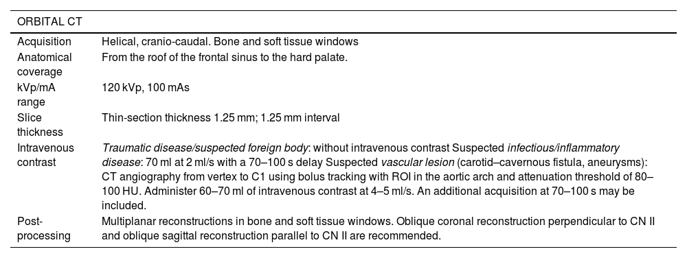
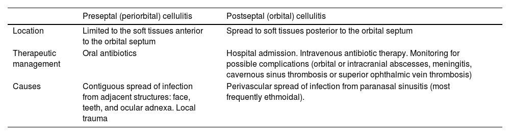
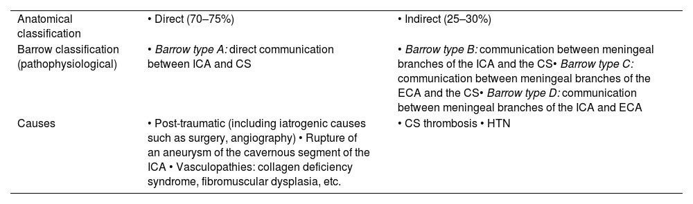
Urgent ophthalmic pathology can be grouped into four categories: infectious, traumatic, vascular, and inflammatory. Imaging techniques play a fundamental role in their diagnosis, positioning the radiologist at the forefront when dealing with these entities. CT is usually the technique of choice within the emergency context, with MRI and ultrasound playing a secondary role. Radiologists must be familiar with the main imaging findings of these pathologies to make a rapid and accurate diagnosis that contributes to a positive outcome for the patient. This article reviews orbital anatomy, imaging techniques and protocols that should be employed, as well as key diagnostic clues that every radiologist should know.

Keywords:
Computed tomography
Protocol
Ocular infection
Retinal detachment
Choroidal detachment
Lens dislocation
Eye injuries
Intraocular foreign bodies
Carotid cavernous fistula
Eye inflammation
Resumen

La patología oftálmica urgente se puede agrupar en cuatro categorías: infecciosa, traumática, vascular e inflamatoria. Las técnicas de imagen juegan un papel fundamental en su diagnóstico, lo que posiciona al radiólogo en el primer eslabón de estas entidades. La TC suele ser la técnica de elección dentro del contexto urgente, presentando la RM y la ecografía un papel secundario. Los radiólogos debemos estar familiarizados con los principales hallazgos de imagen de estas patologías para poder realizar un diagnóstico rápido y preciso que contribuya a la buena evolución del paciente. Este artículo repasa la anatomía orbitaria, las técnicas y protocolos de imagen que se deben emplear, así como las claves diagnósticas que todo radiólogo debe conocer.

Palabras clave:
Tomografía computarizada
Protocolo
Infección ocular
Desprendimiento de retina
Desprendimiento coroideo
Luxación cristalino
Daño ocular
Cuerpos extraños intraoculares
Fístula carótido-cavernosa
Inflamación ocular

Article

These are the options to access the full texts of the publication Radiología (English Edition)
Subscriber
Subscriber

If you already have your login data, please click here .

If you have forgotten your password you can you can recover it by clicking here and selecting the option “I have forgotten my password”
Purchase
Purchase article

Purchasing article the PDF version will be downloaded

Purchase now
Contact
Phone for subscriptions and reporting of errors
From Monday to Friday from 9 a.m. to 6 p.m. (GMT + 1) except for the months of July and August which will be from 9 a.m. to 3 p.m.
Calls from Spain
932 415 960
Calls from outside Spain
+34 932 415 960
E-mail
Article options
Tools