


We aimed to analyze the relationship between the initial chest X-ray findings in patients with severe acute respiratory syndrome due to infection with SARS-CoV-2 and eventual clinical worsening and to compare three systems of quantifying these findings.
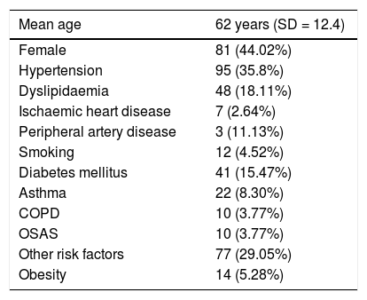
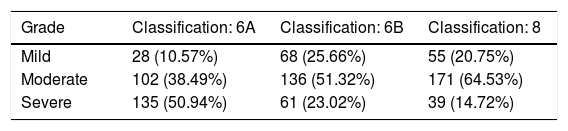
Material and methodsThis retrospective study reviewed the clinical and radiological evolution of 265 adult patients with COVID-19 attended at our center between March 2020 and April 2020. We recorded data related to patients’ comorbidities, hospital stay, and clinical worsening (admission to the ICU, intubation, and death). We used three scoring systems taking into consideration 6 or 8 lung fields (designated 6A, 6B, and 8) to quantify lung involvement in each patient’s initial pathological chest X-ray and to classify its severity as mild, moderate, or severe, and we compared these three systems. We also recorded the presence of alveolar opacities and linear opacities (fundamentally linear atelectasis) in the first chest X-ray with pathologic findings.
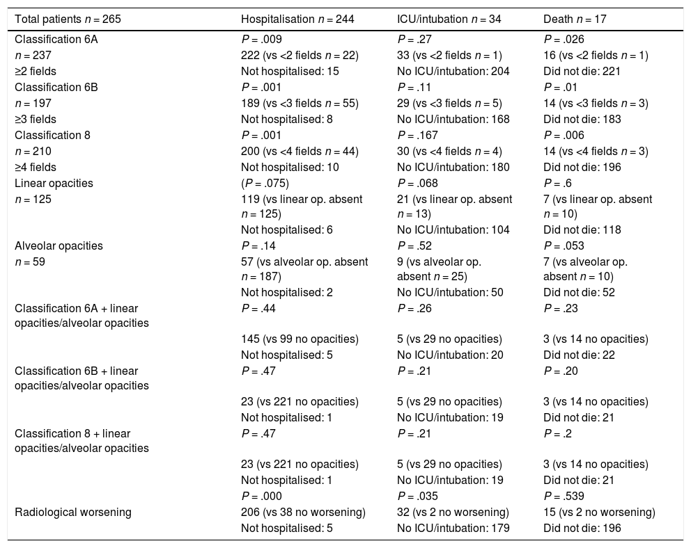
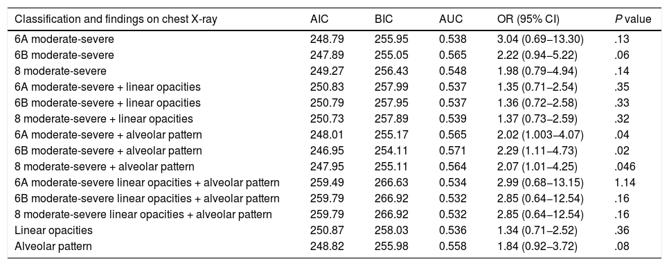
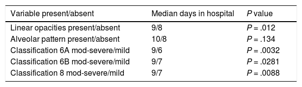
ResultsIn the χ2 analysis, moderate or severe involvement in the three classification systems correlated with hospital admission (P = .009 in 6A, P = .001 in 6B, and P = .001 in 8) and with death (P = .02 in 6A, P = .01 in 6B, and P = .006 in 8). In the regression analysis, the most significant associations were 6B with alveolar involvement (OR 2.3; 95%CI 1.1.–4.7; P = .025;) and 8 with alveolar involvement (OR 2.07; 95% CI 1.01.–4.25; P = .046). No differences were observed in the ability of the three systems to predict clinical worsening by classifications of involvement in chest X-rays as moderate or severe.
ConclusionModerate/severe extension in the three chest X-ray scoring systems evaluating the extent of involvement over 6 or 8 lung fields and the finding of alveolar opacities in the first pathologic X-ray correlated with mortality and the rate of hospitalization in the patients studied. No significant difference was found in the predictive ability of the three classification systems proposed.
El propósito de este estudio en 265 pacientes con síndrome respiratorio agudo severo por coronavirus 2 (SARS-CoV-2) es establecer una relación entre la cuantificación inicial de los hallazgos en la radiografía de tórax en pacientes con COVID-19 y un eventual empeoramiento clínico, así como comparar tres sistemas de cuantificación que se proponen.
Material y métodosEn este estudio retrospectivo se revisó la evolución clínica y radiológica de 265 pacientes adultos con infección por COVID-19 atendidos en nuestro centro entre marzo y abril de 2020, registrándose la presencia de comorbilidades, así como datos de estancia hospitalaria y empeoramiento clínico (ingreso en unidad de cuidados intensivos/intubación y fallecimiento). La afectación en la radiografía de tórax inicial patológica de cada paciente se cuantificó siguiendo tres sistemas de puntuación diferentes sobre 6 u 8 campos pulmonares (denominados 6 A, 6 B y 8) para determinar una afectación leve, moderada o grave y establecer comparación entre dichos sistemas. También se registró la presencia de opacidades alveolares y opacidades lineales (fundamentalmente atelectasia laminar) en la primera radiografía patológica.
ResultadosLa afectación moderada o grave en los tres sistemas de cuantificación (6 A, 6 B y 8) mostró correlación en el estudio de χ2 con la variable ingreso hospitalario (P = 0,009, 0,001, 0,001, respectivamente) y fallecimiento de los pacientes (P = 0,02, 0,01, 0,006, respectivamente). El estudio de regresión mostró como más significativas las asociaciones 6 B + afectación alveolar [odds ratio (OR) 2,3, P = 0,025, intervalo de confianza (IC) al 95% 1,1–4,7) y clasificación 8 + afectación alveolar (OR 2,07, P = 0,046, IC 95% 1,01–4,25). La afectación moderada o grave en la radiografía de tórax no mostró diferencias significativas entre los tres sistemas de cuantificación en el estudio predictivo de empeoramiento clínico.
ConclusiónLa extensión moderada/grave en los tres sistemas de puntuación de la radiografía de tórax valorando la extensión de enfermedad sobre 6 y 8 campos pulmonares y la existencia de opacidades alveolares en la primera radiografía patológica se correlacionó con la mortalidad y la tasa de hospitalización en los pacientes estudiados. No se apreció una diferencia significativa en la capacidad predictiva de los tres sistemas de cuantificación propuestos.

