Buscar en
Radiología (English Edition)
Toda la web
Inicio Radiología (English Edition) High resolution (3T) magnetic resonance neurography of the sciatic nerve
Journal Information
Vol. 55. Issue 3.
Pages 195-202 (May - June 2013)
Share
Share
Download PDF
More article options
Visits
3841
Vol. 55. Issue 3.
Pages 195-202 (May - June 2013)
Update in radiology
High resolution (3T) magnetic resonance neurography of the sciatic nerve
Neurografía por resonancia magnética de alta resolución (3Tesla) del nervio ciático
Visits
3841
C. Cejas
Corresponding author
ccejas@fleni.org.ar

Corresponding author.
, M. Aguilar, L. Falcón, N. Caneo, M.C. Acuña
Servicio de Resonancia Magnética, Departamento de Diagnóstico por Imágenes, Fundación para la Lucha de las Enfermedades Neurológicas de la Infancia Dr. Raúl Carrea (FLENI), Buenos Aires, Argentina
This item has received
Article information
Abstract
Full Text
Bibliography
Download PDF
Statistics
Figures (7)
Show moreShow less
Tables (2)
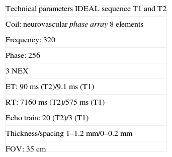
Table 1. Technical parameters. IDEAL sequence.
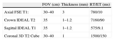
Table 2. Sciatic nerve neurography protocol.
Show moreShow less
Abstract

Magnetic resonance (MR) neurography refers to a set of techniques that enable the structure of the peripheral nerves and nerve plexuses to be evaluated optimally. New two-dimensional and three-dimensional neurographic sequences, in particular in 3T scanners, achieve excellent contrast between the nerve and perineural structures. MR neurography makes it possible to distinguish between the normal fascicular pattern of the nerve and anomalies like inflammation, trauma, and tumor that can affect nerves. In this article, we describe the structure of the sciatic nerve, its characteristics on MR neurography, and the most common diseases that affect it.

Keywords:
Sciatic nerve
Sciatic neuropathy
Neurography
Magnetic resonance imaging
Resumen

La neurografía por resonancia magnética (RM) hace referencia a un conjunto de técnicas con capacidad para valorar óptimamente la estructura de los nervios periféricos y de los plexos nerviosos. Las nuevas secuencias neurográficas 2D y 3D, en particular en equipos de 3Tesla, consiguen un contraste excelente entre el nervio y las estructuras perineurales. La neurografía por RM permite distinguir el patrón fascicular normal del nervio y diferenciarlo de las anomalías que lo afectan, como inflamaciones, traumas y tumores. En este artículo se describe la estructura del nervio ciático, sus características en la neurografía por RM y las dolencias que lo afectan con mayor frecuencia.

Palabras clave:
Nervio ciático
Neuropatía ciática
Neurografía
Imagen por resonancia magnética

Article

These are the options to access the full texts of the publication Radiología (English Edition)
Subscriber
Subscriber

If you already have your login data, please click here .

If you have forgotten your password you can you can recover it by clicking here and selecting the option “I have forgotten my password”
Purchase
Purchase article

Purchasing article the PDF version will be downloaded

Price 19.34 €

Purchase now
Contact
Phone for subscriptions and reporting of errors
From Monday to Friday from 9 a.m. to 6 p.m. (GMT + 1) except for the months of July and August which will be from 9 a.m. to 3 p.m.
Calls from Spain
932 415 960
Calls from outside Spain
+34 932 415 960
E-mail
Article options
Tools
es en pt

¿Es usted profesional sanitario apto para prescribir o dispensar medicamentos?

Are you a health professional able to prescribe or dispense drugs?

Você é um profissional de saúde habilitado a prescrever ou dispensar medicamentos

Quizás le interese:
10.1016/j.rxeng.2019.11.007
No mostrar más