


La ecocardiografía 3D (E3D) es más precisa que la técnica 2D en la valoración de los volúmenes y la fracción de eyección (FE) del ventrículo izquierdo (VI). Este estudio compara, con respecto a cardiorresonancia magnética (cRM), 2sistemas de E3D: E3D en un solo latido con detección automática de bordes endocárdicos (Siemens SC2000;A) frente a E3D con adquisición de varios latidos y detección semiautomática de bordes endocárdicos (Philips iE33;B).
MétodosA 22 pacientes remitidos para estudio mediante cRM se les realizó E3D con sistema de adquisición en un solo latido y detección automática de bordes (A) y con adquisición en varios latidos y detección semiautomática de bordes (B).
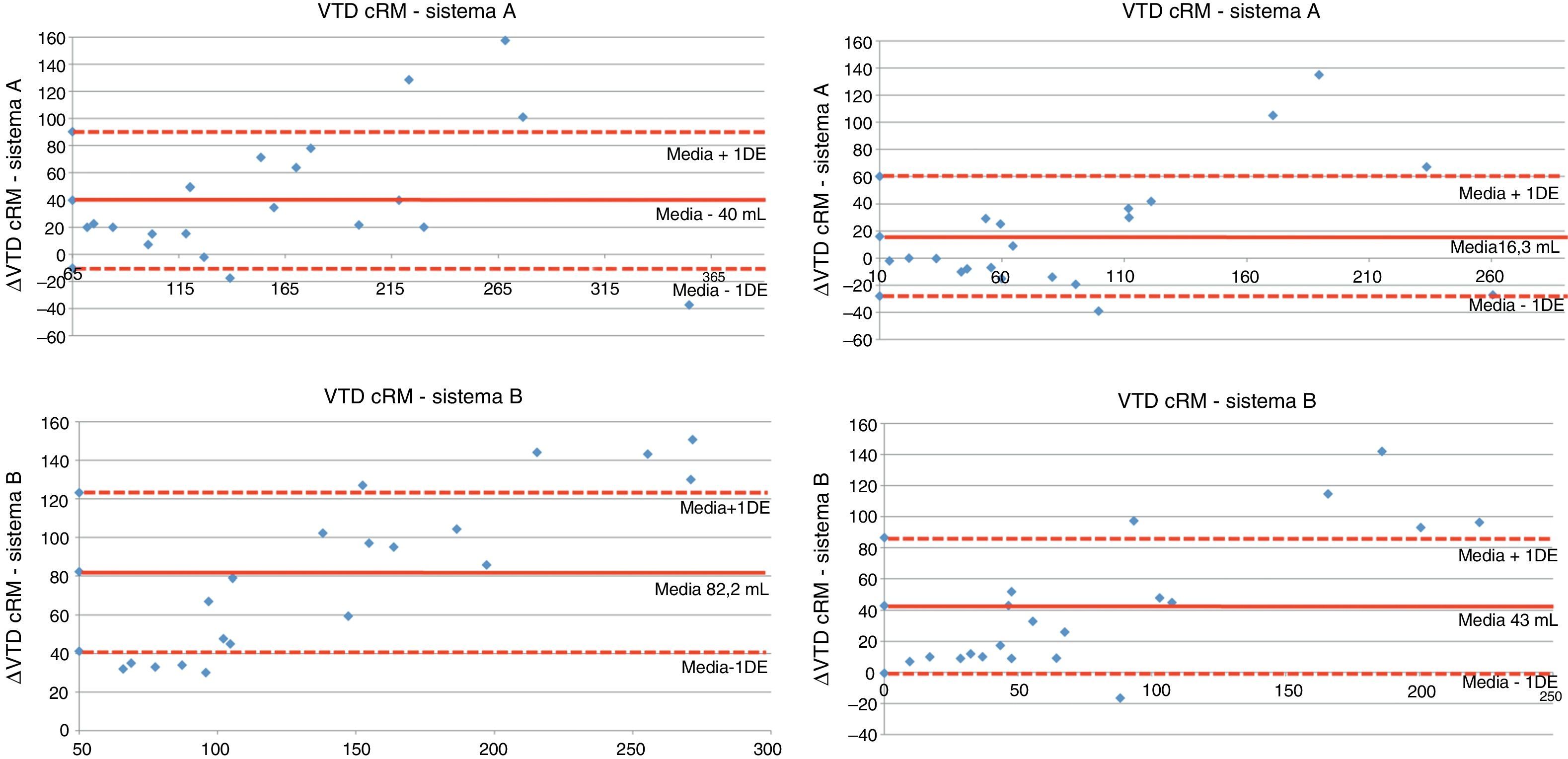
ResultadosLos volúmenes y la FE medios por cRM fueron: VTD: 211,07±85,4ml; VTS: 119,2±89,25ml; FE: 48,67±20,07%. La correlación entre la cRM y los 2sistemas de E3D fue excelente para el VTD (rhoA 0,912; rhoB 0,907; p<0,0001), VTS (rhoA 0,877; rhoB 0,945; p<0,0001) y FE (rhoA 0,974; rhoB 0,951; p<0,0001). El análisis mediante Bland-Altman reveló infraestimación de los volúmenes del VI con ambos sitemas de E3D, siendo esta menor con el sistemaA queB (–40±50,2 vs –82,2±40,9ml, respectivamente, para VTD; –16±44,4 vs –43±43,5ml, respectivamente, para VTS; p=0,001). No hubo diferencias en la medida de la FE entre las 3técnicas de imagen.
ConclusiónLa E3D infraestima los volúmenes del VI. En nuestra serie esta infraestimación fue menor con el sistema de adquisición en único latido y detección automática de bordes.
3-D echocardiography (3DE) is superior to the 2-D technique in assessing left ventricular (LV) volumes and ejection fraction (EF). This study compares two 3DE systems with cardiac magnetic resonance imaging (cMRI): single-beat 3DE with automated endocardiac border detection (Siemens SC2000;A) and 3DE with multi-beat acquisition and semi-automated endocardiac borders detection (Philips iE33;B).
MethodsA 3DE with single-beat acquisition and automated border detection (A) and 3DE with multi-beat acquisition and semi-automated border detection (B) was performed on 22patients referred for a study using cMRI.
ResultsThe mean volumes and EF measured by cMRI were: DTV: 211.07±85.4mL; STV: 119.2±89.25mL; EF: 48.67±20.07%. The correlation between cMRI and the two 3DE systems was excellent for the DTV (rhoA 0.912; rhoB 0.907; P<.0001), STV (rhoA 0.877; rhoB 0.945; P<.0001) and EF (rhoA 0.974; rhoB 0.951; P<.0001). The Bland-Altman analysis showed an underestimation of the LV volumes with both 3DE systems, with the difference being lower in systemA than in systemB (–40±50.2 vs –82.2±40.9mL, respectively for DTV; –16±44.4 vs –43±43.5mL, respectively for STV; P=.001). There were no differences in the measurement of EF between the 3imaging techniques.
Conclusion3DE underestimates LV volumes. In our series, this underestimate was lower in the system with single-beat acquisition and automated border detection.
Los volúmenes y la fracción de eyección (FE) del ventrículo izquierdo (VI) son parámetros de un gran valor en las enfermedades cardiovasculares y con un significativo impacto en la toma de decisiones clínicas. El método más extendido para la determinación de estos parámetros es generalmente el método de Simpson modificado mediante ecocardiografía 2D. No obstante, la exactitud de este método es limitada debido a que aplica asunciones geométricas y es conocida su infraestimación de los volúmenes ventriculares. La ecocardiografía 3D en tiempo real (E3D), en cambio, no realiza dichas asunciones geométricas, logrando de este modo una valoración más exacta de los volúmenes del VI1.
Existen en el mercado diversos sistemas para adquisición del VI por E3D. Unos realizan la adquisición del volumen del VI adquiriendo sincronizadamente con el ECG varios subvolúmenes (al menos4) a lo largo de varios latidos. Este método tiene varias limitaciones, ya que para evitar artefactos requiere un ritmo cardíaco regular, mantener una posición estable de la sonda y contener la respiración por parte del paciente. Con la intención de superar estas limitaciones surgieron los sistemas de E3D con adquisición en un solo latido.
Otra de las ventajas de la E3D respecto a la ecocardiografía 2D es la incorporación rutinaria de la detección más o menos automática del borde endocárdico. Los distintos sistemas de E3D emplean metodologías diferentes para esta tarea2. El propósito de este estudio es comparar, con respecto a cardiorresonancia magnética (cRM), en una misma población de sujetos 2sistemas distintos de E3D: por una parte la E3D en un solo latido con detección completamente automática de bordes endocárdicos (Siemens Acuson), y por otra la E3D con adquisición en varios latidos y detección semiautomática de bordes endocárdicos (Philips iE33).
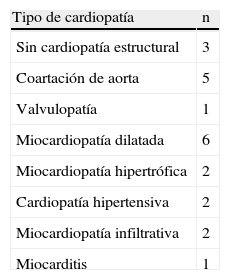
MétodosSe incluyeron 22pacientes consecuivos (54,5% varones; 44,2±19,3años) remitidos para la realización de cRM (Siemens, Viewform). En la tabla 1 se muestra la enfermedad cardíaca que indicó el estudio de cRM. A todos los individuos se les realizó E3D el mismo día de la realización de la cRM, tras concluir la misma. En cada individuo las imágenes fueron adquiridas con 2sistemas de E3D de forma consecutiva: Siemens Acuson SC2000 (Siemens, Erlangen, Alemania) (sistemaA) y Philips iE33 (Philips, California, EE.UU.) (sistemaB). La adquisición con el sistemaB se llevó a cabo en 4latidos. No se utilizó contraste en ningún estudio. Los estudios fueron analizados por 2observadores independientes ciegos al resultado de la cRM. Los estudios fueron asignados a 3categorías según la necesidad de corrección manual del borde endocárdico detectado de forma automática o semiautomática: no necesidad de corrección, necesidad de corrección en <3segmentos y necesidad de corrección en ≥3segmentos. Para comprobar la variabilidad inter e intraobservador en las medidas del ecocardiograma 9estudios fueron cuantificados con ambos sistemas de E3D por los 2operadores independientes y a su vez por el mismo operador en un intervalo de tiempo superior a 15días. La concordancia se midió mediante el coeficiente de correlación interclases, obteniendo un valor de 0,91.
Las variables continuas se expresaron como media±desviación estándar y las categóricas, como frecuencias en porcentajes. Las variables cuantitativas se compararon mediante prueba de la t de Student. Todos los valores de p fueron calculados mediante tests de 2colas, y se consideró significativo un valor de p<0,05. Para el análisis estadístico se utilizó el programa SPSS versión 19.0.
ResultadosEl análisis de los volúmenes y de la FE del VI con ambos sistemas de E3D fue posible en 20 de los pacientes (90,9%). En 2 (9,1%) no fue posible realizar el análisis con ninguno de los 2sistemas por mala ventana acústica. De los 20pacientes analizados, 18 (81,8%) se encontraban en ritmo sinusal. Los volúmenes y la FE medios obtenidos con cRM fueron: VTD: 211,07±85,4ml; VTS: 119,2±89,25ml; FE: 48,67±20,07%. La correlación entre la cRM y los 2sistemas de E3D fue excelente para el VTD (rhoA 0,912; rhoB 0,907; p<0,0001), VTS (rhoA 0,877, rhoB 0,945; p<0,0001) y FE (rhoA 0,974; rhoB 0,951; p<0,0001). Sin embargo, el análisis de concordancia mediante Bland-Altman reveló infraestimación de los volúmenes del VI con ambos sistemas de E3D respecto a cRM, siendo esta infraestimación menor con el sistema A (–40±50,2ml para VTD; –16±44,4ml para VTS) vs sistema B (–82,2±41ml para VTD; –43±43,5ml para VTS); p=0,001 (fig. 1).
.")
Con el sistemaA se llevó a cabo la corrección manual en <3segmentos en 3pacientes (15%); en ≥3segmentos en 5pacientes (25%), no siendo necesaria en el resto (12pacientes, 60%). Con el sistemaB, la corrección manual no fue necesaria en 7pacientes (35%), fue necesaria en <3segmentos en 4pacientes (20%), siendo necesaria en ≥3segmentos en 9pacientes (45%).
El cálculo de la FE fue igualmente exacto con ambos sistemas de E3D sin variaciones significativas respecto a los valores obtenidos con cRM.
DiscusiónAunque en menor grado que con el ecocardiograma 2D, la E3D también presenta una infraestimación de los volúmenes del VI respecto al patrón oro de la cRM3. En este trabajo la infraestimación fue menor con el sistema de adquisición en un solo latido y detección totalmente automática de bordes. Se ha postulado que la infraestimación del E3D se debería a que, a diferencia de la cRM, la detección de bordes realizada por el ecocardiograma incluiría a las trabéculas en la pared ventricular, considerando menos volumen de cavidad4. En nuestro trabajo observamos que la infraestimación fue menor con el sistemaA que con elB. Creemos que la explicación a esta diferencia podría radicar en 2 hechos:
- 1.
El sistemaB es semiautomático y requiere ajustar los planos de corte del VI, seleccionar las imágenes telediastólica y telesistólica y señalar en cada una de ellas 5puntos. Con estas referencias el seguimiento del borde endocárdico se realiza mediante el patrón de speckle, lo cual podría suponer la exclusión sistemática de las trabéculas del volumen del VI, explicando la obtención de volúmenes menores. Sin embargo, para la detección de bordes el sistemaA emplea un modelo estadístico basado en una base de datos que tiene en cuenta la forma y la apariencia endocárdica en telediástole y telesístole. El contorno es seguido a lo largo de la secuencia completa a partir de ambas imágenes, fundiendo el movimiento estimado en ellas con un modelo dinámico basado en una base de datos poblacional. Este método, al no considerar el patrón de speckle, podría suponer un mejor ajuste de la delimitación al verdadero endocardio, evitando así la inclusión de la trabécula en la pared miocárdica.
- 2.
La cuantificación de volúmenes con el sistemaB requirió de un mayor número de ajustes manuales para un mejor seguimiento del ápex. En muchos casos estos retoques tuvieron lugar en el ápex. Aunque el resultado final de seguimiento fue visualmente satisfactorio, puede que este hecho tuviera consecuencias en una mayor infraestimación de volúmenes por parte de este sistema.
Como se puede apreciar en el análisis Bland-Altman, hubo casos donde el volumen fue superior con el sistemaA que con la cRM. Este hecho sucedió especialmente para el volumen telesistólico (9pacientes). Aunque estas diferencias tuvieron poca significación en cuanto valor absoluto, es de mencionar que este hecho sucedió especialmente en pacientes con VI no dilatado. Desconocemos la explicación a este hallazgo, que requeriría de una población mayor para su confirmación. Si así fuera, esto podría ser debido a un seguimiento automático más allá del borde endocárdico en la telesístole de ventrículos no dilatados por parte del sistemaA.
El sistemaA, al realizar una adquisición en un único latido, aportaría una ventaja teórica en pacientes con arritmias, como fibrilación auricular, al evitar el artefacto de pegamiento que sucedería en estos pacientes en la adquisición de múltiples latidos con el sistemaB. Este hecho no pudo ser estudiado en nuestro trabajo, ya que los casos con fibrilación auricular estuvieron escasamente representados (solo 2 de 20).
La principal limitación de este estudio es el pequeño tamaño muestral, por lo que sus resultados habrían de ser confirmados en una población mayor de pacientes.
ConclusionesLa E3D infraestima los volúmenes del VI, en relación con los obtenidos mediante cRM. En nuestra serie esta infraestimación fue menor con el sistema de adquisición en un único latido y detección automática de bordes.
Conflicto de interesesLos autores declaran no tener ningún conflicto de intereses.
