Buscar en
Archivos de la Sociedad Española de Oftalmología (English Edition)
Toda la web
Inicio Archivos de la Sociedad Española de Oftalmología (English Edition) Mapping the thickness changes on retinal layers segmented by spectral-domain opt...
Journal Information
Vol. 93. Issue 6.
Pages 263-273 (June 2018)
Share
Share
Download PDF
More article options
Visits
6
Vol. 93. Issue 6.
Pages 263-273 (June 2018)
Original article
Mapping the thickness changes on retinal layers segmented by spectral-domain optical coherence tomography using the posterior pole program in glaucoma
Mapeo de los cambios de grosor en el glaucoma de las capas retinianas maculares segmentadas usando el programa de polo posterior de la tomografía de coherencia óptica de dominio espectral
Visits
6
J.J. García-Medinaa,b,c,d,e,f,
Corresponding author
, M. del-Rio-Vellosillog, A. Palazón-Cabanesa, M. Tudela-Molinoa, C. Gómez-Molinaa, A. Guardiola-Fernándeza, M.P. Villegas-Péreza,c,d,e
a Departamento de Oftalmología, Hospital General Universitario Reina Sofía, Murcia, Spain
b Departamento de Oftalmología, Hospital General Universitario Morales Meseguer, Murcia, Spain
c Departamento de Oftalmología, Optometría, Otorrinolaringología y Anatomía Patológica, Facultad de Medicina, Universidad de Murcia, Murcia, Spain
d Red Temática de Patología Ocular OFTARED, Instituto de Salud Carlos III, Madrid, Spain
e Instituto Murciano de Investigación Biosanitaria, Hospital Virgen de la Arrixaca (IMIB-Virgen de la Arrixaca), Murcia, Spain
f Unidad de Investigación Oftalmológica Santiago Grisolía, Valencia, Spain
g Departamento de Anestesiología, Hospital Universitario Virgen de la Arrixaca, Murcia, Spain
This item has received
Article information
Abstract
Full Text
Bibliography
Download PDF
Statistics
Figures (13)
Show moreShow less
Tables (4)
Table 1. Demographic and clinical characteristics of study subjects.
Table 2. Mean superior hemisphere thickness comparisons in the studied layers between the control and glaucoma groups.
Table 3. Mean inferior hemisphere thickness comparisons in the studied layers between the control and glaucoma groups.
Table 4. Thickness comparisons of macular retinal layers between the superior and inferior hemisphere in each group.
Show moreShow less
Abstract
Objectives

To evaluate changes in retinal layers of the macula (mRLs) using OCT posterior pole program (PPP) in primary open-angle glaucoma (POAG).
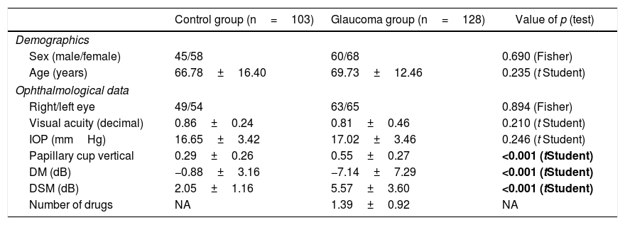
Material and methods

The study included 128 patients with POAG and 103 healthy controls who had PPP maps (macular grid 8×8) drawn by SD-OCT. Only one eye per patient was studied. The 9 mRLs were automatically segmented by prototype software, obtaining: a macular retinal nerve fiber layer (mRNFL), ganglion cell layer (GCL), inner plexiform layer (IPL), inner nuclear layer (INL), outer plexiform+nuclear layer, photoreceptor layer, retinal pigment epithelium (RPE), outer retina and RPE+outer retina. Thickness values were obtained on 64 cells of the grid for each mRL, and mean thickness of superior and inferior hemispheres were calculated. Comparisons of mean thickness of these hemispheres and thickness of each cell between groups were determined. Differences in the cell by cell comparisons were represented quantitatively by heat maps for each mRL.
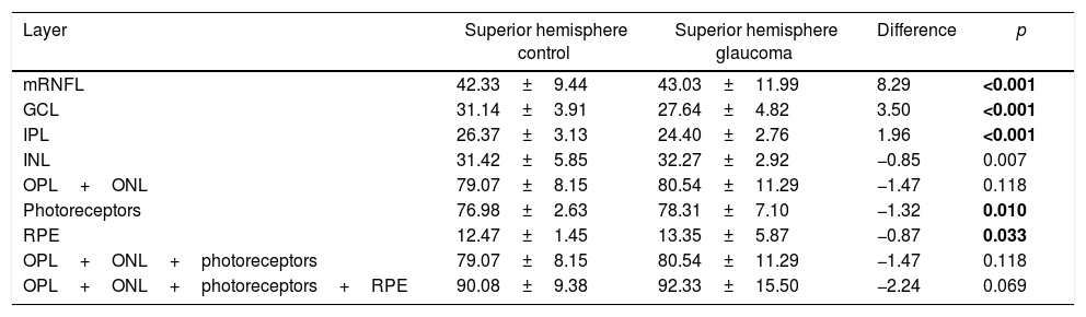
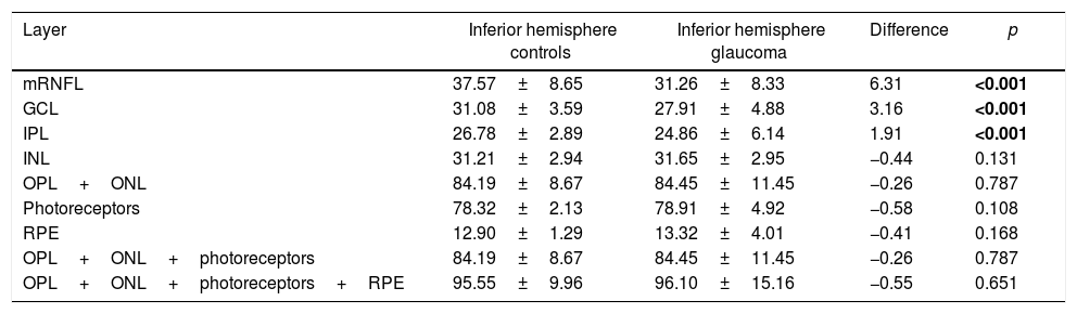
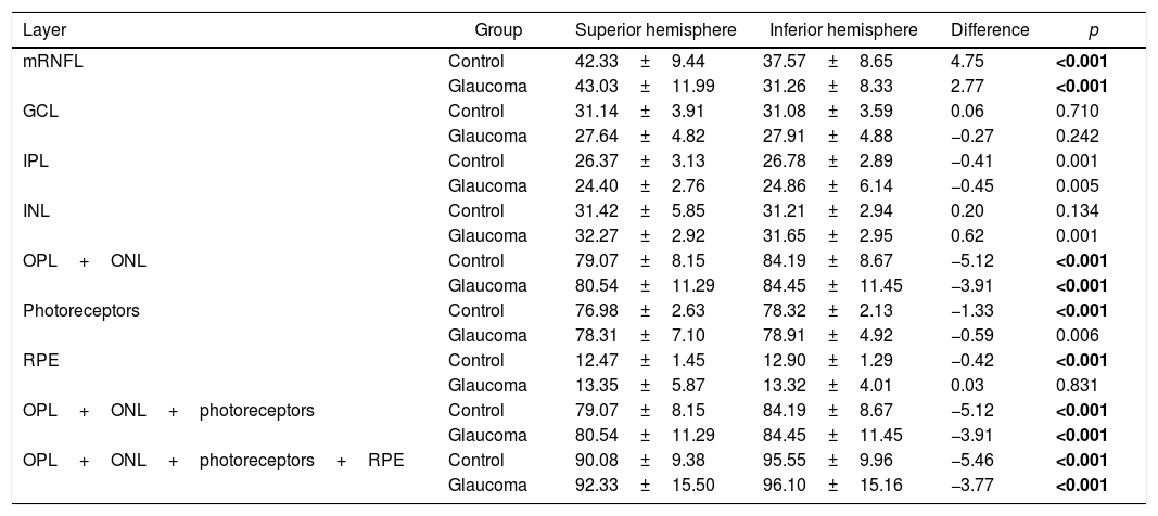
Results

Photoreceptors and RPE were found in POAG group when comparing thickness of hemispheres, thinning of mRNFL, GCL, IPL, and thickening of INL. Heat maps showed symmetrical thinning patters between superior and inferior hemispheres in inner retinal layers (except for INL) and asymmetrical thickening patters in outer retinal layers in GPAA group.

Conclusions

There are thickness changes in all mRLs in POAG, when studied by PPP. Thinning of inner layers (except for INL), and thickening of outer layers in POAG show different symmetry patterns in relation to horizontal meridian.

Keywords:
Glaucoma
Thickness
Layer
Macula
Posterior pole
Segmentation
Mapping
8×8
Resumen
Objetivos

Evaluar los cambios de las capas retinianas maculares (CRM) usando el programa de OCT de polo posterior (PP) en el glaucoma primario de ángulo abierto (GPAA).

Material y métodos

Ciento veintiocho pacientes con GPAA y 103 controles sanos con mapas de PP (rejilla macular 8×8) obtenidos mediante SD-OCT fueron incluidos. Solo un ojo por paciente fue considerado. Entonces 9 CRM se segmentaron automáticamente mediante un software prototipo obteniendo: capas de fibras nerviosas maculares, capa de células ganglionares (GCL), plexiforme interna, nuclear interna (INL), plexiforme+nuclear externa, capa de fotorreceptores, epitelio pigmentario de la retina (RPE) y retina externa completa. Se obtuvieron los valores de grosor de las 64 celdas de la rejilla para cada una de las CRM y se calcularon los grosores promedio de los hemisferios superior e inferior. Se realizó una comparación de los grosores promedio de dichos hemisferios y de los grosores celda a celda entre los 2 grupos. Las diferencias en comparaciones celda a celda fueron representadas mediante mapas de calor para cada CRM.

Resultados

Al comparar los grosores de los hemisferios se encontraron adelgazamientos en capas de fibras nerviosas maculares, capa de células ganglionares y plexiforme interna y engrosamientos en INL, fotorreceptores y RPE en GPAA. Los mapas de calor mostraron patrones de adelgazamiento simétricos entre ambos hemisferios (superior e inferior) en capas de la retina interna (excepto INL) y patrones de engrosamiento asimétricos en las CRM externas en GPAA.

Conclusiones

Existen patrones de cambio en el grosor en todas las CRM en el GPAA estudiadas mediante el programa PP. Los adelgazamientos de las capas internas (excepto INL) y los engrosamientos de las externas en el GPAA presentan diferentes patrones de simetría respecto al meridiano horizontal.

Palabras clave:
Glaucoma
Grosor
Capa
Mácula
Polo posterior
Segmentación
Mapeado
8×8

Article

These are the options to access the full texts of the publication Archivos de la Sociedad Española de Oftalmología (English Edition)
Subscriber
Subscriber

If you already have your login data, please click here .

If you have forgotten your password you can you can recover it by clicking here and selecting the option “I have forgotten my password”
Subscribe
Subscribe to

Archivos de la Sociedad Española de Oftalmología (English Edition)

Purchase
Purchase article

Purchasing article the PDF version will be downloaded

Price 19.34 €

Purchase now
Contact
Phone for subscriptions and reporting of errors
From Monday to Friday from 9 a.m. to 6 p.m. (GMT + 1) except for the months of July and August which will be from 9 a.m. to 3 p.m.
Calls from Spain
932 415 960
Calls from outside Spain
+34 932 415 960
E-mail
Article options
Tools
es en pt

¿Es usted profesional sanitario apto para prescribir o dispensar medicamentos?

Are you a health professional able to prescribe or dispense drugs?

Você é um profissional de saúde habilitado a prescrever ou dispensar medicamentos

Quizás le interese:
10.1016/j.oftale.2020.09.015
No mostrar más