


Climatic droplets keratopathy (CDK) is closely associated with superficial corneal erosions and lack of protective mechanisms against the harmful effects of ultraviolet radiation (UVR) during a prolonged period of time. One of the difficulties in studying the pathogenic mechanisms involved in this human disease is the lack of an experimental animal model. In this paper, a study is conducted on the effects of 4 types of lasers at various powers and time conditions on the normal guinea pig corneas in order to select only one laser condition that reversibly injures the epithelium and superficial stroma, without leaving scarring.
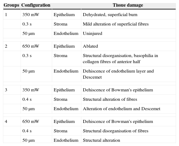
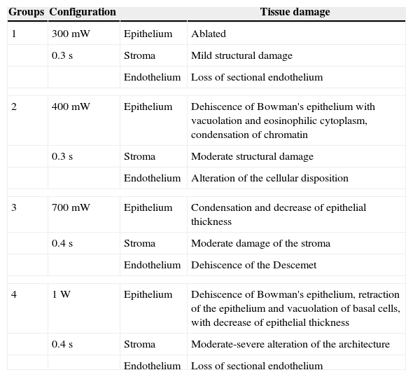
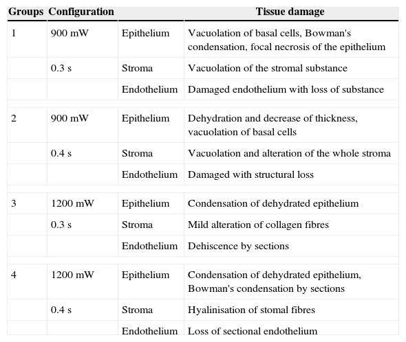
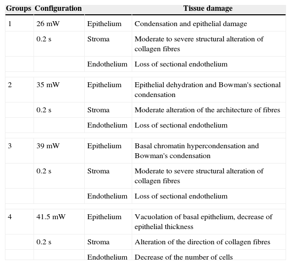
MethodsDamage was induced in the cornea of guinea pigs using different powers and exposure times of 4 types of laser: argon, CO2, diode and Nd–Yag, and any injuries were evaluated by biomicroscopy (BM) and optical microscopy. Corneas from other normal animals were exposed to argon laser (350mW, 0.3s, 50μm of diameter), and the induced alterations were studied at different times using BM, optical coherence tomography (OCT) and transmission electron microscopy (TEM).
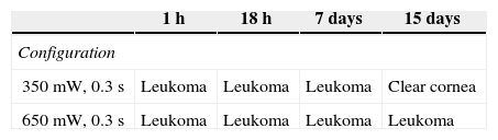
ResultsOnly argon laser at 350mW, 0.3s and 50μm of diameter produced epithelium and superficial stroma lesions. Some leukomas were observed by BM, and they disappeared by day 15. Corneal thickness measured by OCT decreased in the eyes treated with argon laser during the first week. Using TEM, different ultrastructural alterations in corneal epithelium and stroma were observed during the early days, which disappeared by day 15.
ConclusionsIt was possible to develop reproducible corneal epithelium and anterior stroma injuries using argon laser at 350mW, 0.3s and 50μm of diameter. In vivo and in vitro studies showed that injured corneas with these laser conditions did not leave irreversible microscopic or ultrastructural alterations. This protocol of corneal erosion combined with exposure to UVR and partial deficiency of ascorbate in the diets of the animals for an extended period of time has been used to try to develop an experimental model of CDK.
La queratopatía climática esferoidea (QCE) está íntimamente asociada a erosiones corneales superficiales y a carencia de mecanismos protectores contra los efectos nocivos de la radiación ultravioleta (RUV) durante muchos años. Debido a que una de las dificultades en el estudio de los mecanismos patogénicos de esta enfermedad humana es la ausencia de un modelo experimental, en este trabajo quisimos identificar cuál es el mejor método para estudiar una de las variables involucradas en su génesis (erosiones superficiales de la córnea). A tal fin investigamos los efectos producidos por 4 tipos de láser con diferentes condiciones de potencia y tiempo en la córnea de cobayos normales con el fin de seleccionar un láser y condición que lesione solamente el epitelio y estroma superficial, de manera reversible, sin dejar cicatrices.
MétodosSe indujeron daños en la córnea de cobayos utilizando distintas potencias y tiempos con 4 tipos de láser: argón, CO2, diodo y Nd-Yag en distintos grupos de animales y se evaluaron dichas lesiones por biomicroscopia (BM) y microscopia óptica. Córneas de otros animales normales fueron expuestas a láser de argón (350 mW, 0,3 s, 50μm de diámetro) y las alteraciones inducidas se estudiaron en diferentes tiempos utilizando BM, tomografía de coherencia óptica (TCO) y microscopia electrónica (ME).
ResultadosSolo el láser argón a una potencia de 350 mW, 0,3 s, 50μm de diámetro produjo lesiones de epitelio y estroma superficial, manteniéndose indemne el endotelio. Por BM se observaron algunos leucomas que desaparecieron hacia el día 15. Mediante TCO se observó un adelgazamiento del espesor corneal en los ojos tratados con esas condiciones de láser argón durante la primera semana. Mediante ME, se observaron diferentes alteraciones ultraestructurales en epitelio y estroma corneal durante los primeros días, las cuales desaparecieron hacia el día 15.
ConclusionesFue posible desarrollar lesiones corneales en epitelio y estroma anterior de cobayos de manera reproducible mediante el uso de láser argón. Los estudios in vivo e in vitro demostraron que las córneas lesionadas con este láser y en esas condiciones no dejaron alteraciones microscópicas ni ultraestructurales irreversibles. Este modo de erosión corneal combinado con exposición a RUV y déficit parcial de ascorbato en la dieta de los animales durante un período prolongado de tiempo está siendo utilizado con el fin de intentar desarrollar un modelo experimental de QCE.
