Buscar en
Archivos de la Sociedad Española de Oftalmología (English Edition)
Toda la web
Inicio Archivos de la Sociedad Española de Oftalmología (English Edition) Dome shaped macula associated with myopic retinochoroidosis. Use of spironolacto...
Journal Information
Vol. 92. Issue 10.
Pages 499-506 (October 2017)
Share
Share
Download PDF
More article options
Visits
61
Vol. 92. Issue 10.
Pages 499-506 (October 2017)
Short communication
Dome shaped macula associated with myopic retinochoroidosis. Use of spironolactone
Mácula en cúpula asociada a la retinocoroidosis miópica. Utilidad de la espironolactona
Visits
61
P. Rocha Cabreraa,
Corresponding author
procha975@yahoo.es

Corresponding author.
, L. Cordovés Dortaa, D. Ángel Pereiraa, J.A. Abreu Reyesa, M.J. Losada Castilloa, J. Lorenzo Moralesb, M.Á. Serrano Garcíaa
a Servicio de Oftalmología, Hospital Universitario de Canarias, La Laguna, Tenerife, Spain
b Instituto de Enfermedades Tropicales y Salud Pública, Universidad de La Laguna, La Laguna, Tenerife, Spain
This item has received
Article information
Abstract
Full Text
Bibliography
Download PDF
Statistics
Figures (3)
Show moreShow less
Abstract
Cases reports

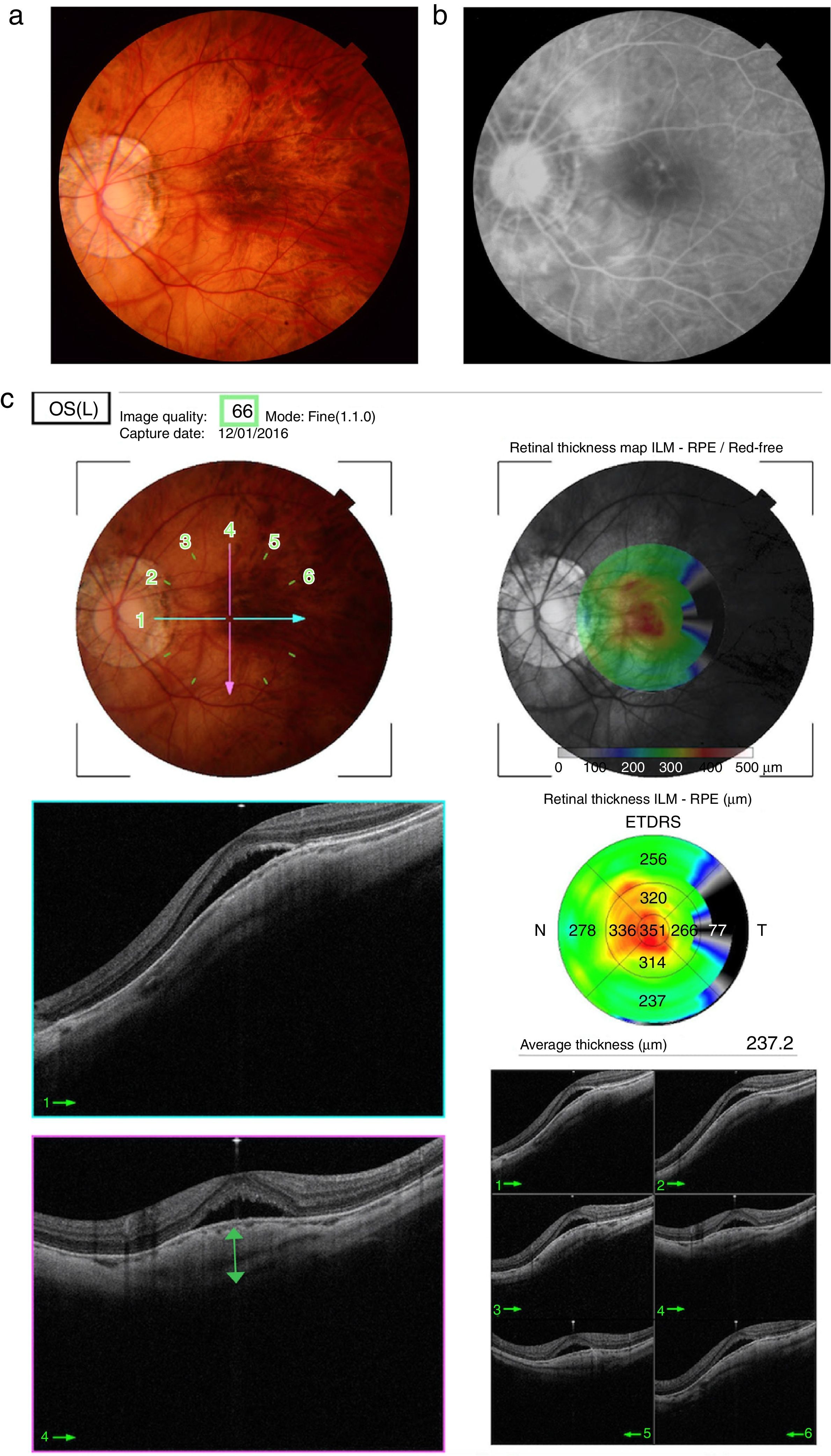
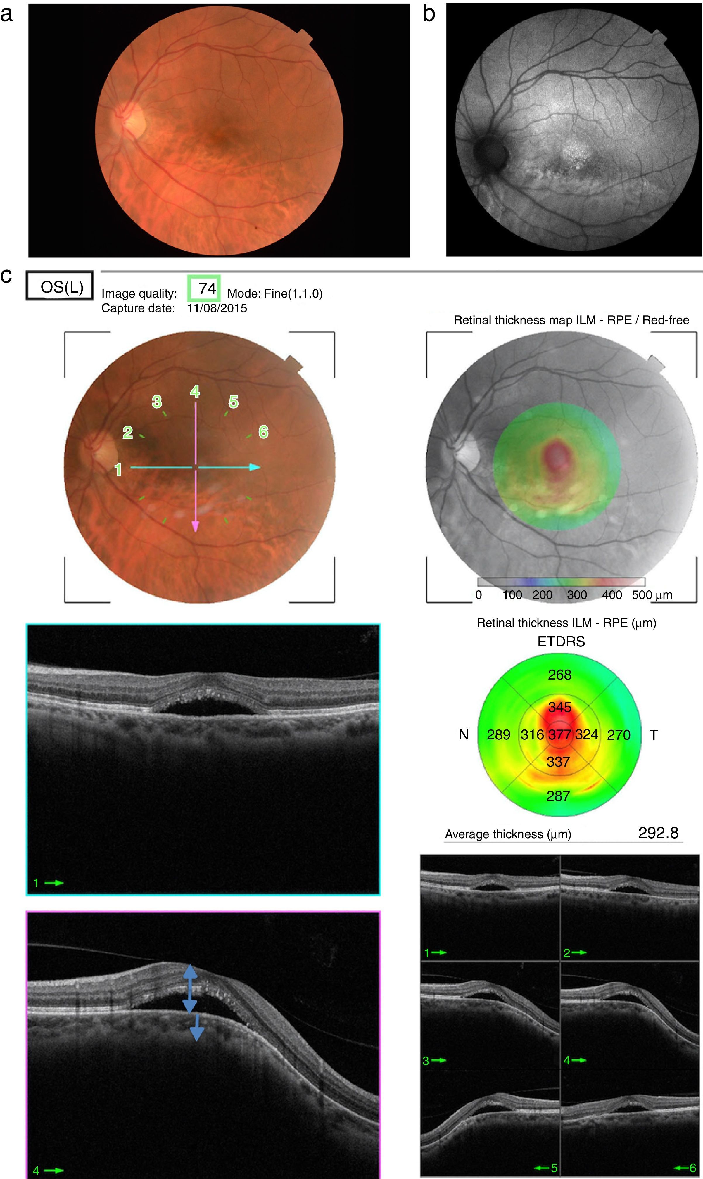
The cases are presented of three women of 22, 36 and 55 years old with bilateral myopic retinochoroidosis. They had unilateral decreased visual acuity, normal bilateral tonometry, and biomicroscopy. Funduscopy showed bilateral and unilateral myopic maculopathy, and Optical Coherence Tomography (OCT) showed a dome shaped macula with neurosensory detachment. Treatment was started with spironolactone and an improvement by OCT was shown in all cases.

Discussion

The etiopathogenic mechanism of the dome shaped macula is discussed. OCT demonstrated to be the fundamental test in the follow-up of this condition. After the evidence shown, initial treatment with spironolactone is suggested.

Keywords:
Macula
Dome
Etiopathogenic mechanism
Treatment
Spironolactone
Resumen
Casos clínicos

Se presentan los casos de 3 mujeres de 22, 36 y 55 años de edad con retinocoroidosis miópica bilateral. Las pacientes presentan disminución de agudeza visual unilateral, tonometría y biomicroscopia bilateral normal. En la funduscopia se evidencia maculopatía unilateral, y en la tomografía de coherencia óptica (OCT), mácula en cúpula con desprendimiento neurosensorial (DNS). Iniciado tratamiento con espironolactona, en todos los casos se comprueba mejoría por OCT.

Discusión

Se discute el mecanismo etiopatogénico de la mácula en cúpula. La OCT se demuestra como técnica fundamental en el seguimiento de esta patología. Tras la evidencia mostrada, se postula tratamiento inicial con espironolactona.

Palabras clave:
Mácula
Cúpula
Mecanismo etiopatogénico
Tratamiento
Espironolactona

Article

These are the options to access the full texts of the publication Archivos de la Sociedad Española de Oftalmología (English Edition)
Subscriber
Subscriber

If you already have your login data, please click here .

If you have forgotten your password you can you can recover it by clicking here and selecting the option “I have forgotten my password”
Subscribe
Subscribe to

Archivos de la Sociedad Española de Oftalmología (English Edition)

Purchase
Purchase article

Purchasing article the PDF version will be downloaded

Price 19.34 €

Purchase now
Contact
Phone for subscriptions and reporting of errors
From Monday to Friday from 9 a.m. to 6 p.m. (GMT + 1) except for the months of July and August which will be from 9 a.m. to 3 p.m.
Calls from Spain
932 415 960
Calls from outside Spain
+34 932 415 960
E-mail
Article options
Tools
es en pt

¿Es usted profesional sanitario apto para prescribir o dispensar medicamentos?

Are you a health professional able to prescribe or dispense drugs?

Você é um profissional de saúde habilitado a prescrever ou dispensar medicamentos