Buscar en
Archivos de la Sociedad Española de Oftalmología (English Edition)
Toda la web
Inicio Archivos de la Sociedad Española de Oftalmología (English Edition) Clinical, fundoscopic, tomographic and angiographic characteristics of dome shap...
Journal Information
Vol. 92. Issue 10.
Pages 458-463 (October 2017)
Share
Share
Download PDF
More article options
Visits
6
Vol. 92. Issue 10.
Pages 458-463 (October 2017)
Original article
Clinical, fundoscopic, tomographic and angiographic characteristics of dome shaped macula classified by bulge height
Estudio de las características clínicas, fundoscópicas, tomográficas y angiográficas de la mácula en domo según la altura de protrusión macular
Visits
6
J. Fajardo Sáncheza,
Corresponding author
juliafajardo7@gmail.com

Corresponding author.
, C.E. Chau Ramosa, J.A. Roca Fernándezb, J.L. Urcelay Seguraa
a Servicio de Oftalmología, Hospital General Universitario Gregorio Marañón, Instituto Provincial de Oftalmología, Madrid, Spain
b Clínica Ricardo Palma, Lima, Peru
This item has received
Article information
Abstract
Full Text
Bibliography
Download PDF
Statistics
Figures (7)
Show moreShow less
Tables (1)
Table 1. Ophthalmological examination: clinic, funduscopic, tomographic and angiographic characteristics of each eye included in the study.
Abstract
Objective

To classify dome shaped macula cases by their bulge height (BH). To analyze the characteristics associated with the groups formed by this classification.
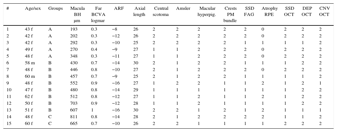
Methods

Observational, descriptive and cross-sectional study on 15 selected eyes with dome shaped macula and high myopia. Using Caillaux method and optical coherence tomography images, 3 groups were determined by their BH: low (50–350μm), medium (351–650μm), and high (>650μm), and a study of visual acuity, axial length, presence of subfoveal serous detachment, and images by fluorescein angiography and optic coherence tomography, as main variables. The confidence interval was 95%.

Results

By using the chi-squared test, the study showed that a BH higher than 400μm was associated with lower visual acuity, presence of subfoveal serous detachment, and greater atrophy of the retinal pigment epithelium measured by disk diameters (p<0.05).

Conclusions

The medium and high BH showed a positive correlation with the presence of foveal serous detachment and a lower visual acuity.

Keywords:
Dome shaped macula
Bulge height
Foveal serous detachment
High myopia
Pathological myopia
Fluorescein angiography
Optical coherence tomography
Resumen
Objetivo

Clasificar la mácula en domo según la altura de protrusión macular (AP) y analizar las características asociadas a cada grupo.

Métodos

Estudio observacional, descriptivo y transversal. Selección de 15 ojos con mácula en domo y miopía magna. Utilizando imágenes por tomografía de coherencia óptica (OCT) y el método de Caillaux, se clasificó la AP en baja (50-350μ), media (351-650μ) y alta (>650μ) y se estudió la agudeza visual, longitud axial, presencia de desprendimiento seroso subfoveal y las imágenes obtenidas por angiografía fluoresceínica y OCT como variables principales. El nivel de confianza utilizado fue de 95%.

Resultados

Utilizando el test de chi cuadrado, el estudio observó que una AP mayor a 400μ se asoció con menor agudeza visual, mayor presencia de desprendimiento seroso subfoveal y mayor presencia de atrofia del epitelio pigmentario de la retina medida en diámetros de papila (p<0,05).

Conclusiones

Las AP media y alta mostraron una asociación positiva con la presencia de desprendimiento seroso subfoveal y disminución de laagudeza visual.

Palabras clave:
Mácula en domo
Altura de protrusión macular
Miopía magna
Desprendimiento seroso foveal
Angiografía fluoresceínica
Tomografía de coherencia óptica

Article

These are the options to access the full texts of the publication Archivos de la Sociedad Española de Oftalmología (English Edition)
Subscriber
Subscriber

If you already have your login data, please click here .

If you have forgotten your password you can you can recover it by clicking here and selecting the option “I have forgotten my password”
Subscribe
Subscribe to

Archivos de la Sociedad Española de Oftalmología (English Edition)

Purchase
Purchase article

Purchasing article the PDF version will be downloaded

Price 19.34 €

Purchase now
Contact
Phone for subscriptions and reporting of errors
From Monday to Friday from 9 a.m. to 6 p.m. (GMT + 1) except for the months of July and August which will be from 9 a.m. to 3 p.m.
Calls from Spain
932 415 960
Calls from outside Spain
+34 932 415 960
E-mail
Article options
Tools
es en pt

¿Es usted profesional sanitario apto para prescribir o dispensar medicamentos?

Are you a health professional able to prescribe or dispense drugs?

Você é um profissional de saúde habilitado a prescrever ou dispensar medicamentos