Buscar en
Archivos de Cardiología de México
Toda la web
Inicio Archivos de Cardiología de México Cardiac resynchronization therapy: evaluation of ventricular dysynchrony and pat...
Información de la revista
Vol. 80. Núm. 4.
Páginas 289-300 (Octubre 2010)
Compartir
Compartir
Descargar PDF
Más opciones de artículo
Vol. 80. Núm. 4.
Páginas 289-300 (Octubre 2010)
Acceso a texto completo
Cardiac resynchronization therapy: evaluation of ventricular dysynchrony and patient selection
Terapia de resincronización cardiaca: evaluación de la disincronía ventricular y selección de pacientes
Visitas
6204
Giselle Baqueroa, Javier E. Banchsa, William R. Davidson Jra, Erica D. Penny-Petersona, Soraya M. Samiia, Deborah L. Wolbrettea, Gerald V. Naccarellia, Mario D. Gonzaleza
a Penn State Hershey Heart & Vascular Institute; Penn State College of Medicine.
Este artículo ha recibido
Información del artículo
Resumen
Texto completo
Bibliografía
Descargar PDF
Estadísticas
Figuras (3)
Mostrar másMostrar menos
Cardiac resynchronization therapy (CRT) is an established treatment modality for systolic heart failure. Aimed to produce simultaneous biventricular stimulation and correct the lack of ventricular synchrony in selected patients with congestive heart failure, CRT has shown to improve mortality and reduce hospital admissions when compared to medical treatment. At present, the indication criteria for the implantation of a CRT device include an ejection fraction of less than 35%, heart failure symptoms consistent with NYHA functional class III-IV and a QRS complex duration equal or longer than 120 milliseconds. It has been reported that 30% of patients who meet those criteria still may not derive clinical benefit from CRT. Due to the existing diversity of imaging modalities and resources for their process and analysis, a great expectation in terms of more accurate diagnosis of ventricular dyssynchrony has been raised. Reliable identification of dyssynchrony could allow us to better predict the favorable response of an individual patient to CRT and therefore offer this procedure to those individuals most likely to benefit. We review the available techniques for the study of ventricular dyssynchrony for CRT patient selection and the results of its application in clinical trials. Despite tremendous progress in the imaging technology available for the assessment and diagnosis of ventricular dyssynchrony, an ideal method has not been identified and the duration of the QRS complex in the surface ECG remains the accepted criteria of dyssynchrony in the selection of patients for CRT.
Keywords:
Terapia de resincronización cardiaca; Asincronía; Ecocardiografía; Insuficiencia cardiaca; EEUU
La terapia de resincronización cardiaca es una modalidad de tratamiento bien establecida para la insuficiencia sistólica cardiaca. Dirigida a producir una estimulación biventricular simultánea y a corregir la falta de sincronía ventricular en pacientes seleccionados con insuficiencia cardiaca congestiva, la terapia de resincronización cardiaca ha mostrado ser capaz de mejorar los índices de mortalidad y reducir las admisiones hospitalarias cuando se compara con el tratamiento médico. Actualmente, los criterios para la implantación de un dispositivo de terapia de resincronización cardiaca incluyen una fracción de eyección menor a 35%, síntomas de insuficiencia cardiaca consistentes con la clase funcional NYHA III-IV, y una duración del complejo QRS igual o mayor de 120 milisegundos. Se ha reportado que 30% de los pacientes que cumplen con estos criterios pueden inclusive no obtener beneficio clínico de la terapia de resincronización cardiaca. Debido a la diversidad existente de los estudios de imagenología y de los recursos para su proceso y análisis, ha surgido una gran expectativa en términos de un diagnóstico más exacto de la asincronía ventricular. La identificación confiable de la asincronía nos podría permitir predecir mejor la respuesta favorable de un paciente en particular a la terapia de resincronización cardiaca y así ofrecer este procedimiento a aquellos pacientes con mayores probabilidades de beneficiarse de dicha terapia. Hacemos una revisión de las técnicas disponibles para el estudio de la asincronía ventricular para la selección de pacientes para esta terapia y los resultados de su aplicación en pruebas clínicas. A pesar de los grandes progresos alcanzados en la tecnología de imágenes disponibles para la evaluación y diagnóstico de la asincronía ventricular, no se ha identificado un método ideal y la duración del complejo QRS en el ECG de superficie sigue siendo el criterio aceptado de asincronía en la selección de pacientes para terapia de resincronización cardiaca.
Palabras clave:
Cardiac resynchronization therapy; Dyssynchrony; Echocardiography; Heart failure; USA
Texto completo

Introduction

Cardiac resynchronization therapy (CRT) creates simultaneous or near simultaneous biventricular stimulation aimed at correcting the lack of ventricular synchrony found in some patients with congestive heart failure and, most commonly, those with left bundle branch block. CRT is among the most important contemporary advances in the treatment of heart failure.1 Several randomized clinical trials have demonstrated benefits in survival, hospitalizations for heart failure, functional capacity and improvement in left ventricular function and architecture among patients with advanced systolic dysfunction (ejection fraction equal to or less than 35% and New York Heart Association functional class III) and QRS duration equal or greater to 120 milliseconds. The benefit is additional to that of optimal medical therapy.2-11 Two recent clinical studies have suggested similar benefits, although more modest, in patients with low ejection fraction (35% -40%) and wide QRS and less severe heart failure, class II (NYHA).12-15 Although there is still no consensus on the routine use of CRT in this population, it is very possible that in the near future these indications expand to include less symptomatic patients as a therapeutic or prophylactic measure to avoid the progression of the disease. Based on the large volume of existent clinical evidence, CRT has acquired a solid position in the treatment of heart failure. However, clinical trials have also shown that only 60% - 80% of patients receiving these devices experience clinical or electrocardiographic improvement.3,16,17

At the present time, a QRS complex with duration greater than 120 milliseconds, regardless of the underlying conduction defect, is considered sufficient electrocardiographic criteria for patients with an ejection fraction of less than 35% and symptoms consistent with class III (NYHA) heart failure to be candidates for the implantation of a resynchronization cardiac device.18-21 These criteria are based on the clinical trials mentioned above. The scientific community has explored persistently newer techniques for the assessment of ventricular dyssynchrony to optimize the selection of patients for this therapy. Taking into account the diversity among imaging modalities that are currently available and the existing resources for their process and analysis, it is expected that imaging methods would make possible the most accurate diagnosis of ventricular dyssynchrony. The reliable identification of dyssynchrony should allow us to predict whether a patient will respond positively to CRT, regardless of the QRS duration, in order to offer the procedure to those patients who are more likely to benefit and likewise prevent unnecessary implantation in patients with low likelihood of clinical improvement. Mechanical dyssynchrony has been demonstrated in patients with normal QRS duration and can be absent in up to 20% of patients with left bundle branch block, as shown recently by magnetic resonance imaging.22-27 With all its benefits, CRT is not exempt of risks. The implantation of CRT devices include the risks inherent to the implantation process such as pneumothorax, cardiac perforation, dissection of the coronary sinus, contrast induced nephropathy, bleeding and infection of the device, among others. Additionally, there is subsequent risk of paradoxical worsening of heart failure if the biventricular stimulation exacerbates the degree of dyssynchrony, causes diaphragmatic stimulation, or complicated by lead dislocation.28-31

This manuscript aims to review available techniques for the study of ventricular dyssynchrony for patient selection and the results of its application in clinical trials.

Dyssynchrony

Electromechanical ventricular dyssynchrony is the pathophysiologic abnormality targeted by CRT. In the case of interventricular dyssynchrony the focus is the temporal difference in activation between both ventricles, as compared to intraventricular dyssynchrony, which refers to the relative delay of activation among different segments of the left ventricle. Complete electrical activation occurs in less than 80 milliseconds in the normal heart, by fast conduction through the His-Purkinje system.32 Delay in the activation of a large enough segment of the left ventricle (electrical dyssynchrony) may result in an asymmetrical and inefficient systolic contraction (mechanical dyssynchrony) that eventually compromises cardiac pump function, with subsequent adverse remodeling of the left ventricle and development or progression of heart failure. This phenomenon is critical in patients with low myocardial contractile reserve as in ischemic and dilated cardiomyopathies.

The best model of ventricular dyssynchrony is due to left bundle branch block, in which the left ventricle is activated later and slower than normal following activation of the right ventricle. Once the right ventricle has been activated through the right bundle, depolarization spreads through the interventricular septum resulting in delayed activation of the posterior and lateral walls of the left ventricle.33 The sequence of depolarization determines the sequence of mechanical contraction. When the right ventricle and the septum are activated before the left ventricle, the septum is pulled away from the lateral wall of the left ventricle, which ends up contracting late, at a time when the septum is shifted to the opposite direction. At first glance, this phenomenon is responsible for the characteristic paradoxical septal motion observed in the echocardiogram of patients with left bundle branch block (apical four-chamber view). It has been demonstrated that the sequence of conduction and ventricular activation can shown individual variations among different patients with similar degree of left bundle branch block.34,35

The time elapsed between the time of maximal impulse in the septum and the lateral wall of the left ventricle is one of the most common measures of ventricular dys-synchrony and the normal value varies depending on the method used and its validation. It is understood that left intraventricular dyssynchrony is more important in the pathophysiology of heart failure than interventricular dyssynchrony. Most clinical studies have focused on the activation and mechanical function of the left ventricle during the evaluation of dyssynchrony in patients with heart failure.

Left bundle branch block is present in about 15-20% of patients with heart failure due to systolic dysfunction and confers a negative prognosis, determined by decreased survival and accelerated progression of the disease.36-40 Even in patients that lack systolic dysfunction but have increase risk of cardiovascular disease, presence of left bundle branch block represents an increased risk for heart failure and mortality.41-43 Similarly, the mechanical dys-synchrony induced by iatrogenic left bundle branch block, which is characteristic of artificial stimulation of the right ventricle in the case of traditional pacemakers, has been associated with increased risk of impaired left ventricular function in patients with or without heart failure, and favorable response to biventricular stimulation with CRT.44-48 It has been accepted that dyssynchrony associated with left bundle branch block is a factor that contributes negatively to the pathophysiology of heart failure. Excessive prolongation of the QRS duration in patients with advanced heart failure may also be a passive marker of the severity of the disease and not only an active factor in the process. Rosenmbaum, Elizari and Lazzari described the variants of bundle branch blocks.49 These include block at the level of the branches "bundle branch block"; blockade in the major subdivisions known as "segmental block"; blockade in the Purkinje network called "reticular" or "Purkinje block" and "parietal blocks" that occurs due to delayed conduction in the myocardium itself. This classification although frequently ignored, is essential to understand the variety of possible patho-physiological scenarios involved in the pattern and duration of the QRS and the complexity of the clinical problem.

The QRS duration as a measure of electrical dyssynchrony and the pattern of the left bundle branch block, suggest the presence of mechanical dyssynchrony and are often associated with it. However, the delay in conduction, as might be inferred, does not necessarily correspond consistently with the measures of mechanical dys-synchrony obtained with imaging modalities. As discussed above, some patients presenting with left bundle branch block and advanced heart failure do not show meaningful evidence of mechanical dyssynchrony by echocardiography. Likewise, evidence of ventricular dyssynchrony has been found in patients with conduction defects other than left bundle branch block or in patients with normal QRS duration. Based on these electro-mechanical inconsistencies, some researchers have reported that mechanical resynchronization and its benefits can be achieved with CRT regardless of QRS duration.50 This finding, adds to the arguments against the exclusive use of QRS duration as a measure of mechanical dyssynchrony and strict criteria in patient selection for CRT.

Since not every patient with prolonged QRS duration have mechanical dyssynchrony demonstrable with imaging techniques, it is understandable that with the current selection criteria for CRT, there will be a percentage of patients that do not benefit from the treatment. Similarly it is conceivable that some patients with "normal" QRS duration and currently considered not candidates for CRT, will exhibit mechanical dyssynchrony with advanced imaging modalities, enough to justify a CRT device and show clinical improvement. These observations have turned the study of ventricular dyssynchrony into a rapidly expanding field in medical research.

Diagnosis of dyssynchrony by echocardiography

The need to identify an accurate method for the selection of the ideal patient, the one with the highest probability to respond to CRT, has fueled the development of imaging techniques to study ventricular dyssynchrony. Most of these techniques are based in echocardiography because of its convenience, availability and relatively low cost. Clinical studies mostly from single centers, with relatively small samples, initially yielded promising results.51-64

The simplest echocardiographic technique for the study of ventricular dyssynchrony is the analysis of M mode ultrasound that registers the delay between the left ventricular septum and the posterior wall. In the parasternal window, longitudinal or transverse axis, the M-mode ultrasound cursor is positioned at the level of the mid ventricle where papillary muscles may be observed and with the record speed set at 50-100 mm/s, the time delay between the point of maximum displacement of the septum and the posterior wall is measured (Figure 1). More than 130 milliseconds of difference is considered the cutoff value for the diagnosis of dyssynchrony.52,53 The addition of tissue color Doppler to M-mode echocardiography may help to more precisely identify the time of maximum displacement of the ventricular walls. Although this is a very practical technique, the variability between successive measurements and the lack of predictability in the response to CRT in more recent studies suggest that it cannot be used as a single reliable measure in the selection of patients for CRT.65,66

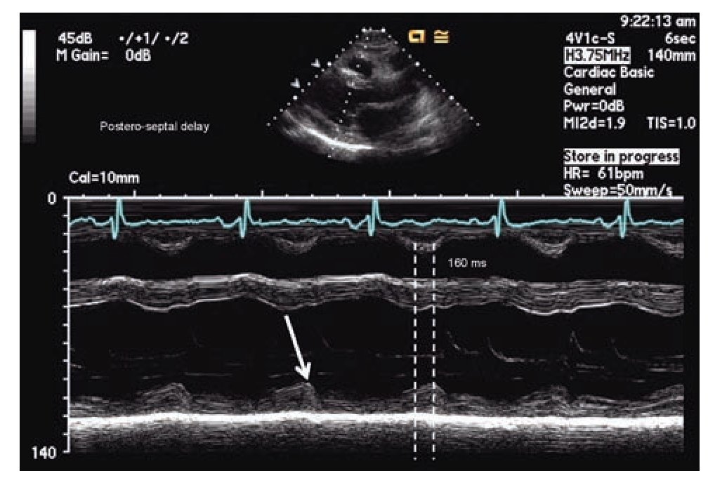
Figure 1. M-mode echocardiography in para septal long axis demonstrating a delay of 160 milliseconds between the point of maximum contraction in the antero-septum and the posterior wall of the left ventricle, consistent with mechanical dyssynchrony (≥ 130 milliseconds).

Tissue Doppler is the technique most frequently used in the study of ventricular dyssynchrony and is currently by consensus the recommended method.67 Longitudinal and radial shortening velocities can be recorded with tissue Doppler in different segments from different windows to assess the time of contraction of each segment and quantify the difference among individual segments. Time intervals are measured in reference to the electrocardiogram using the beginning of the QRS complex and the peak systolic velocity recorded in selected regions of interest on basal and mid segments of opposing walls wherever the maximal systolic velocity is found to be stable. The good quality measurements from different beats for a segment on the same plane are usually averaged.67 Several authors have evaluated the data obtained by this method using different combinations of measures including the use of the septal and lateral basal segments in the apical four-chamber view (dyssynchrony present when the delay in activation of opposite walls is more than 65 msec) or the use of a dyssynchrony index that incorporates measurements in 12 segments in all three standard planes of the apical window (basal anterior, inferior, septal and lateral in the two and four chamber and longitudinal views).54,58,61,68 The index corresponds to the standard deviation of the times of maximum velocity of the 12 segments, with a normal cutoff value 33 ms.69 A feature known as tissue synchronization imaging allows some computers display color-coded images of automated speed times superimposed on 2D images, providing a quick visual guide to identify segments of interest.70,71 (Figure 2)

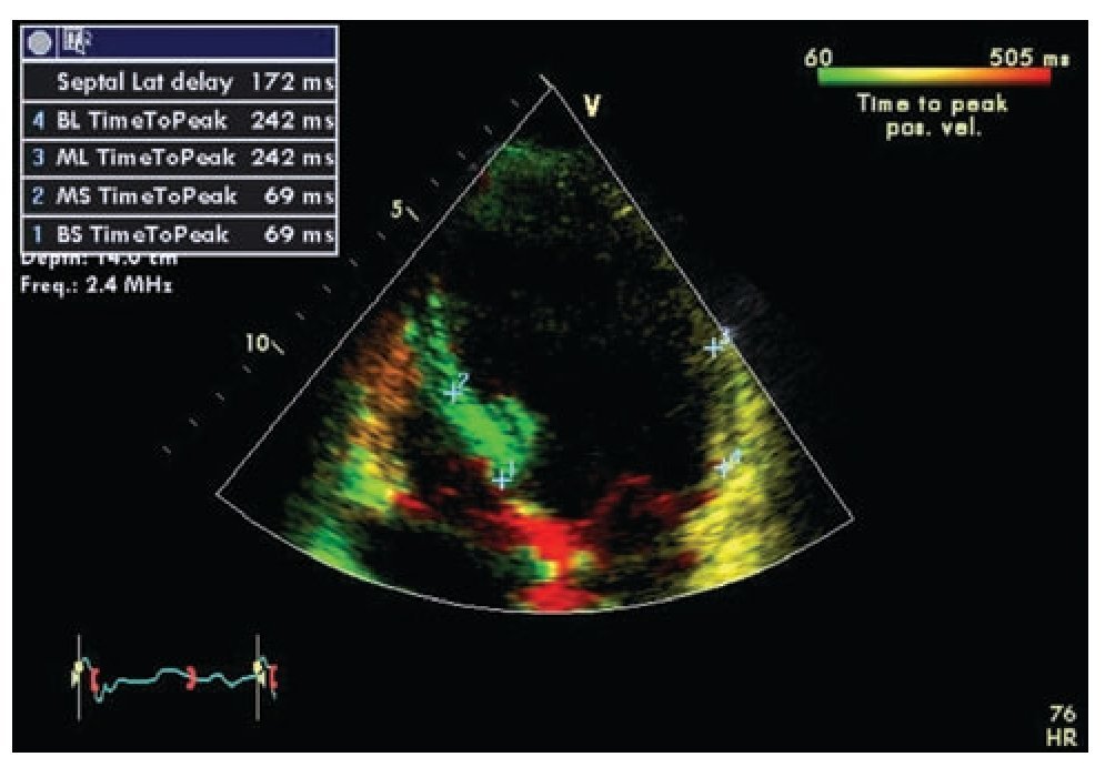
Figure 2. Tissue synchronization imaging of the left ventricle from the apical four-chamber view showing color-coded tissue Doppler consistent with maximal delay at the base and mid lateral wall (yellow). The top left frame shows records of maximum systolic velocity for basal ("BL" and "BS") and mid ("ML" and "MS") segments of the lateral wall and septum. A TSI (tissue synchronization imaging) index can be calculated based on these measurements.

Pulsed tissue Doppler imaging without color can also be used in the evaluation of dyssynchrony. The cursor should be located in the areas where the signal is reproducible. The measurement is done from the beginning or peak of the QRS and the onset of the systolic velocity signal corresponding to the segment of interest. A difference of 60 msec or more on opposite walls is considered diagnostic for dyssynchrony.56,67,72 (Figure 3)

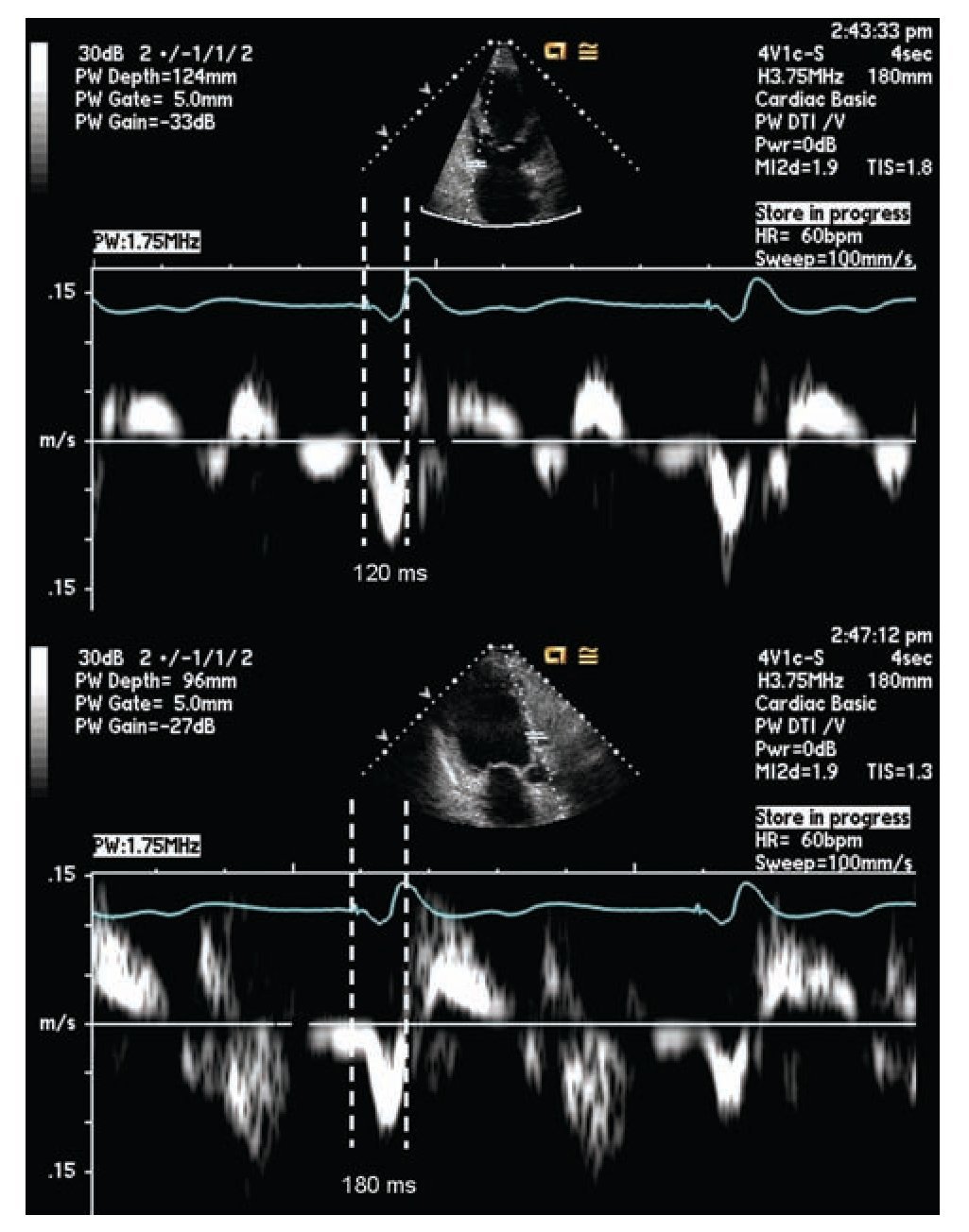
Figure 3. Pulsed tissue Doppler imaging demonstrating mechanical delay between the base of the left anterior and inferior walls, from the apical long axis view. The measurement is taken between the pacing artifact and the beginning of displacement of the evaluated segment determined by tissue Doppler. This measurement is dependent on technology, good signal quality is essential for a reliable measurement. The example shows a delay of 60 milliseconds in the lateral wall consistent with mechanical dyssynchrony between these segments (≥ 60 milliseconds).

Another approach has been developed around the concept of myocardial deformation. Instead of using the Doppler registered tissue velocity, the myocardial deformation is calculated as a proportionate measure of the sampled tissue shortening along the Doppler cursor line. The proposed advantage of this technique is the accuracy to register myocardial contraction independently of passive motion. Unfortunately this method has technical limitations because it is dependent on the Doppler's angle of incidence; the acquisition of images is complicated by an increased incidence of noise and low reproducibility. Initial studies suggested that deformation measures corresponded better with mechanical dyssynchrony than myocardial velocity.73 However, it showed little correlation with the echocardiographic response to CRT.58,64,74 Some of the limitations of this technique may have improved with the addition of speckle tracking. Speckle tracking is an additional resource that eliminates the influence of the Doppler's angle of incidence, improving the diagnostic value of the deformation parameters, in both short and long axis.75-78 The presence of dyssynchrony in the two modes of longitudinal and radial deformation has shown to correspond with a greater likelihood of response to CRT.79,80 Ventricular displacement can also be obtained from tissue Doppler and its association with response to CRT has been demonstrated.26,81-82 However the advantages and diagnostic superiority shown with speckle tracking,74 has popularized this technique; further perfection and inclusion in larger clinical trials would probably be necessary for its adoption as a preferred technique in the diagnosis of dyssynchrony.

Other Imaging Modalities

Three-dimensional echocardiography with various imaging processing techniques, stress echocardiography, magnetic resonance and nuclear myocardial perfusion imaging are other diagnostic modalities used in the assessment of ventricular dyssynchrony and resynchronization.

The initial application of three-dimensional echo-cardiography in the study of CRT was to evaluate the effects of biventricular pacing in volume and ventricular function.83,84 Using tissue Doppler, the systolic dyssynchrony index may be calculated which has shown good correlation with the response to CRT with a sensitivity of 88-96% and specificity of 85-88%, according to different authors using slightly different cutoff values (5.6% -10%).85-89 Although there is evidence that the measurement of the systolic dyssynchrony index with this technique is reproducible,88,90 when compared with the measurement of dyssynchrony using 2D echocardiography tissue Doppler, the correlation and agreement between them is poor.89,91 Preliminary studies suggest that identifying the more delayed segment of the left ventricle using three-dimensional echocardiography, allows determining the ideal segment for the placement of the left ventricular lead.92-94 At present, the use of three-dimensional echocardiography is limited by its availability, technical complexity, and lower temporal-spatial resolution.95

Several studies with small sample sizes (42-71 patients) and different designs have explored the value of exercise or dobutamine pharmacological stress echocardiography. Overall, they show that the presence of contractile reserve or myocardial viability is an independent predictor of clinical improvement and increased ejection fraction in patients with CRT with sensitivity between 70% and 100% and specificity between 43.8% and 88%.96-100 The correlation is kept regardless of the presence of demonstrable dyssynchrony at rest suggesting an additional prognostic value conferred by the presence of contrac-tile reserve.97 The demonstration of dyssynchrony during exercise has also shown to have superior predictive value than the assessment made at rest with 89% of positive predictive value compared to 70% in a study involving 64 patients.98 The presence of contractile reserve in the segment corresponding to the location of the left ventricular lead has shown to be of particular importance, and could be useful especially in the planning of the procedure to increase the likelihood of clinical response.99-100 The results of a prospective multicenter clinical trial designed to compare the predictive value of pharmacological stress echocardiography with low doses of dobutamine in patients with conventional indications for CRT have not been published. In this study follow-up was planned for one year with clinical response parameters and evaluation of left ventricular reverse remodeling, it will also include a comparison of dyssynchrony measurements using conventional echocardiographic methods.101

Single photon emission computed tomography (SPECT) of myocardial perfusion, as well as magnetic resonance imaging is useful in defining areas of scar and myocar-dial viability. Patients with perfusion defects or extensive areas of scar are less likely to experience clinical improvement, reduction of left ventricular dimension or increased ejection fraction in response to CRT102-109. Analysis protocols for gated perfusion "SPECT " have been developed for the assessment of ventricular dyssynchrony and validated with tissue Doppler.110,111 Subsequent studies have confirmed its predictive value for clinical response in patients receiving CRT devices.112-115 A percentage of total scar less than 15% has been associated with an increased likelihood of clinical improvement.116,117 Demonstration of mechanical dyssynchrony by different magnetic resonance techniques may also help predict the response to CRT independently of the presence of electrical dyssynchrony or scar.106,108,109,116-118 More studies and larger samples are needed to determine the role of this technology in clinical practice. Cost, technical complexity, equipment availability, time to complete the studies and its relative incompatibility with pacemakers and defibrillators, besides referral bias are some of the advantages of the echocardiographic methods over MRI.

Clinical Trials and dyssynchrony measurements

The use of echocardiographic parameters in identifying suitable patients for CRT was studied in a large multicenter clinical trial, the study of predictors of response to CRT (PROSPECT).66 The study involved 53 centers in the United States, Europe and China. A total of 498 patients with standard indications for CRT were studied with twelve echocardiographic parameters and were followed for six months to evaluate clinical response and ventricular endsystolic volume. The ability of echocardiographic parameters to predict clinical, anatomical or both responses was the study objective. The authors found wide variability in the analysis of the echocardiographic parameters regardless of the training provided to operators at each center. The parameters studied in PROSPECT included two M-mode echocardiography measurements, one combined with pulsed Doppler; three parameters based on pulsed Doppler and seven using tissue Doppler including confirmation by longitudinal deformation rate in one of them. Clinical improvement, determined with a standard instrument, occurred in 69% of patients and reduction of end-systolic volume of over 15% was registered in 56% of the patients in whom this data could be analyzed. The sensitivity and specificity from the 12 echocardiographic parameters evaluated to predict response to CRT significantly varied between 6-77% and 31-93% respectively. The authors concluded that none of the parameters studied could be recommended to improve patient selection for CRT. A subsequent study analyzed the factors associated with clinical improvement and decrease left ventricular end systolic volume in the patient population of PROSPECT. Patients with higher percentage of volumetric improvement (reduction greater than or equal to 30%) were female with nonischemic cardiomyopathy, wider QRS complex and with more evidence of dyssynchrony. Advanced Class IV heart failure in the NYHA classification and history of ventricular tachycardia were associated with a negative response characterized by increased left ventricular systolic volume.119

In another recent clinical trial including 239 patients undergoing CRT, the predictive value of the time to peak systolic velocity and peak systolic deformation using tissue Doppler in the four basal segments, was evaluated. Median follow up was 20 months and mortality was 33%. The maximum delay in reaching the peak systolic velocity was found to be an independent predictor value of lower cardiac mortality after CRT with a relative risk of 0.46.120 PROMISE-CRT was a prospective multicenter study of nine centers in Minnesota (United States) in which 71 patients were studied with tissue Doppler and speckle tracking prior to CRT. Improvement in mechanical dyssynchrony was not associated with clinical response but changes in radial dyssynchrony determined with speckle tracking were associated with reduction in ventricular volume, suggesting reverse remodeling.121

The RethinQ study the role of CRT in patients with narrow QRS (defined in the study as less than 130 milliseconds) and evidence of mechanical dyssynchrony. The technique included M-mode echocardiography and delay in opposite walls determined with pulsed tissue Doppler. The results were disappointing. The improvement in peak oxygen consumption during cardiopulmonary exercise testing, mortality, quality of life and 6 min walk were evaluated showing no difference between patients receiving CRT and controls. While the idea of resynchronizing a heart that shows no electrical dyssynchrony could be considered unreasonable, measurements of mechanical dyssynchrony and its previous validation, even among patients with heart failure and narrow QRS has maintained interest on this population.122

Discussion

While there is continuous search for novel techniques 0f assessing ventricular dyssynchrony, the complexity of the patho-physiological process of heart failure, have made the practical and clinical application of these techniques a big puzzle to be solved. Judging the clinical response to CRT has been nothing less than controversial. While it is true that a considerable proportion of patients do not show significant clinical improvement to CRT, they still might be receiving a less obvious benefit. The definition of favorable clinical response is particularly difficult to assess in heart failure. Clinical evaluation systems that take into account the patient's responses to standarized quality of life questionnaires, 6 minute walk, NYHA functional class, recurrent hospital admissions, ejection fraction, measures of left ventricular geometry and survival are some of the frequently used criteria to assess response to treatment in heart failure.122 Slowing or stopping the progression of heart failure, measurements of systemic inflammation, neurohumoral activity and changes at the genetic and histological levels among others, are also potential benefits that have been suggested as a result from CRT but more difficult to quantify with precision and reproducibility in a large-scale clinical trial.123-126 These more subtle effects may lead to a decrease or delay in long term morbidity and mortality without the patient necessarily experiencing subjective clinical improvement in the short term. So far there is no clear consensus on the best way to measure the patient's response to a therapeutic modality and what parameters have definitive prognostic value. Patients who are considered treatment failures by the criteria of some clinical studies could still received benefits from the treatment that goes unnoticed due to the design of the study.

Multiple comorbidities and the dynamic nature of its progression, often present in patients with advanced systolic failure may confuse the interpretation of the clinical response to CRT. These include cardiac arrhythmias, anemia, lung disease, pulmonary hypertension, valvular heart disease, right ventricular failure and renal insufficiency, among others. Many of these conditions have been identified as factors that reduce the clinical response to CRT.127-134 At the present time we do not have adequate tools to accurately predict the progression of the disease in a particular patient. The point in the progression of the disease at which the resynchronization device is implanted and the rate of the disease progression can be decisive in the patient's clinical "response".

Technical difficulties implanting the coronary sinus lead is a major limiting factor in the clinical evaluation of CRT devices. The anatomy of the coronary sinus is often a determinant factor in the position of the lead. Although the operator could accurately identify the ideal segment to stimulate, the anatomy of the coronary sinus may not necessarily provide an appropriate branch for a stable lead in that area. Furthermore, there is risk of phrenic nerve stimulation, which runs in close proximity to the lateral free wall of the left ventricle, where quite often a suitable ideal branch for the lead placement is found.135-138

Right bundle branch block has been recognized as another negative prognostic factor for response to CRT,139-142 however at least two retrospective studies with limited number of patients found that patients with right bundle branch block and left anterior fascicular block are more likely to receive a benefit from these devices.143,144 Just like not all patterns of left bundle branch block reflect the same conduction system defect,49 right bundle branch block may also be present in hearts with different degrees of dyssynchrony; in some cases enough to benefit from resynchronization. While the widening of the QRS complex is usually the result of a conduction defect, in an individual patient it may correspond to myocardial disease per se, and thus the assessment of myocardial reserve and viability appear important in predicting the response to CRT.

All the techniques discussed in this manuscript have shown in a variety of studies their value and ability to predict clinical response and prognosis in patients who are considered candidates for CRT. We have intentionally not included in this work AV or interventricular optimization, a subject which in itself is a vast field of controversial research and clinical practice that could be considered another factor in CRT response.145-153 The task would be to create a combination of techniques and parameters with the best reproducibility, lowest cost and highest prognostic value. An ideal method should identify and characterize the presence and quality of electrical and mechanical dyssynchrony and their correspondence; at the same time be able to identify the presence, extent and distribution of myocardial viability and scar tissue, yielding the highest positive predictive value for the reverse remodeling process, clinical response and survival in a prospective multicenter clinical trial including a large patient population. Additional benefits of this method would include its ability to identify the ideal position for the electrode catheter, and the availability of target veins.

Conclusions

While it is true that a proportion of patients receiving CRT devices do not appear to respond clinically to this mode of treatment and even a smaller proportion could worsen, the clinical results obtained applying the current selection criteria are impressive considering the relatively crude technique of implantation and the overwhelming volume of limitations and adverse clinical situations. Despite tremendous progress in the technology available for the assessment and diagnosis of ventricular dyssynchrony, the ideal method has not been identified. At present, the duration of the QRS complex in the surface ECG remains the most reliable criteria for selecting patients who may respond to CRT. Inclusion or exclusion criteria based on images of dyssynchrony will have to go through the slow and costly process of prospective multicenter clinical trials before being widely accepted and recommended as clinically useful. Meanwhile, the use of these technologies in clinical practice should be framed in the process of gathering information in the form of databases or clinical trials in order to generate knowledge or as an additional tool for clinical judgment in particularly difficult cases looking for very specific answers; otherwise its routine use at present would be no more than an exercise of good will with good intentions and uncertain results.


Corresponding author: Mario D. Gonzalez.

Professor of Medicine. Director, Clinical Electrophysiology 500 University Drive, P.O. Box 850. Hershey,

PA 17033-0850.FAX: (717) 531-4077.

E-mail: mgonzalez@hmc.psu.edu

Received on August 30, 2010;

accepted on October 6, 2010.

Bibliography
[1]
Tang WH, Francis GS..
The year in heart failure..
, 16 (J Am Coll Cardiol 2010), pp. 688-696
[2]
Cazeau S, Leclercq C, Lavergne T, et al..
Multisite Stimulation in Cardiomyopathies (MUSTIC) Study Investigators..
Effects of multisite biventricular pacing in patients with heart failure and intraventricular conduction delay, 344 (N Engl J Med 2001), pp. 873-880
[3]
Abraham WT, Fisher WG, Smith AL, et al..
MIRACLE Study Group. Multicenter InSync Randomized Clinical Evaluation..
Cardiac resynchronization in chronic heart failure, 346 (N Engl J Med 2002), pp. 1845-1853
[4]
Auricchio A, Stellbrink C, Sack S, et al..
Pacing Therapies in Congestive Heart Failure (PATH-CHF) Study Group..
Long-term clinical effect of hemodynamically optimized cardiac resynchronization therapy in patients with heart failure and ventricular conduction delay, 39 (J Am Coll Cardiol 2002), pp. 2026-2033
[5]
Linde C, Leclercq C, Rex S, et al..
Long-term benefits of biventricular pacing in congestive heart failure: results from the MUltisite STimulation in cardiomyopathy (MUSTIC) study..
, 40 (J Am Coll Cardiol 2002), pp. 111-118
[6]
Young JB, Abraham WT, Smith AL, et al..
Multicenter InSync ICD Randomized Clinical Evaluation (MIRACLE ICD) Trial Investigators..
Combined cardiac resynchronization and implantable cardioversion defibrillation in advanced chronic heart failure: the MIRACLE ICD Trial, 289 (JAMA 2003), pp. 2685-2694
[7]
Higgins SL, Hummel JD, Niazi IK, et al..
Cardiac resynchronization therapy for the treatment of heart failure in patients with intraventricular conduction delay and malignant ventricular tachyarrhythmias..
, 42 (J Am Coll Cardiol 2003), pp. 1454-1459
[8]
Bradley DJ, Bradley EA, Baughman KL, et al..
Cardiac resynchronization and death from progressive heart failure: a meta-analysis of randomized controlled trials..
, 289 (JAMA 2003), pp. 730-740
[9]
Young JB, Abraham WT, Smith AL, et al..
Multicenter InSync ICD Randomized Clinical Evaluation (MIRACLE ICD) Trial Investigators..
Combined cardiac resynchronization and implantable cardioversion defibrillation in advanced chronic heart failure: the MIRACLE ICD Trial, 289 (JAMA 2003), pp. 2685-2694
[10]
Bristow MR, Saxon LA, Boehmer J, et al..
Comparison of Medical Therapy, Pacing, and Defibrillation in Heart Failure (COMPANION) Investigators..
Cardiac-resynchronization therapy with or without an implantable defibrillator in advanced chronic heart failure, 350 (N Engl J Med 2004), pp. 2140-2150
[11]
Cleland JG, Daubert JC, Erdmann E, et al..
Tavazzi L and Cardiac Resynchronization-Heart Failure (CARE-HF) Study Investigators, The effect of cardiac resynchronization on morbidity and mortality in heart failure, N Engl J Med 2005..
, 352 pp. 1539-1549
[12]
Linde C, Abraham WT, Gold MR, et al..
REVERSE (REsynchronization reVErses Remodeling in Systolic left vEntricular dys-function) Study Group..
Randomized trial of cardiac resynchronization in mildly symptomatic heart failure patients and in asymptomatic patients with left ventricular dysfunction and previous heart failure symptoms, 52 (J Am Coll Cardiol 2008), pp. 1834-1843
[13]
Daubert C, Gold MR, Abraham WT, et al..
REVERSE Study Group..
Prevention of disease progression by cardiac resynchronization therapy in patients with asymptomatic or mildly symptomatic left ventricular dysfunction: insights from the European cohort of the REVERSE (Resynchronization Reverses Remodeling in Systolic Left Ventricular Dysfunction) trial, 10 (J Am Coll Cardiol 2009), pp. 1837-1846
[14]
Moss AJ, Hall WJ, Cannom DS, Klein et al..
MADIT-CRT Trial Investigators..
Cardiac-resynchronization therapy for the prevention of heart-failure events, 361 (N Engl J Med 2009), pp. 1329-1338
[15]
St John Sutton M, Ghio S, Plappert T, et al..
REsynchronization reVErses Remodeling in Systolic left vEntricular dysfunction (REVERSE) Study Group..
Cardiac resynchronization induces major structural and functional reverse remodeling in patients with New York Heart Association class I/II heart failure, 10 (Circulation 2009), pp. 1858-1865
[16]
Bax JJ, Gorcsan J 3rd..
Echocardiography and noninvasive imaging in cardiac resynchronization therapy: results of the PROSPECT (Predictors of Response to Cardiac Resynchronization Therapy) study in perspective..
, 26 (J Am Coll Cardiol 2009), pp. 1933-1943
[17]
Yu CM, Fung JW, Zhang Q, et al..
Tissue Doppler imaging is superior to strain rate imaging and postsystolic shortening on the prediction of reverse remodeling in both ischemic and nonischemic heart failure after cardiac resynchronization therapy..
, 110 (Circulation 2004), pp. 66-73
[18]
Hunt SA, Abraham WT, Chin MH, et al..
American College of Cardiology, American Heart Association Task Force on Practice Guidelines, American College of Chest Physicians, International Society for Heart and Lung Transplantation, Heart Rhythm Society..
ACC/AHA 2005 guideline update for the diagnosis and management of chronic heart failure in the adult: a report of the American College of Cardiology/American Heart Association Task Force on Practice Guidelines (Writing Committee to Update the 2001 Guidelines for the Evaluation and Management of Heart Failure), 112 (Circulation 2005), pp. e154 e235
[19]
Swedberg K, Cleland J, Swedberg K, et al..
Task Force for the Diagnosis and Treatment of Chronic Heart Failure of the European Society of Cardiology..
Guidelines for the diagnosis and treatment of chronic heart failure: executive summary (update 2005), 26 (Eur Heart J 2005), pp. 1115-1140
[20]
Heart Failure Society of America..
Executive summary: HFSA 2006 comprehensive heart failure practice guideline..
, 12 (J Card Fail 2006), pp. 10-38
[21]
Strickberger SA, Conti J, Daoud EG, et al..
Patient selection for cardiac resynchronization therapy: from the Council on Clinical Cardiology Subcommittee on Electrocardiography and Arrhythmias and the Quality of Care and Outcomes Research Interdisciplinary Working Group, in collaboration with the Heart Rhythm Society..
, 111 (Circulation 2005), pp. 2146-2150
[22]
Ghio S, Constantin C, Klersy C, et al..
Interventricular and intraventricular dyssynchrony are common in heart failure patients, regardless of QRS duration..
, 25 (Eur Heart J 2004), pp. 571-578
[23]
Turner MS, Bleasdale RA, Vinereanu D, et al..
Electrical and mechanical components of dyssynchrony in heart failure patients with normal QRS duration and left bundle-branch block: impact of left and biventricular pacing..
, 109 (Circulation 2004), pp. 2544-2549
[24]
Schuster P, Faerestrand S, Ohm OJ..
Color Doppler tissue velocity imaging can disclose systolic left ventricular asynchrony independent of the QRS morphology in patients with severe heart failure..
, 27 (Pacing Clin Electrophysiol 2004), pp. 460-467
[25]
Ghio S, Constantin C, Klersy C, et al..
Interventricular and intraventricular dyssynchrony are common in heart failure patients, regardless of QRS duration..
, 25 (Eur Heart J 2004), pp. 571-578
[26]
Sade LE, Kanzaki H, Severyn D, et al..
Quantification of radial mechanical dyssynchrony in patients with left bundle branch block and idiopathic dilated cardiomyopathy without conduction delay by tissue displacement imaging..
, 94 (Am J Cardiol 2004), pp. 514-518
[27]
Han Y, Chan J, Haber I, et al..
Circumferential myocardial strain in cardiomyopathy with and without left bundle branch block..
, 12 (J Cardiovasc Magn Reson 2010), pp. 2
[28]
McAlister FA, Ezekowitz JA, Wiebe N, et al..
Systematic review: cardiac resynchronization in patients with symptomatic heart failure..
, 141 (Ann Intern Med 2004), pp. 381-390
[29]
McAlister FA, Ezekowitz J, Hooton N, et al..
Cardiac resynchronization therapy for patients with left ventricular systolic dys-function: a systematic review..
, 297 (JAMA 2007), pp. 2502-2514
[30]
Fauchier L, Poret P, Robin I, et al..
Different criteria of cardiac resynchronization therapy and their prognostic value for worsening heart failure or major arrhythmic events in patients with idiopathic dilated cardiomyopathy..
, 97 (Am J Cardiol 2006), pp. 393-399
[31]
Kleemann T, Becker T, Strauss M, et al..
Impact of left ventricular lead position on the incidence of ventricular arrhythmia and clinical outcome in patients with cardiac resynchronization therapy..
, 28 (J Interv Card Electrophysiol 2010), pp. 109-116
[32]
Durre.r, Dir.k, Van Da.m, et al..
Total Excitation of the Isolated Human Heart..
, 41 (Circulation 1970), pp. 899-912
[33]
Grines CL, Bashore TM, Boudoulas H, et al..
Functional abnormalities in isolated left bundle branch block..
The effect of interventricular asynchrony, 79 (Circulation 1989), pp. 845-853
[34]
Fung JW, Yu CM, Yip G, et al..
Variable left ventricular activation pattern in patients with heart failure and left bundle branch block..
, 90 (Heart 2004), pp. 17-19
[35]
Auricchio A, Fantoni C, Regoli F, et al..
Characterization of left ventricular activation in patients with heart failure and left bundle-branch block..
, 109 (Circulation 2004), pp. 1133-1139
[36]
McCullough PA, Hassan SA, Pallekonda V, et al..
Bundle branch block patterns, age, renal dysfunction, and heart failure mortality..
, 102 (Int J Cardiol 2005), pp. 303-308
[37]
Tabrizi F, Englund A, Rosenqvist M, et al..
Influence of left bundle branch block on long-term mortality in a population with heart failure..
, 28 (Eur Heart J 2007), pp. 2449-2455
[38]
Zannad F, Huvelle E, Dickstein K, et al..
Left bundle branch block as a risk factor for progression to heart failure..
, 9 (Eur J Heart Fail 2007), pp. 7-14
[39]
Hawkins NM, Wang D, McMurray JJ, et al..
CHARM Investigators and Committees..
Prevalence and prognostic impact of bundle branch block in patients with heart failure: evidence from the CHARM programme, 9 (Eur J Heart Fail 2007), pp. 510-517
[40]
Stephenson K, Skali H, McMurray JJ, et al..
Long-term outcomes of left bundle branch block in high-risk survivors of acute myocardial infarction: the VALIANT experience..
, 4 (Heart Rhythm 2007), pp. 308-313
[41]
Eriksson P, Wilhelmsen L, Rosengren A..
Bundle-branch block in middle-aged men: risk of complications and death over 28 years..
The primary prevention study in Göteborg, Sweden, 26 (Eur Heart J 2005), pp. 2300-2306
[42]
Sumner G, Salehian O, Yi Q, et al..
HOPE Investigators..
The prognostic significance of bundle branch block in high-risk chronic stable vascular disease patients: a report from the HOPE trial, 20 (J Cardiovasc Electrophysiol 2009), pp. 781-787
[43]
Dhingra R, Pencina MJ, Wang TJ, et al..
Electrocardiographic QRS duration and the risk of congestive heart failure: the Framingham Heart Study..
, 47 (Hypertension 2006), pp. 861-867
[44]
Horwich T, Foster E, De Marco T, et al..
Effects of resynchronization therapy on cardiac function in pacemaker patients "upgraded" to biventricular devices..
, 15 (J Cardiovasc Electrophysiol 2004), pp. 1284-1289
[45]
Eldadah ZA, Rosen B, Hay I, et al..
The benefit of upgrading chronically right ventricle-paced heart failure patients to resynchronization therapy demonstrated by strain rate imaging..
, 3 (Heart Rhythm 2006), pp. 435-442
[46]
Shimano M, Tsuji Y, Yoshida Y, et al..
Acute and chronic effects of cardiac resynchronization in patients developing heart failure with long-term pacemaker therapy for acquired complete atrioventricular block..
, 9 (Europace 2007), pp. 869-874
[47]
Foley PW, Muhyaldeen SA, Chalil S, et al..
Long-term effects of upgrading from right ventricular pacing to cardiac resynchronization therapy in patients with heart failure..
, 11 (Europace 2009), pp. 495-501
[48]
Nazeri A, Massumi A, Rasekh A, et al..
Cardiac Resynchronization Therapy in Patients with Right Ventricular Pacing-Induced Cardiomyopathy..
, 33 (Pacing Clin Electrophysiol 2010), pp. 37-40
[49]
Rosenbaum MB, Elizari MV, Lazzari JO..
The Hemiblocks..
Oldsmar, Fla, 2-3 (, Tampa Tracings 1970),
[50]
Leclercq C, Faris O, Tunin R, et al..
Systolic improvement and mechanical resynchronization does not require electrical synchrony in the dilated failing heart with left bundle-branch block..
, 106 (Circulation 2002), pp. 1760-1763
[51]
Sogaard P, Egeblad H, Kim WY, et al..
Tissue Doppler imaging predicts Improved systolic performance and reversed left ventricular remodeling during long-term cardiac resynchronization therapy..
, 40 (J Am Coll Cardiol 2002), pp. 723-730
[52]
Pitzalis MV, Iacoviello M, Romito R, et al..
Cardiac resynchronization therapy tailored by echocardiographic evaluation of ventricular asynchrony..
, 40 (J Am Coll Cardiol 2002), pp. 1615-1622
[53]
Yu CM, Chau E, Sanderson JE, et al..
Tissue Doppler echocardiographic evidence of reverse remodeling and improved synchronicity by simultaneously delaying regional contraction after biventricular pacing therapy in heart failure..
, 105 (Circulation 2002), pp. 438-445
[54]
Yu CM, Fung WH, Lin H, et al..
Predictors of left ventricular reverse remodeling after cardiac resynchronization therapy for heart failure secondary to idiopathic dilated or ischemic cardiomyopathy..
, 91 (Am J Cardiol 2003), pp. 684-688
[55]
Bax JJ, Molhoek SG, van Erven L, et al..
Usefulness of myocardial tissue Doppler echocardiography to evaluate left ventricular dyssynchrony before and after biventricular pacing in patients with idiopathic dilated cardiomyopathy..
, 91 (Am J Cardiol 2003), pp. 94-97
[56]
Bax JJ, Marwick TH, Molhoek SG, et al..
Left ventricular dyssynchrony predicts benefit of cardiac resynchronization therapy in patients with end-stage heart failure before pacemaker implantation..
, 92 (Am J Cardiol 2003), pp. 1238-1240
[57]
Sogaard P, Hassager C..
Tissue Doppler imaging as a guide to resynchronization therapy in patients with congestive heart failure..
, 19 (Curr Opin Cardiol 2004), pp. 447-451
[58]
Yu CM, Fung JW, Zhang Q, et al..
Tissue Doppler imaging is superior to strain rate imaging and postsystolic shortening on the prediction of reverse remodeling in both ischemic and nonischemic heart failure after cardiac resynchronization therapy..
, 110 (Circulation 2004), pp. 66-73
[59]
Sun JP, Chinchoy E, Donal E, et al..
Evaluation of ventricular synchrony using novel Doppler echocardiographic indices in patients with heart failure receiving cardiac resynchronization therapy..
, 17 (J Am Soc Echocardiogr 2004), pp. 845-850
[60]
Notabartolo D, Merlino JD, Smith AL, et al..
Usefulness of the peak velocity difference by tissue Doppler imaging technique as an effective predictor of response to cardiac resynchronization therapy..
, 94 (Am J Cardiol 2004), pp. 817-820
[61]
Bax JJ, Bleeker GB, Marwick TH, et al..
Left ventricular dys-synchrony predicts response and prognosis after cardiac resynchronization therapy..
, 44 (J Am Coll Cardiol 2004), pp. 1834-1840
[62]
Bleeker GB, Bax JJ, Schalij MJ, et al..
Tissue Doppler imaging to assess left ventricular dyssynchrony and resynchronization therapy..
, 6 (Eur J Echocardiogr 2005), pp. 382-384
[63]
Pitzalis MV, Iacoviello M, Romito R, et al..
Ventricular asynchrony predicts a better outcome in patients with chronic heart failure receiving cardiac resynchronization therapy..
, 45 (J Am Coll Cardiol 2005), pp. 65-69
[64]
Yu CM, Gorcsan J II.I, Bleeker GB, et al..
Usefulness of tissue Doppler velocity and strain dyssynchrony for predicting left ventricular reverse remodeling response after cardiac resynchronization therapy..
, 100 (Am J Cardiol 2007), pp. 1263-1270
[65]
Marcus GM, Rose E, Viloria EM, et al..
Septal to posterior wall motion delay fails to predict reverse remodeling or clinical improvement in patients undergoing cardiac resynchronization therapy..
, 46 (J AmColl Cardiol 2005), pp. 2208-2214
[66]
Chung ES, Leon AR, Tavazzi L, et al..
Results of the Predictors of Response to CRT (PROSPECT) trial..
, 117 (Circulation 2008), pp. 2608-2616
[67]
Gorcsan J 3r.d, Abraham T, Agler DA, et al..
American Society of Echocardiography Dyssynchrony Writing Group..
Echocardiography for cardiac resynchronization therapy: recommendations for performance and reporting--a report from the American Society of Echocardiography Dyssynchrony Writing Group endorsed by the Heart Rhythm Society, 21 (J Am Soc Echocardiogr 2008), pp. 191-213
[68]
Bleeker GB, Bax JJ, Fung JW, et al..
Clinical versus echocardio-graphic parameters to assess response to cardiac resynchronization therapy..
, 97 (Am J Cardiol 2006), pp. 260-263
[69]
Yu CM, Fung JW, Chan CK, et al..
Comparison of efficacy of reverse remodeling and clinical improvement for relatively narrow and wide QRS complexes after cardiac resynchronization therapy for heart failure..
, 15 (J Cardiovasc Electrophysiol 2004), pp. 1058-1065
[70]
Gorcsan J II.I, Kanzaki H, Bazaz R, et al..
Usefulness of echo-cardiographic tissue synchronization imaging to predict acute response to cardiac resynchronization therapy..
, 93 (Am J Cardiol 2004), pp. 1178-1181
[71]
Edner M, Kim Y, Hansen KN, et al..
Prevalence and inter-relationship of different Doppler measures of dyssynchrony in patients with heart failure and prolonged QRS: a report from CARE-HF..
, 7 (Cardiovasc Ultrasound 2009), pp. 1
[72]
Penicka M, Bartunek J, De Bruyne B, et al..
Improvement of left ventricular function after cardiac resynchronization therapy is predicted by tissue Doppler imaging echocardiography..
, 109 (Circulation 2004), pp. 978-83
[73]
Breithardt OA, Stellbrink C, Herbots L, et al..
Cardiac resynchronization therapy can reverse abnormal myocardial strain distribution in patients with heart failure and left bundle branch block..
, 42 (J Am Coll Cardiol 2003), pp. 486-494
[74]
Yu CM, Zhang Q, Chan YS, et al..
Tissue Doppler velocity is superior to displacement and strain mapping in predicting left ventricular reverse remodeling response after cardiac resynchronization therapy..
, 92 (Heart 2006), pp. 1452-1456
[75]
Suffoletto MS, Dohi K, Cannesson M, et al..
Novel speckle-tracking radial strain from routine black-and-white echocardiographic images to quantify dyssynchrony and predict response to cardiac resynchronization therapy..
, 113 (Circulation 2006), pp. 960-968
[76]
Tanaka H, Hara H, Saba S, et al..
Prediction of response to cardiac resynchronization therapy by speckle tracking echocardiography using different software approaches..
, 22 (J Am Soc Echocardiogr 2009), pp. 677-684
[77]
Seo Y, Ishizu T, Sakamaki F, et al..
Mechanical dyssynchrony assessed by speckle tracking imaging as a reliable predictor of acute and chronic response to cardiac resynchronization therapy..
, 22 (J Am Soc Echocardiogr 2009), pp. 839-846
[78]
Carasso S, Rakowski H, Witte KK, et al..
Left ventricular strain patterns in dilated cardiomyopathy predict response to cardiac resynchronization therapy: timing is not everything..
, 22 (J Am Soc Echocardiogr 2009), pp. 242-250
[79]
Gorcsan J II.I, Tanabe M, Bleeker GB, et al..
Combined longitudinal and radial dyssynchrony predicts ventricular response after resynchronization therapy..
, 50 (J Am Coll Cardiol 2007), pp. 1476-1483
[80]
Bertola B, Rondano E, Sulis M, et al..
Cardiac dyssynchrony quantitated by time-to-peak or temporal uniformity of strain at longitudinal, circumferential, and radial level: implications for resynchronization therapy..
, 22 (J Am Soc Echocardiogr 2009), pp. 665-671
[81]
Sun JP, Chinchoy E, Donal E, et al..
Evaluation of ventricular synchrony using novel Doppler echocardiographic indices in patients with heart failure receiving cardiac resynchronization therapy..
, 17 (J Am Soc Echocardiogr 2004), pp. 845-850
[82]
Tada H, Toide H, Okaniwa H, et al..
Maximum ventricular dys-synchrony predicts clinical improvement and reverse remodeling during cardiac resynchronization therapy..
, 1 (Pacing Clin Electrophysiol 2007), pp. S13-S18
[83]
Sogaard P, Kim WY, Jensen HK, et al..
Impact of acute biventricular pacing on left ventricular performance and volumes in patients with severe heart failure: a tissue Doppler and three-dimensional echocardiographic study..
, 95 (Cardiology 2001), pp. 173-182
[84]
Kim WY, Sogaard P, Mortensen PT, et al..
Three-dimensional echocardiography documents haemodynamic improvement by biventricular pacing in patients with severe heart failure..
, 85 (Heart 2001), pp. 514-520
[85]
Kapetanakis S, Kearney MT, Siva A, et al..
Real-time three-dimensional echocardiography: a novel technique to quantify global left ventricular mechanical dyssynchrony..
, 112 (Circulation 2005), pp. 992-1000
[86]
Marsan NA, Bleeker GB, Ypenburg C, et al..
Real-time three-dimensional echocardiography permits quantification of left ventricular mechanical dyssynchrony and predicts acute response to cardiac resynchronization therapy..
, 19 (J Cardiovasc Electrophysiol 2008), pp. 392-399
[87]
Marsan NA, Bleeker GB, Ypenburg C, et al..
Real-time three-dimensional echocardiography as a novel approach to assess left ventricular and left atrium reverse remodeling and to predict response to cardiac resynchronization therapy..
, 5 (Heart Rhythm 2008), pp. 1257-1264
[88]
Soliman OI, Geleijnse ML, Theuns DA, et al..
Usefulness of left ventricular systolic dyssynchrony by real-time three-dimensional echocardiography to predict long-term response to cardiac resynchronization therapy..
, 103 (Am J Cardiol 2009), pp. 1586-1591
[89]
Kleijn SA, van Dijk J, de Cock CC, et al..
Assessment of intraventricular mechanical dyssynchrony and prediction of response to cardiac resynchronization therapy: comparison between tissue Doppler imaging and real-time three-dimensional echocardiography..
, 22 (J Am Soc Echocardiogr 2009), pp. 1047-1054
[90]
Soliman OI, van Dalen BM, Nemes A, et al..
Quantification of left ventricular systolic dyssynchrony by real-time three-dimensional echocardiography..
, 22 (J Am Soc Echocardiogr 2009), pp. 232-2339
[91]
Faletra FF, Conca C, Klersy C, et al..
Comparison of eight echo-cardiographic methods for determining the prevalence of mechanical dyssynchrony and site of latest mechanical contraction in patients scheduled for cardiac resynchronization therapy..
, 103 (Am J Cardiol 2009), pp. 1746-1752
[92]
Becker M, Hoffmann R, Schmitz F, et al..
Relation of optimal lead positioning as defined by three-dimensional echocardiography to long-term benefit of cardiac resynchronization..
, 100 (Am J Cardiol 2007), pp. 1671-1676
[93]
Tanaka H, Hara H, Saba S, et al..
Usefulness of three-dimensional speckle tracking strain to quantify dyssynchrony and the site of latest mechanical activation..
, 105 (Am J Cardiol 2010), pp. 235-242
[94]
Yu CM, Bax JJ, Monaghan M, et al..
Echocardiographic evaluation of cardiac dyssynchrony for predicting a favorable response to cardiac resynchronisation therapy..
, 90 Suppl 6 (Heart 2004), pp. vi17-22
[95]
Da Costa A, Thévenin J, Roche F, et al..
Prospective validation of stress echocardiography as an identifier of cardiac resynchronization therapy responders..
, 3 (Heart Rhythm 2006), pp. 406-413
[96]
Tuccillo B, Muto C, Iengo R, et al..
Presence of left ventricular contractile reserve, evaluated by means of dobutamine stress-echo test, is able to predict response to cardiac resynchronization therapy..
, 23 (J Interv Card Electrophysiol 2008), pp. 121-126
[97]
Ciampi Q, Pratali L, Citro R, et al..
Identification of responders to cardiac resynchronization therapy by contractile reserve during stress echocardiography..
, 11 (Eur J Heart Fail 2009), pp. 489-496
[98]
Rocchi G, Bertini M, Biffi M, et al..
Exercise stress echocardiography is superior to rest echocardiography in predicting left ventricular reverse remodelling and functional improvement after cardiac resynchronization therapy..
, 30 (Eur Heart J 2009), pp. 89-97
[99]
Lancellotti P, Senechal M, Moonen M, et al..
Myocardial contrac-tile reserve during exercise predicts left ventricular reverse remodelling after cardiac resynchronization therapy..
, 10 (Eur J Echocardiogr 2009), pp. 663-668
[100]
Sénéchal M, Lancellotti P, Magne J, et al..
Contractile Reserve Assessed Using Dobutamine Echocardiography Predicts Left Ventricular Reverse Remodeling after Cardiac Resynchronization Therapy: Prospective Validation in Patients with Left Ventricular Dyssynchrony..
, 27 (Echocardiography 2010), pp. 668-676
[101]
Muto C, Gasparini M, Iacopino S, et al..
Efficacy of LOw-dose DObutamine stress-echocardiography to predict cardiac resynchronization therapy response (LODO-CRT) multicenter prospective study: design and rationale..
, 156 (Am Heart J 2008), pp. 656-661
[102]
Sciagrà R, Giaccardi M, Porciani MC, et al..
Myocardial perfusion imaging using gated SPECT in heart failure patients undergoing cardiac resynchronization therapy..
, 45 (J Nucl Med 2004), pp. 164-168
[103]
Adelstein EC, Saba S..
Scar burden by myocardial perfusion imaging predicts echocardiographic response to cardiac resynchronization therapy in ischemic cardiomyopathy..
, 153 (Am Heart J 2007), pp. 105-112
[104]
Adelstein EC, Saba S..
Baseline scintigraphic abnormalities by myocardial perfusion imaging predict echocardiographic response to cardiac resynchronization therapy in nonischemic cardiomyopathy..
, 31 (Clin Cardiol 2008), pp. 217-224
[105]
Ypenburg C, Schalij MJ, Bleeker GB, et al..
Impact of viability and scar tissue on response to cardiac resynchronization therapy in ischemic heart failure patients..
, 28 (Eur Heart J 2007), pp. 33-41
[106]
Bilchick KC, Dimaano V, Wu KC, et al..
Cardiac magnetic resonance assessment of dyssynchrony and myocardial scar predicts function class improvement following cardiac resynchronization therapy..
, 1 (JACC Cardiovasc Imaging 2008), pp. 561-568
[107]
Bleeker GB, Kaandorp TA, Lamb HJ, et al..
Effect of postero-lateral scar tissue on clinical and echocardiographic improvement after cardiac resynchronization therapy..
, 113 (Circulation 2006), pp. 969-976
[108]
Marsan NA, Westenberg JJ, Ypenburg C, et al..
Magnetic resonance imaging and response to cardiac resynchronization therapy: relative merits of left ventricular dyssynchrony and scar tissue..
, 30 (Eur Heart J 2009), pp. 2360-2367
[109]
Taylor AJ, Elsik M, Broughton A, et al..
Combined dyssynchrony and scar imaging with cardiac magnetic resonance imaging predicts clinical response and long-term prognosis following cardiac resynchronization therapy..
, 12 (Europace 2010), pp. 708-713
[110]
Henneman MM, Chen J, Ypenburg C, et al..
Phase analysis of gated myocardial perfusion single-photon emission computed tomography compared with tissue Doppler imaging for the assessment of left ventricular dyssynchrony..
, 49 (J Am Coll Cardiol 2007 24), pp. 1708-1714
[111]
Trimble MA, Borges-Neto S, Smallheiser S, et al..
Evaluation of left ventricular mechanical dyssynchrony as determined by phase analysis of ECG-gated SPECT myocardial perfusion imaging in patients with left ventricular dysfunction and conduction disturbances..
, 14 (J Nucl Cardiol 2007), pp. 298-307
[112]
Henneman MM, Chen J, Dibbets-Schneider P, et al..
Can LV dyssynchrony as assessed with phase analysis on gated myocardial perfusion SPECT predict response to CRT? J Nucl Med 2007..
, 48 pp. 1104-1111
[113]
Boogers MM, Van Kriekinge SD, Henneman MM, et al..
Quantitative gated SPECT-derived phase analysis on gated myocar-dial perfusion SPECT detects left ventricular dyssynchrony and predicts response to cardiac resynchronization therapy..
, 50 (J Nucl Med 2009), pp. 718-725
[114]
Atchley AE, Trimble MA, Samad Z, et al..
Use of phase analysis of gated SPECT perfusion imaging to quantify dyssynchrony in patients with mild-to-moderate left ventricular dysfunction..
, 16 (J Nucl Cardiol 2009), pp. 888-894
[115]
Westenberg JJ, Lamb HJ, van der Geest RJ, et al..
Assessment of left ventricular dyssynchrony in patients with conduction delay and idiopathic dilated cardiomyopathy: head-to-head comparison between tissue doppler imaging and velocity-encoded magnetic resonance imaging..
, 47 (J Am Coll Cardiol 200616), pp. 2042-2048
[116]
White JA, Yee R, Yuan X, et al..
Delayed enhancement magnetic resonance imaging predicts response to cardiac resynchronization therapy in patients with intraventricular dyssynchrony..
, 48 (J Am Coll Cardiol 2006 21), pp. 1953-1960
[117]
Jansen AH, Bracke F, van Dantzig JM, et al..
The influence of myocardial scar and dyssynchrony on reverse remodeling in cardiac resynchronization therapy..
, 9 (Eur J Echocardiogr 2008), pp. 483-488
[118]
Rüssel IK, Zwanenburg JJ, Germans T, et al..
Mechanical dyssynchrony or myocardial shortening as MRI predictor of response to biventricular pacing? J Magn Reson Imaging 2007..
, 26 pp. 1452-1460
[119]
van Bommel RJ, Bax JJ, Abraham WT, et al..
Characteristics of heart failure patients associated with good and poor response to cardiac resynchronization therapy: a PROSPECT (Predictors of Response to CRT) sub-analysis..
, 30 (Eur Heart J 2009), pp. 2470-2477
[120]
Zhang Q, van Bommel RJ, Fung JW, et al..
Tissue Doppler velocity is superior to strain imaging in predicting long-term cardiovascular events after cardiac resynchronisation therapy..
, 95 (Heart 2009), pp. 1085-1090
[121]
Bank AJ, Kaufman CL, Kelly AS, et al..
PROMISE-CRT Investigators..
Results of the Prospective Minnesota Study of ECHO/TDI in Cardiac Resynchronization Therapy (PROMISE-CRT) study, 15 (J Card Fail 2009), pp. 401-409
[122]
Hawkins NM, Petrie MC, Burgess MI, et al..
Selecting patients for cardiac resynchronization therapy: the fallacy of echocardio-graphic dyssynchrony..
, 53 (J Am Coll Cardiol 2009 26), pp. 1944-1959
[123]
Mullens W, Verga T, Grimm RA, et al..
Persistent hemodynamic benefits of cardiac resynchronization therapy with disease progression in advanced heart failure..
, 53 (J Am Coll Cardiol 2009 17), pp. 600-607
[124]
St John Sutton M, Ghio S, Plappert T, et al..
REsynchronization reVErses Remodeling in Systolic left vEntricular dysfunction (REVERSE) Study Group..
Cardiac resynchronization induces major structural and functional reverse remodeling in patients with New York Heart Association class I/II heart failure, 120 (Circulation 2009 10), pp. 1858-1865
[125]
Daubert C, Gold MR, Abraham WT, et al..
REVERSE Study Group..
Prevention of disease progression by cardiac resynchronization therapy in patients with asymptomatic or mildly symptomatic left ventricular dysfunction: insights from the European cohort of the REVERSE (Resynchronization Reverses Remodeling in Systolic Left Ventricular Dysfunction) trial, 54 (J Am Coll Cardiol 2009), pp. 1837-1846
[126]
Aiba T, Hesketh GG, Barth AS, et al..
Electrophysiological consequences of dyssynchronous heart failure and its restoration by resynchronization therapy..
[128]
Buck S, Rienstra M, Maass AH, et al..
Cardiac resynchronization therapy in patients with heart failure and atrial fibrillation: importance of new-onset atrial fibrillation and total atrial conduction time..
, 10 (Europace 2008), pp. 558-65
[129]
Borleffs CJ, Ypenburg C, van Bommel RJ, et al..
Clinical importance of new-onset atrial fibrillation after cardiac resynchronization therapy..
, 6 (Heart Rhythm 2009), pp. 305-310
[130]
Boriani G, Gasparini M, Landolina M, et al..
InSync/InSync ICD Italian Registry Investigators. Effectiveness of cardiac resynchronization therapy in heart failure patients with valvular heart disease: comparison with patients affected by ischaemic heart disease or dilated cardiomyopathy..
The InSync/InSync ICD Italian Registry, 30 (Eur Heart J 2009), pp. 2275-2283
[131]
Tabereaux PB, Doppalapudi H, Kay GN, et al..
Limited response to cardiac resynchronization therapy in patients with concomitant right ventricular dysfunction..
, 21 (J Cardiovasc Electrophysiol 2009), pp. 431-435
[132]
Bernard A, Donal E, Leclercq C, et al..
Impact of right ventricular contractility on left ventricular dyssynchrony in patients with chronic systolic heart failure..
Int J Cardiol 2009, ([Epub ahead of print]),
[133]
Fung JW, Szeto CC, Chan JY, et al..
Prognostic value of renal function in patients with cardiac resynchronization therapy..
, 122 (Int J Cardiol 2007), pp. 10-16
[134]
Shalaby A, El-Saed A, Voigt A, et al..
Elevated serum creati-nine at baseline predicts poor outcome in patients receiving cardiac resynchronization therapy..
, 31 (Pacing Clin Electrophysiol 2008), pp. 575-579
[135]
Duray GZ, Hohnloser SH, Israel CW..
Coronary sinus side branches for cardiac resynchronization therapy: prospective evaluation of availability, implant success, and procedural determinants..
, 19 (J Cardiovasc Electrophysiol 2008), pp. 489-494
[136]
Gras D, Böcker D, Lunati M, et al..
CARE-HF Study Steering Committee and Investigators..
Implantation of cardiac resynchronization therapy systems in the CARE-HF trial: procedural success rate and safety, 9 (Europace 2007), pp. 516-522
[137]
Bax JJ, Abraham T, Barold SS, et al..
Cardiac resynchronization therapy: Part 2--issues during and after device implantation and unresolved questions..
, 46 (J Am Coll Cardiol 2005), pp. 2168-2182
[138]
León AR, Abraham WT, Curtis AB, et al..
MIRACLE Study Program..
Safety of transvenous cardiac resynchronization system implantation in patients with chronic heart failure: combined results of over 2,000 patients from a multicenter study program, 46 (J Am Coll Cardiol 2005), pp. 2348-2356
[139]
Reynolds MR, Joventino LP, Josephson ME; Miracle ICD Investigators..
Relationship of baseline electrocardiographic characteristics with the response to cardiac resynchronization therapy for heart failure..
, 27 (Pacing Clin Electrophysiol 2004), pp. 1513-1518
[140]
Egoavil CA, Ho RT, Greenspon AJ, et al..
Cardiac resynchronization therapy in patients with right bundle branch block: analysis of pooled data from the MIRACLE and Contak CD trials..
, 2 (Heart Rhythm 2005), pp. 611-615
[141]
Byrne MJ, Helm RH, Daya S, et al..
Diminished left ventricular dyssynchrony and impact of resynchronization in failing hearts with right versus left bundle branch block..
, 50 (J Am Coll Cardiol 2007), pp. 1484-1490
[142]
Adelstein EC, Saba S..
Usefulness of baseline electrocardiographic QRS complex pattern to predict response to cardiac resynchronization..
, 103 (Am J Cardiol 2009 15), pp. 238-42
[143]
Aranda JM Jr, Conti JB, Johnson JW, et al..
Cardiac resynchronization therapy in patients with heart failure and conduction abnormalities other than left bundle-branch block: analysis of the Multicenter InSync Randomized Clinical Evaluation (MIRACLE)..
, 27 (Clin Cardiol 2004), pp. 678-682
[144]
Chandra R, Zolty R, Palma E..
A left hemiblock improves cardiac resynchronization therapy outcomes in patients with a right bundle branch block..
, 33 (Clin Cardiol 2010), pp. 89-93
[145]
Barold SS, Ilercil A, Herweg B..
Echocardiographic optimization of the atrioventricular and interventricular intervals during cardiac resynchronization..
, 10 Suppl 3 (Europace 2008), pp. iii88-95
[146]
Sawhney NS, Waggoner AD, Garhwal S, et al..
Randomized prospective trial of atrioventricular delay programming for cardiac resynchronization therapy..
Heart Rhythm, 1 (2004), pp. 562-7
[147]
Scharf C, Li P, Muntwyler J, et al..
Rate-dependent AV delay optimization in cardiac resynchronization therapy..
, 28 (Pacing Clin Electrophysiol 2005), pp. 279-284
[148]
Kerlan JE, Sawhney NS, Waggoner AD, et al..
Prospective comparison of echocardiographic atrioventricular delay optimization methods for cardiac resynchronization therapy..
, 3 (Heart Rhythm 2006), pp. 148-154
[149]
Vernooy K, Verbeek XA, Cornelussen RN, et al, Prinzen FW..
Calculation of effective VV interval facilitates optimization of AV delay and VV interval in cardiac resynchronization therapy..
Heart Rhythm, 4 (2007), pp. 75-82
[150]
Gold MR, Niazi I, Giudici M, et al..
A prospective comparison of AV delay programming methods for hemodynamic optimization during cardiac resynchronization therapy..
J Cardiovasc Electro-physiol, 18 (2007), pp. 490-496
[151]
Boriani G, Biffi M, Müller CP, et al..
Resynchronization for Hemo-dYnamic Treatment for Heart Failure Management II (RHYTHM II) Investigators..
A prospective randomized evaluation of VV delay optimization in CRT-D recipients: echocardiographic observations from the RHYTHM II ICD study, 32 (Pacing Clin Electro-physiol 2009), pp. S120-125
[152]
Gras D, Gupta MS, Boulogne E, et al..
Optimization of AV and VV delays in the real-world CRT patient population: an international survey on current clinical practice..
, 32 (Pacing Clin Electrophysiol 2009), pp. S236-239
[153]
Mokrani B, Lafitte S, Deplagne A, et al..
Echocardiographic study of the optimal atrioventricular delay at rest and during exercise in recipients of cardiac resynchronization therapy systems..
, 6 (Heart Rhythm 2009), pp. 972-977
Opciones de artículo
Herramientas
es en pt

¿Es usted profesional sanitario apto para prescribir o dispensar medicamentos?

Are you a health professional able to prescribe or dispense drugs?

Você é um profissional de saúde habilitado a prescrever ou dispensar medicamentos