


Merkel cell carcinoma was first described by Toker in 1972. It is an uncommon, primary neuroendocrine skin carcinoma which appears in the dermoepidermic area, grows fast, is very aggressive and has a poor prognosis. The aim of this work is to highlight the importance of this tumour, which develops mainly in the skin of the head and neck area, and whose prevalence has increased in recent years.
Material and methodWe gathered data on 16 patients suffering cutaneous neuroendocrine carcinoma treated at our hospital between September 12, 1991 and July 13, 2012. We indicated the age and gender of patients. We described the area where the tumour was located, indicating the size in millimetres, according to the major axis of the lesion.
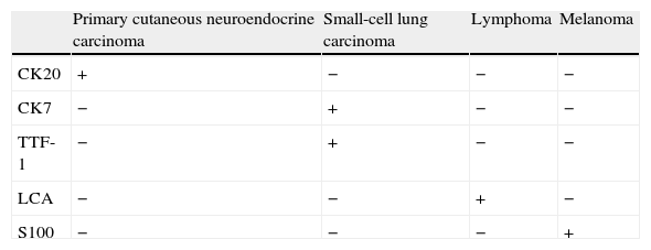
ResultsMost of the patients studied were over 70 years old, except for one who was 55. The highest frequency of cases appeared among patients aged over 80 years. In the cases studied, when the tumour appeared in the head and neck region (10/16), its location could be nasal-lateronasal, cheek-malar, upper eyelid, frontal or mandibular. The major axis of the lesion ranged between 7 and 35mm. Unlike with epidermoid or basocellular carcinomas, recurrence and ganglionar metastases were common. Immunohistochemical (CK20) tests are essential for a correct diagnosis. Treatment is usually surgical and occasionally followed by radiotherapy and chemotherapy.
ConclusionThis carcinoma is not a very common skin tumour. It appears in old age, in the head and neck region in 50% of cases and often leads to exitus.
El llamado clásicamente carcinoma de células de Merkel fue descrito por Toker en 1972, se trata de un carcinoma neuroendocrino primario de la piel. Aparece en la unión dermoepidérmica, es poco frecuente, de crecimiento rápido, agresivo y de mal pronóstico. El objetivo de este trabajo es dar a conocer este carcinoma que se implanta preferentemente en la piel de la cabeza y del cuello, y que aumenta su prevalencia en los últimos años.
Material y métodoRecogemos 16 pacientes afectados por el carcinoma neuroendocrino primario de la piel, tratados en nuestro centro entre 12/09/91 y 13/07/12. Se precisa la edad y el sexo. Se describe la zona de implantación del tumor. Su tamaño lo expresamos en milímetros según el eje mayor de la lesión.
ResultadosNuestros pacientes son mayores de 70 años, excepto la última incluida que contaba 55, la mayor frecuencia es en mayores de 80. Los casos recogidos, cuando asientan en la piel de cabeza y cuello (10/16) tienen localización: nasal-lateronasal, mejilla-malar, párpado superior, frontal, mandibular. El eje mayor de la lesión oscila entre 7 y 35mm. A diferencia de lo que ocurre en los carcinomas espinocelulares o basocelulares son frecuentes las recurrencias y las metástasis. Para el diagnóstico es imprescindible la inmunohistoquímica con citoqueratina 20. El tratamiento es quirúrgico, ocasionalmente seguido de radioterapia y quimioterapia.
ConclusiónSe trata de un carcinoma poco frecuente de la piel, aparece en la edad avanzada, asienta en cabeza y cuello en más del 50% de los casos y conduce con frecuencia al exitus.