


To analyze the MRI characteristics of uterine sarcomas (mainly carcinosarcomas) and to compare them with those of adenocarcinomas to define the findings that would be useful for the differential diagnosis.
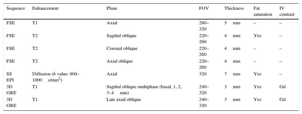
Materials and methodsWe retrospectively reviewed the MRI studies of 13 patients with histologically diagnosed uterine sarcoma. We analyzed tumor size, signal in T2-weighted, unenhanced and gadolinium-enhanced T1-weighted, and diffusion-weighted sequences. We compared the data obtained with those of another series of 30 consecutive cases of adenocarcinomas studied with MRI.
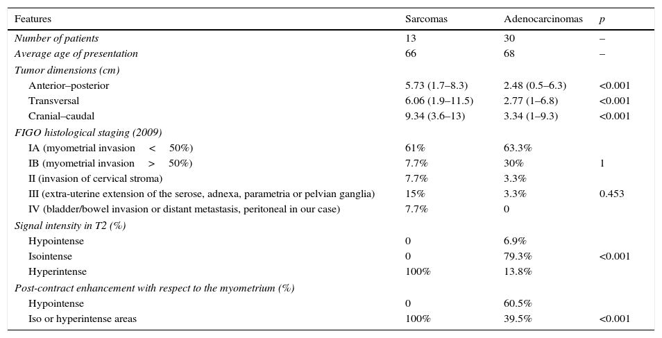
ResultsThe sarcomas (>9cm in 77% of cases) were considerably larger than the adenocarcinomas (p<0.001). There were no differences in FIGO staging by MRI or surgery: both tumor types were diagnosed in early stages. The signal intensity in T2-weighted images differed significantly between the two tumor types: all the sarcomas were heterogeneous and predominantly hyperintense with respect to the myometrium in T2-weighted sequences (p<0.001). In postcontrast studies, all the sarcomas showed enhancement greater than or equal to the myometrium; this finding was significantly different from the adenocarcinomas (p<0.001). In diffusion-weighted sequences, we found no significant differences in ADC values in the areas with greatest restriction, but the ADC map was more heterogeneous in the sarcomas.
ConclusionUterine sarcomas do not have specific characteristics on MRI, but some findings can indicate the diagnosis. In our study, we found significant differences between sarcomas and adenocarcinomas. Sarcomas were larger, had more hyperintense and heterogeneous signal intensity in T2-weighted sequences, and enhanced more than or at least as much as the myometrium.
Analizar las características radiológicas en resonancia magnética (RM) de sarcomas uterinos (principalmente carcinosarcomas) y compararlas con las de adenocarcinomas para definir hallazgos útiles para el diagnóstico diferencial.
Material y métodosRevisamos retrospectivamente los estudios de RM de 13 pacientes con diagnóstico histológico de sarcoma uterino. Analizamos el tamaño tumoral, la señal en secuencias T1, T2, T1 con gadolinio y difusión. Comparamos los datos obtenidos con otra serie de 30 casos consecutivos de adenocarcinomas estudiados mediante RM.
ResultadosLos sarcomas presentaron un tamaño considerablemente mayor que los adenocarcinomas (p<0,001), y midieron más de 9cm en el 77% de los casos. No hubo diferencias en la estadificación tumoral según la FIGO valorada mediante RM y cirugía, y ambos tumores se diagnosticaron en estadios tempranos. La intensidad de señal en T2 fue significativamente diferente (p<0,001), y el 100% de los sarcomas heterogéneos fueron predominantemente hiperintensos en T2 respecto al miometrio. El 100% de los sarcomas presentó un realce igual o mayor que el miometrial en el estudio poscontraste, con una diferencia significativa (p<0,001) con los adenocarcinomas. En difusión no evidenciamos diferencias en los valores de ADC (apparent diffusion coefficient) en las áreas con mayor restricción, pero el mapa de ADC fue más heterogéneo en los sarcomas.
ConclusiónLos sarcomas uterinos no presentan características específicas en RM, pero algunos hallazgos pueden indicar este diagnóstico. En nuestro estudio mostraron diferencias significativas con respecto a los adenocarcinomas, presentándose como tumores de mayor tamaño, de señal hiperintensa y heterogénea en T2, y con un realce mayor o igual que el miometrial.

