


Transcranial Doppler ultrasound (TDU) is useful in cerebrovascular patients. TDU findings are operator-dependent; they can also be influenced by anatomical and physiological variables as well as by the altitude at which the study is done.
ObjectiveTo report the cerebral hemodynamic parameters measured by TDU in subjects who live in Quito, Ecuador (altitude 2850m).
Material and methodsWe recruited 47 volunteers with no history or clinical evidence of stroke, hypertension, metabolic disorders, or hematologic disorders; 2 patients were excluded because they did not have a viable cranial window for TDU study. Thus, we recorded mean cerebral blood flow velocity, peak systolic flow velocity, end-diastolic flow velocity, and pulsatility indices in 45 patients (28 (62.2%) women; mean age, 35.9 years). We recorded patients’ age, sex, and hematocrit. We analysed cerebrovascular hemodynamic parameters by sex and age group.
ResultsNo significant differences between hemispheres were observed in mean flow velocities, except in the anterior cerebral arteries with right predominance. Flow velocities were higher in women and in the youngest age group. No significant differences in the pulsatility indices were found between sexes or between age groups. The flow velocities in this series are lower than those reported for other series.
ConclusionsThe hemodynamic parameters in this series are lower than in other series and are influenced by the altitude, age, and sex.
El Doppler transcraneal (DTC) es una herramienta útil en la atención del paciente cerebrovascular. Sus resultados, además de ser dependientes del operador, pueden estar influenciados por variables fisiológicas y anatómicas, así como por la altura a la que se encuentra el sujeto estudiado.
ObjetivoDescribir los parámetros hemodinámicos cerebrales medidos mediante DTC en sujetos que habitan en la ciudad de Quito.
Material y métodosSe reclutaron para el estudio 47 voluntarios, sin antecedentes ni evidencia clínica de ictus, hipertensión arterial, enfermedades metabólicas o hematológicas. Fueron excluidos dos individuos por ausencia de ventana craneal útil para el ultrasonido. En 45 casos se registraron las velocidades medias de flujo, de pico sistólico, de fin de diástole e índices de pulsatilidad. Se recogieron la edad, el sexo y los niveles de hematocrito. Los datos anteriores se analizaron por sexo y según el grupo de edad.
ResultadosPredominaron las mujeres (28; 62,2%) y la media de edad fue de 35,9 años. No se encontraron diferencias interhemisféricas significativas en las velocidades medias de flujo, excepto en las arterias cerebrales anteriores con predominio derecho. Se registraron mayores velocidades de flujo en las mujeres y en el subgrupo de menor edad. No de demostraron diferencias significativas entre los dos subgrupos de edad ni por sexo en el índice de pulsatilidad. En relación con otras series, las velocidades de flujo son inferiores.
ConclusionesEn el grupo de personas examinadas, los parámetros hemodinámicos registrados son menores que los publicados en otras series y están influenciados por la altura, la edad y el sexo.
Ultrasound began to be used in the study of cerebral vascular diseases in the early 1980s,1–3 when the hemodynamic parameters of the arteries making up the circle of Willis were first studied using transcranial Doppler (TCD).3 Since then, this technique has made great steps forwards, and is now considered essential to the study and treatment of cerebrovascular disease.4
Analysis of the hemodynamic characteristics of each segment makes it possible to monitor the changes that occur in each clinical situation and arrive at a specific diagnosis. For this, knowledge of each artery's parameters, possible anatomical variations and hemodynamic characteristics is essential.4–6
Many studies have used TCD to look at the particular velocimetric features of the cerebral vessels.3,6–10 However, their results are varied and, in trained hands, the methodology of the examination or the ultrasound device used matters less than the characteristics of the population group examined3,6 and the influence of physiological variables such as age, sex and metabolic, endocrine or haematological factors, etc.6,11–15
In Latin America, and especially in high-altitude populations, research referring to the characteristics of cerebral blood flow, measured by TCD, is scarce.9,10 The aim of our study is to describe the cerebral hemodynamic parameters using TCD in a group of subjects living at an altitude of 2850m above sea level.
Material and methodsPatientsThe TCD assessment was performed between the months of January and February 2018. Participants were selected from among healthcare personnel and patients’ families, the purpose of the research was explained to them and they voluntarily agreed to participate.
Inclusion criteriaPeople over 15 years of age who had been living in Quito for more than six months and had no history and/or clinical evidence of disease were selected.
Exclusion criteriaSubjects with a history of stroke, hypertension, metabolic or haematological disease and those with cranial windows not suitable for ultrasound.
Transcranial Doppler examination procedureThe investigation was carried out using a digital TCD system (ST3, made in Seattle, Washington, USA). The study began by placing the person to be examined in the supine decubitus position. After 10min at rest, the physician took up position behind the volunteer's head, in a comfortable position to place the 2mHz Doppler probe over the acoustic cranial windows, in pulsed Doppler mode, applying sufficient gel to guarantee ultrasound transmission. Before assessing intracranial circulation, the extracranial carotid arteries were examined using the 4mHz probe in continuous Doppler mode, in order to rule out significant stenoses (over 50%). The sample volume was set at 10mm and the sweep speed at 3s.
Through the temporal window, introducing a slight angulation to the transducer, the first vessel to be identified is the middle cerebral artery (MCA), between 30 and 60mm, with a flow signal towards the probe. Following the path of the MCA to a depth of 60–65mm, a bidirectional flow pattern corresponding to the bifurcation of the internal carotid artery (ICA) was sought. At this point, directing the ultrasound beam slightly in the cephalic direction and forward, the proximal segment (A1) of the anterior cerebral artery (ACA), in which the direction of blood flow is away from the probe, was examined. Next, decreasing the depth until a bidirectional pattern, corresponding to the carotid bifurcation, was encountered and directing the ultrasound beam slightly rearward and in a caudal direction, at a depth between 65 and 70mm, the precommunicating segment (P1) of the posterior cerebral artery (PCA), in which the direction of flow is towards the probe, was located. At this point, inclining the transducer slightly rearward and deepening to 70mm, the postcommunicating segment (P2), in which the direction of flow is away from the probe, was located (Fig. 1A).
 Ultrasound of each arterial segment of the circle of Willis, represented on cranial computed tomography angiography in an axial plane, showing the inclination of the ultrasound probe through the temporal window. (B) Ultrasounds of the vessels of the posterior circulation through the transforaminal window.")
(A) Ultrasound of each arterial segment of the circle of Willis, represented on cranial computed tomography angiography in an axial plane, showing the inclination of the ultrasound probe through the temporal window. (B) Ultrasounds of the vessels of the posterior circulation through the transforaminal window.
The extracranial internal carotid artery (eICA) was identified by ultrasound through the submandibular window, placing the 2mHz probe within the sternocleidomastoid muscle at a depth of 50mm. The study was completed by determining the flow velocities (FV) in the V4 segments of the vertebral arteries and the basilar artery through the transforaminal window (Fig. 1B). The peak systolic velocity (PSV), end diastolic velocity (EDV), mean flow velocity (MFV) and Gosling's pulsatility index (PI) were recorded for all arterial segments. The TCD examination was performed by the first author, who has prior training in neurosonology techniques and more than five years of experience.
Statistical analysisAge, sex and haematocrit were recorded in all cases. The following hemodynamic variables were recorded in the TCD examination: MFV, PSV, EDV and PI (Fig. 2).

The data were collected prospectively in a database designed in Excel 2016. The absolute and relative frequencies, mean and standard deviations of the variables considered were described. Student's t-test was used for the statistical analysis of the numerical variables. Each patient's hemispheres were considered separately and by sidedness in order to be able to identify whether any significant interhemispheric asymmetries were present. The hemodynamic parameters were analysed by sex and age (dichotomised into two groups: under 40 years and 40 years or over). All statistical tests were performed with the program SPSS (v. 20.0); p-values <0.05 were considered significant.
Ethical considerationsThe research was conducted with the approval of the hospital's research department. The studies were performed after explaining to each volunteer the aim and nature of the examination and obtaining their consent. No personal data relating to the subjects’ identities is revealed in the research.
FindingsForty-seven (47) volunteers were recruited; two subjects were excluded due to not having optimal acoustic cranial windows (4.2%). In the remainder, 100% of the arterial segments were successfully examined (basilar artery, n=45; for the other segments assessed bilaterally, n=90). The mean age of the 45 subjects included was 35.9±11.7 (range 15–58 years); subjects under 40 years of age were in the majority (27; 60%). More than half, 28, were women (62.2%) and 17 (37.8%) were men. Average haematocrit levels were 43.9±3.7 (range 39–59.7); in women, haematocrit figures were significantly lower (women 42.9±2.02 vs. men 45.6±5.08) (p 0.001).
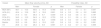
The grouped hemodynamic parameters are shown in Table 1. The highest MFVs were recorded in the MCAs (49.3±14.7cm/s), followed by the ACA (39.9±12.6cm/s) and the basilar artery (36.7±10.1cm/s). The average MFV in the vessels of the anterior circulation was 41.3cm/s; lower MFV values were recorded in the vessels of the vertebrobasilar circulation, PCAs P1 segment (34.6±12.2cm/s), basilar artery (36.7±10.1cm/s).
General results of the transcranial Doppler ultrasound.
| Parameters (SD) | MCA | ACA | eICA | P1 | P2 | Basilar | V4 |
|---|---|---|---|---|---|---|---|
| PSV | 74.4±21.1 | 60.5±18.1 | 53.9±9.9 | 51.9±19.1 | 52.1±12.6 | 54.1±14.8 | 36.7±9.3 |
| EDV | 35.6±10.2 | 28±9.1 | 24.5±4.2 | 24.7±8.9 | 24.3±7.5 | 25.4±6.9 | 18.0±4.0 |
| MFV | 49.3±14.7 | 39.9±12.6 | 34.6±6.5 | 34.6±12.2 | 34.6±8.8 | 36.7±10.1 | 26.0±6.6 |
| PI | 0.8±0.12 | 0.8±0.19 | 0.8±0.2 | 0.8±0.1 | 0.8±0.3 | 0.8±0.2 | 0.7±0.2 |
Values in cm/s except pulsatility index (PI).
ACA: anterior cerebral artery; eICA: extracranial internal carotid artery; MCA: middle cerebral artery; SD: standard deviation; P1, P2: posterior cerebral artery; V4: intracranial vertebral artery; EDV: end diastolic velocity; MFV: mean flow velocity; PSV: peak systolic velocity.
On comparing the MFVs of homologous arterial segments, significant asymmetry was only found in the A1 segments (right 42.7±14.4cm/s vs. left 37.2±10.0cm/s) (p 0.034). For the remaining arteries, the following flow velocities were measured: MCA (M1), right 48.6±14.8cm/s vs. left 49.9±14.8cm/s (p 0.542); extracranial ICA, right 34.4±7.9cm/s vs. left 34.8±4.9cm/s (p 0.768; PCA proximal segment (P1) right 36.3±2.1cm/s vs. 32.7±1.5 (p 0.155), distal segment (P2) right 34.5±8.9cm/s vs. 34.6±8.8cm/s (p 0.946); intracranial vertebral arteries (V4), right 25.9±6.5cm/s vs. left 26.2±6.7cm/s (p 0.773).
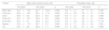
Regarding MFVs and PI by sex (Table 2), in the vessels of the anterior circulation the MFV values recorded were higher in women, except in the ACA. There was only a statistically significant difference in the eICA. There was no difference between the PIs of the arteries assessed, which was between 0.7 and 0.9 in all vessels. In the posterior circulation, the highest MFV values were recorded in women. Regarding the PI, no important differences were detected in any vessels.
Mean flow velocity (cm/s) and pulsatility index by sex.
| Vessel | Mean flow velocity (cm/s), SD | Pulsatility index, SD | ||||
|---|---|---|---|---|---|---|
| Men | Female | p | Men | Female | p | |
| MCA (M1) | 48.7±15.5 | 49.6±14.4 | 0.785 | 0.8±0.1 | 0.7±0.1 | 0.047 |
| ACA (A1) | 40.4±12.9 | 39.7±12.6 | 0.801 | 0.8±0.2 | 0.8±0.2 | 0.682 |
| eICA | 31.5±5.0 | 36.4±6.7 | 0.000 | 0.9±0.2 | 0.8±0.1 | 0.187 |
| PCA (P1) | 34.0±12.3 | 34.9±12.2 | 0.753 | 0.8±0.1 | 0.8±0.1 | 0.516 |
| PCA (P2) | 33.8±9.9 | 35.0±8.2 | 0.556 | 0.8±0.2 | 0.9±0.3 | 0.386 |
| Basilar | 32.5±8.9 | 39.2±10.0 | 0.25 | 0.8±0.2 | 0.8±0.2 | 0.561 |
| V4 | 25.6±6.0 | 26.3±6.9 | 0.591 | 0.7±0.2 | 0.7±0.2 | 0.469 |
Values in cm/s except pulsatility index.
ACA: anterior cerebral artery; ICA: internal carotid artery; MCA: middle cerebral artery; PCA: posterior cerebral artery; SD: standard deviation; MFV: mean flow velocity.
The MFV values recorded were inversely proportional to age, although this was only statistically significant for the MCA (Table 3).
Mean flow velocity (cm/s) and pulsatility index by sex.
| Vessel | Mean flow velocity (cm/s), SD | Pulsatility index, SD | ||||
|---|---|---|---|---|---|---|
| <40 years | >40 years | p | <40 years | >40 years | p | |
| MCA (M1) | 52.7±14.4 | 44.1±13.8 | 0.006 | 0.8±0.1 | 0.7±0.1 | 0.704 |
| ACA (A1) | 42.4±13.0 | 36.1±11.0 | 0.020 | 0.8±0.2 | 0.8±0.1 | 0.720 |
| eICA | 35.3±6.3 | 33.5±6.8 | 0.203 | 0.8±0.1 | 0.8±0.2 | 0.983 |
| PCA (P1) | 35.7±13.7 | 32.9±9.5 | 0.260 | 0.8±0.1 | 0.8±0.1 | 0.303 |
| PCA (P2) | 35.7±9.1 | 32.9±8.4 | 0.143 | 0.8±0.2 | 0.9±0.3 | 0.118 |
| Basilar | 37.4±11.3 | 35.7±8.1 | 0.252 | 0.8±0.1 | 0.8±0.2 | 0.561 |
| V4 | 25.7±6.2 | 26.5±7.1 | 0.569 | 0.7±0.2 | 0.7±0.2 | 0.995 |
Values in cm/s except pulsatility index.
ACA: anterior cerebral artery; ICA: internal carotid artery; MCA: middle cerebral artery; PCA: posterior cerebral artery; SD: standard deviation; MFV: mean flow velocity.
In this study, no significant interhemispheric differences were found in the mean flow velocities in the proximal arterial segments making up the circle of Willis, with the exception of the anterior cerebral arteries, in which greater velocities were found on the right. Higher flow velocities were recorded in women and in the subgroup of participants under 40 years of age. Regarding PI, no significant differences were found between subgroups either by age or sex.
Knowledge of normal FV values in TCD is essential in order to be able to correctly interpret the results in a specific clinical context. However, hemodynamic information from one artery should not be considered in isolation, but together with the results obtained from the other intracranial vessels, after assessing the extracranial arteries. Because TCD results can vary depending on the experience and skill of the operator, and are influenced by physiological and anatomical factors, it is advisable for each laboratory to generate its own reference values, as well as serving as a starting point for adjusting the results of TCD in pathologies such as stenosis, vasospasm, etc.
As altitude above sea level increases, barometric pressure and the partial pressure of gases decrease.16 When assessing the effect of exposure to high altitudes, it is necessary to differentiate between the acute adaptive response that occurs over minutes and hours, and the physiological acclimatisation response that takes place over longer periods. In the first case, the immediate response is expressed as increased frequency and depth of respiration, a process triggered by stimulation of the peripheral chemoreceptors in the carotid and aortic bodies which activate when PO2 drops below normal values (70mmHg); these in turn inform the bulbar respiratory centre to increase the frequency of respiration. Heart rate also increases in the initial stages, therefore raising cardiac output and in turn cerebral oxygenation.17
When a person remains at high altitudes for more or less prolonged periods, they tend to adapt to the hypoxia; this is possible due to changes involving erythropoiesis and iron metabolism.18–22 In populations living at altitude, increased haemoglobin concentration is observed as a result of elevated red blood cell production, with a concomitant decrease in plasma volume22 and increase in blood viscosity.19 This last effect, together with arterial lumen area and vessel length, are the main determiners of blood flow resistance,16 meaning that cerebral blood FV is inversely correlated with haematocrit level.23–25
In Table 4, we compare our results with those of several studies conducted in normal subjects in populations at different altitudes.5,8–10 In the case of the study by Franco et al.,10 in Colombia, we used average FV values as this work does not give overall figures. It should be noted that our results differ from the FV values used as a reference (Aaslid et al.)3 and that as altitude increases, FV values fall in almost all arterial segments. Nevertheless, the velocities we calculated are similar to those published by Segura et al. in a series of 118 healthy subjects assessed in Girona (Spain),8 which could be related to haematocrit levels in those cases, a detail that was not analysed in their work. On the other hand, none of the studies we refer to compare FV with haematocrit levels.3,8–10
Comparison of the velocimetry values obtained with the results of other studies.
| Study | Parameter | MCA (A1) | ACA (A1) | eICA | PCA (P1) | Basilar | Vertebral (V4) |
|---|---|---|---|---|---|---|---|
| Ours (2850m)n=45 | MFV | 49.3 | 39.9 | 34.6 | 34.6 | 36.7 | 26.0 |
| PI | 0.8 | 0.8 | 0.8 | 0.8 | 0.8 | 0.7 | |
| Colombia (995m)Franco et al.10n=51 | MFV | 59.8 | 47.4 | 31.5 | 37.4 | 45.1 | 35.8 |
| PI | 0.74 | 1.01 | 0.80 | 0.77 | 0.78 | 0.76 | |
| Sao Paulo (760m)Fregonesi Barbosa et al.9n=88 | MFV | 62 | 48 | – | 37 | 43 | 32 |
| PI | 0.51 | 0.52 | – | 0.53 | 0.51 | 0.51 | |
| Bern, Switzerland (540m)Aaslid et al.3n=50 | MFV | 62 | 51 | 37 | 44 | – | – |
| PI | – | – | – | – | – | – | |
| Girona, Spain (76m)Segura et al.8n=118 | MFV | 54 | 43 | – | 34 | 37 | 29.1 |
| PI | 0.98 | 1.01 | – | 1.01 | 1.03 | 1.01 |
ACA: anterior cerebral artery; MCA: middle cerebral artery; eICA: extracranial internal carotid artery; PCA: posterior cerebral artery; PI: pulsatility index; V4; intracranial vertebral artery; MFV: mean flow velocity.
Analysing the relationship between MFVs and sex, our results give very similar figures to those obtained in other series.1,8,26,27 We recorded higher FV values for women, a different that appears to be dependent on the lower haematocrit levels found. In keeping with some publications, in addition to the inverse correlation between haematocrit levels and FV,25 there may be a link with oestrogen fluctuations reducing vascular resistance, with a consequent increase in cerebral flow velocity.28,29 In this regard, Isikay et al., in 862 patients with a mean age of 57±16 years, of whom 413 were women (53±17 years) and 449 were men (60±13 years), describe a significant difference in haematocrit concentrations between the sexes (women 39±4 vs. men 42±4, p <0.001) and find an inverse relationship between haematocrit levels and MFV (women 60±18cm/s vs. men 52±15cm/s, p <0.001).30 However, another study with a smaller sample, 63 healthy volunteers (30 men and 33 women, age range 5–69 years), found no difference in flow parameters based on sex, but did find a difference related to increase in age, with greater reductions in the group over 40 years of age.31
Age is the most important factor modifying FVs in TCD.6,8,30–33 Tegeler et al., in a series of 364 healthy subjects between 18 and 80 years of age, conclude that FVs decrease by 4–5% per decade, primarily in the MCAs and ACAs.7 In our study, based on the number of cases, we analysed FVs in two age groups (over and under 40 years) and found a difference of 16.4% for the MCAs and 14.9% for the ACAs between the two groups. In the vessels of the posterior circulation, the difference was less evident (posterior cerebral arteries 7.8%, basilar artery 4.5% and no large difference in the V4 segments of the vertebral arteries).
Analysing whether there was any asymmetry in the FVs of homologous vessels, we found a very small difference in the anterior circulation, with the exception of the A1 segments, where difference was statistically significant (exceeding 5cm/s). The greater asymmetry found in the A1 segment is surely related to the frequency with which hypoplasia is described in these vessels.25 In the posterior circulation, the interhemispheric differences were fewer and not statistically significant. Tegeler et al., in their study, only found a difference in the distal portion of the M1 segment of the MCA (p=0.022) and in the C1 segment of the ICA (p <0.0001), in both cases with slightly higher values on the left.7
Although the FVs in the arteries analysed were lower than those published3,7 and varied based on age and sex, there was no change in the PI. PI, an additional parameter available from TCD,34 indirectly reflects the degree of distal resistance to flow,35 although under conditions of hypocapnia and in patients with subarachnoid haemorrhage it must be interpreted with care.36,37 It can be calculated manually from the difference between the PSV and EDV divided by the MFV, and has the advantage that, being expressed as a proportion, it is not affected by the angle of incidence of the ultrasound beam.34 Its normal value varies between 0.5 and 1.1, and it is very sensitive in the detection of intracranial hemodynamic alterations.38,39 It has been described that PI increases with age,7,33 which was not corroborated in our cases.
The main limitation of our study is its sample size; a greater number of participants could provide more data on the various variables analysed and define their significance. Even so, the results obtained may serve as a reference point for conducting studies in comparable populations from regions at different altitudes.
In conclusion, in the group of people examined, the hemodynamic parameters recorded using TCD ultrasound differ from those published in other series with subjects with no history of disease. The lower flow velocity values recorded appear to be influenced by altitude and haematocrit. Our results could have a clinical impact in the assessment of patients with both extra- and intracranial cerebrovascular conditions, and serve as a starting point for the validation of TCD result in diseases such as stenosis and vasospasm.
Authorship- 1.
Responsible for the integrity of the study: CESM, DRR.
- 2.
Study conception: CESM, DRR.
- 3.
Study design: CESM, DRR.
- 4.
Data collection: CESM.
- 5.
Data analysis and interpretation: CESM, DRR.
- 6.
Statistical processing: CESM, DRR.
- 7.
Literature search: CESM, DRR.
- 8.
Drafting of the article: CESM, DRR.
- 9.
Critical review of the manuscript with intellectually relevant contributions: CESM, DRR.
- 10.
Approval of the final version: CESM, DRR.
The authors declare that they have no conflicts of interest.
Please cite this article as: Scherle Matamoros CE, Rivero Rodríguez D. Parámetros hemodinámicos cerebrales, determinados mediante Doppler transcraneal, en un grupo de voluntarios sanos a 2.850 metros de altura. Radiología. 2019. https://doi.org/10.1016/j.rx.2019.04.003

