


In June 2019 in Seville, at the first course in fetal MRI, endorsed by the Spanish Society of Medical Radiology (SERAM) and the Spanish Society of Pediatric Radiology (SERPE), the Spanish fetal MRI group was founded. To establish this group, a questionnaire was designed for radiologists dedicated to prenatal imaging in Spain and disseminated to the SERAM’s members. The questions were related to the type of hospital, to MRI studies (magnetic field, gestational age, use of sedation, number of studies per year, proportion of fetal neuroimaging studies), and to teaching and research about fetal MRI. A total of 41 responses were received from radiologists in 25 provinces (88% working in public hospitals). Very few radiologists in Spain perform prenatal ultrasonography (7%) or prenatal CT. MRI is done in the second trimester (34%) or in the third trimester (44%). In 95% of centers, fetal brain MRI studies predominate. In 41% of the centers, studies can be done on 3 T MRI scanners. Maternal sedation is used in 17% of centers. The number of fetal MRI studies per year varies widely, being much higher in Barcelona and Madrid than in the rest of Spain.
En junio de 2019 se organizó en Sevilla el primer curso de resonancia magnética (RM) fetal, con el aval de las sociedades españolas de Radiología Médica (SERAM) y Radiología Pediátrica (SERPE), y se fundó el grupo español de RM fetal. Para establecer este grupo, se diseñó un cuestionario para radiólogos que se dediquen a la imagen prenatal en España que anunció la Sociedad Española de Radiología a sus socios. Las preguntas estaban relacionadas con el tipo de hospital, con los estudios de RM (campo magnético, edad gestacional, uso de sedación, número de estudios por año, proporción de estudios de neuroimagen fetal) y con la docencia e investigación de la RM fetal. Recogimos 41 respuestas de 25 provincias (88% hospitales públicos). Muy pocos radiólogos realizan ecografía (7%) o tomografía computarizada prenatal en España. La RM se realiza en el segundo trimestre (34%) o tercer trimestre (44%). En el 95% de los centros predominan los estudios del cerebro fetal. El 41% de los centros tienen la posibilidad de realizar sus estudios en RM 3 Tesla. La sedación materna se usa en el 17% de los centros. El número de estudios de RM fetal por año es muy variable, siendo mucho mayor en Barcelona y Madrid que en el resto de España.
The study of congenital malformations has improved significantly in recent decades with the development of different prenatal diagnosis techniques. Prenatal imaging provides valuable information about malformations and their prognosis, helping to improve pregnancy management, the use of foetal therapies and decision-making, such as pregnancy termination, with more objective information. In addition, they help to plan the birth and the postnatal treatment of many diseases.
Prenatal ultrasound plays a fundamental role in foetal medicine as it is the main screening tool. Foetal magnetic resonance imaging (MRI) is a complementary technique to ultrasound in many cases, to confirm the findings or to more accurately assess the abnormalities detected. With little homogeneity in the number of foetal MRI scans performed in different parts of Spain, the Spanish foetal MRI group was founded in 2019 to standardise and increase the use of this technique. As a starting point, we decided to conduct a survey on the status of foetal MRI in Spain. The aim of this article was to describe the status of foetal MRI in Spain in 2020 and to encourage more centres to use the technique and join this group.
SurveyThe authors designed a questionnaire (Appendix A) on clinical practice in foetal imaging. The Spanish Society of Medical Radiology (SERAM) sent an invitation to complete the questionnaire to all its members in the January 2020 edition of its monthly online journal. This survey was also sent to those attending the first course on foetal MRI held in Seville in June 2019. The questionnaire included questions about the workplace and type of work, multidisciplinary foetal committees, number and type of radiologists reporting foetal MRI, magnetic field of the MRI, number and type of studies, gestational age at the time the studies were performed, and indications, sedation, ultrasound and prenatal computed tomography (CT), research and teaching. Only one response per centre was included. Those who responded and the cities where they worked were not anonymous in order to avoid duplication.
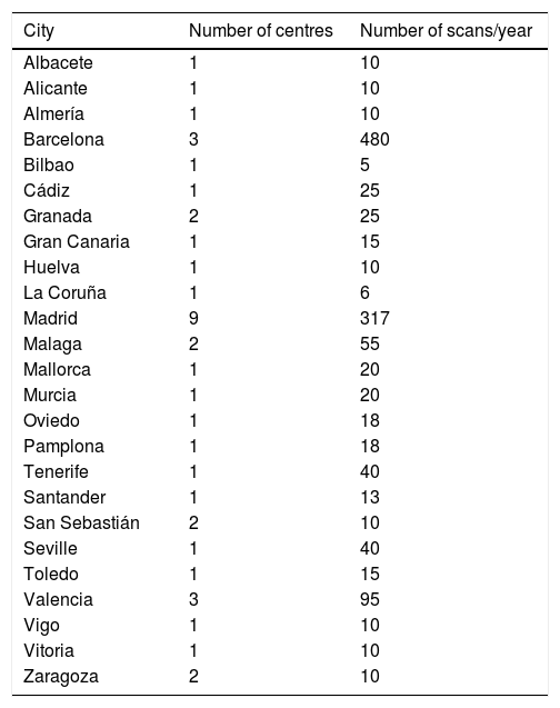
ResultsWe obtained 41 responses from 25 provinces (Fig. 1 and Table 1). There was a response from all the autonomous communities except Castile and León, Extremadura, La Rioja, Ceuta and Melilla. According to this survey, approximately 1,300 foetal MRI studies are performed each year in Spain, the vast majority (approximately 800) in Barcelona and Madrid; most are performed in two hospitals in Barcelona (approximately 200 per year).
.")
Number of foetal magnetic resonance imaging scans per year by province.
| City | Number of centres | Number of scans/year |
|---|---|---|
| Albacete | 1 | 10 |
| Alicante | 1 | 10 |
| Almería | 1 | 10 |
| Barcelona | 3 | 480 |
| Bilbao | 1 | 5 |
| Cádiz | 1 | 25 |
| Granada | 2 | 25 |
| Gran Canaria | 1 | 15 |
| Huelva | 1 | 10 |
| La Coruña | 1 | 6 |
| Madrid | 9 | 317 |
| Malaga | 2 | 55 |
| Mallorca | 1 | 20 |
| Murcia | 1 | 20 |
| Oviedo | 1 | 18 |
| Pamplona | 1 | 18 |
| Tenerife | 1 | 40 |
| Santander | 1 | 13 |
| San Sebastián | 2 | 10 |
| Seville | 1 | 40 |
| Toledo | 1 | 15 |
| Valencia | 3 | 95 |
| Vigo | 1 | 10 |
| Vitoria | 1 | 10 |
| Zaragoza | 2 | 10 |
Thirty-six centres where these studies were carried out were public (88%) and five were private, with patients referred from the public health system and private patients (12%). Most of the hospitals were tertiary (76%), with approximately half (55%) of them having multidisciplinary foetal committees. The indications for performing foetal MRI studies were agreed by consensus between radiologists and gynaecologists in 50% of the centres, by indication of the obstetrician in 40%, and in multidisciplinary committees in 10% of the centres. Very few radiologists perform foetal ultrasound (7%) before foetal MRI; only in Cádiz and Valencia. The vast majority of centres (88%) did not perform foetal CT; only five centres conducted CT as an exception (once a year).
More than half of the foetal MRI studies (51%) were reported on by paediatric radiologists, 22% by radiologists who reported on both adults and children, 20% by neuroradiologists, 5% by paediatric neuroradiologists and 2% by a radiologist who specifically worked in foetal imaging in Valencia. In 59% of the centres, the scanners used were 1.5 Tesla MRI, 39% used both 1.5 and 3 Tesla MRI without distinction and in one centre they used only a 3 Tesla MRI.
The MRI scans were predominantly performed between 18 and 26 weeks’ gestation (second trimester) in 34% of centres, between 27 and 40 weeks' gestation (third trimester) in 44% and indistinctly during the second or third trimester in 22% of the institutions. Most of the foetal MRIs were focused on evaluation of the foetal brain. The proportion of indications to assess the foetal central nervous system ranged from 80% to 100% of cases in 80% of centres and approximately 70% of cases in 15% of centres. Two centres (5%) performed only a third of MRIs focused on the foetal central nervous system.
Half of the surveyed centres never repeated foetal MRI studies, 44% almost never (less than 5% of cases) and the remaining 6% repeated a large percentage of MRIs. In the centres where the scans were repeated, it was mainly to assess cerebral cortical development during the third trimester of pregnancy.
Most centres (83%) did not administer sedation to the mothers prior to foetal MRI. In 17% of the centres, the mothers took benzodiazepines (diazepam) before the MRI to reduce foetal movement.
Four centres (10%) conducted foetal MRI research on the impact of congenital heart disease on the central nervous system, neural tube defects, diaphragmatic hernias, predisposing factors for prematurity, impact of diet on neurodevelopment, placental oxygenation, intrauterine growth retardation and new foetal MRI sequences.
Of those surveyed, 75% expressed their interest in attending annual meetings of the foetal MRI group and 25% every two years. The vast majority of those surveyed expressed their interest in a multidisciplinary group with gynaecologists, geneticists and neonatologists, and in having centres nationally accredited for training in foetal MRI.
Interpretation of the resultsThis article describes the status of foetal MRI in Spain in 2020. Our hope is that it should be a first step to forming a group made up of radiologists and other doctors with an interest in foetal MRI, to popularise and promote the standardisation of this technique here in Spain.
This survey has shown that, as in other European countries, foetal MRI is performed mainly in public hospitals.1 This is because these studies are indicated in many tertiary care centres by multidisciplinary committees for the comprehensive assessment of foetuses with malformations, and such committees are more common in public hospitals. It is also partly due to the fact that these scans take some time and require the physical presence of the radiologist while they are being obtained, at least until the technicians become familiar with the technique, which is less feasible in private centres. The indication for this test comes from the obstetrician in only 40% of cases, in line with the results of a similar European-wide survey conducted.1
This article has also shown that there are few radiologists who perform prenatal ultrasound, similar to the trend in the rest of the world, where this technique is performed by gynaecologists and obstetricians.1,2 In Europe, foetal ultrasound is performed by radiologists only in France, Belgium and Scotland.1 Only 7% of Spanish radiologists perform prenatal ultrasound in conjunction with foetal MRI, a practice that is recommended given that these techniques are complementary.
This survey has also clearly shown that many more scans are performed to assess abnormalities of the CNS than of the foetal body, as previously reported in multiple articles.3,4
This survey also found that the number of foetal MRI scans varies greatly across the different regions of Spain. That may be due in part to the population density of Madrid and Barcelona and to the fact that many patients are referred to these specialised centres from other provinces. Valencia, Malaga and Seville report an intermediate number of scans. We did find striking, however, the low number of foetal MRI scans performed in Galicia, Asturias, Cantabria, the Basque Country, the Balearic Islands, Ceuta and Melilla, given their population densities. This can probably be explained by the fact that centres in these cities did not respond to this study's survey and that there are few hospitals with foetal medicine units and a sufficient volume of disease. Another decisive factor is the lack of knowledge and training in this technique by radiologists and obstetricians here in Spain, where it is not yet included in the training plans for residents of these specialities. In contrast, foetal ultrasound plays a fundamental role in the training of all gynaecologists and, in some cases, radiologists.
This survey found that approximately 40% of the radiologists who perform this technique have access to a 3 Tesla MRI, highlighting the increasing use of these scanners in Spain and across the world thanks to the lack of adverse effects and their greater spatial resolution.5,6
Sedation is used before the scans to reduce the movements of the foetus by 17% of our respondents, a higher rate than reported in Europe.1 There is little scientific literature on this aspect and it is an area that should be assessed in future studies.
The use of foetal CT is very rare here in Spain, despite the limitations of foetal MRI at present in assessing bone structures and the fact that the utility of CT has been described for the study of bone dysplasias.7,8
This survey has revealed the participants' wish to set up a Spanish group that promotes training and research in the field of foetal MRI, asking for annual courses to continue updating their skills in this technique and showing their willingness to participate in multicentre research studies in this field. Training in foetal MRI is particularly important for radiology residents so they are exposed to the technique during their training period and can further develop their experience as their careers progress. The main limitation of this study is that many centres did not respond to the survey, so it does not represent all of Spain; for example, no response was obtained from Castile and León and Extremadura, where they do most likely carry out foetal MRI studies.
ConclusionFoetal MRI in Spain in 2020 was mainly performed in tertiary public hospitals, many of which have multidisciplinary foetal medicine committees. Very few radiologists perform foetal ultrasound and CT, with the vast majority only reporting on MRI scans. Foetal MRI is performed both in the second and third trimesters of pregnancy and most scans are obtained to study the foetal brain. This survey has revealed widespread interest in increasing training and research in this field.
Authorship- 1
Person responsible for the integrity of the study: PCD, LGD and MRP.
- 2
Study conception: PCD, LGD and MRP.
- 3
Study design: PCD, LGD and MRP.
- 4
Data collection: PCD and MRP.
- 5
Data analysis and interpretation: PCD, LGD and MRP.
- 6
Statistical processing: PCD.
- 7
Literature search: PCD, LGD and MRP.
- 8
Drafting of the article: PCD.
- 9
Critical review of the manuscript with intellectually relevant contributions: PCD, LGD and MRP.
- 10
Approval of the final version: PCD, LGD and MRP.
The authors declare that they have no conflicts of interest.
The following is Supplementary data to this article:

