


To determine the frequency of pancreatic metastasis from renal cell carcinoma in patients studied with MDCT during 2007 and to describe the patterns of presentation on MDCT.
Material and methodsWe retrospectively studied 133 patients with renal cell carcinoma who underwent MDCT between January and December 2007. Forty-nine patients presented with disseminated disease. We analyzed the frequency, location, and patterns of presentation of pancreatic metastases.
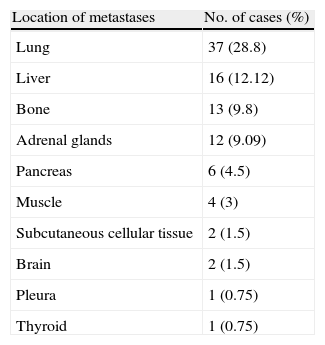
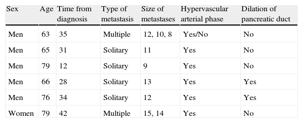
ResultsPancreatic involvement was identified in six patients. Four patients had isolated pancreatic nodules and two presented multiple nodules. A total of nine pancreatic lesions ranging between 8mm and 40mm were detected. All nodules had increased uptake of contrast material in the arterial phase except for one in a patient with multiple nodules, due to necrosis. Two cases were associated with pancreatic duct dilation. Histology was obtained in only one patient.
ConclusionPancreatic involvement of renal cell carcinoma was detected in 4.5% of patients, ranking fifth in frequency in patients with disseminated disease. The arterial phase is necessary to detect pancreatic involvement of renal cell carcinoma. The pattern of presentation is nearly constant, helping differentiate pancreatic metastasis from primary pancreatic adenocarcinoma.
Determinar la frecuencia y las formas de presentación radiológica de las metástasis pancreáticas en los pacientes con carcinoma de células renales estudiados mediante tomografía computarizada multidetector (TCMD) en nuestro hospital durante el año 2007.
Material y métodosEstudio retrospectivo de 133 pacientes con carcinoma de células renales a los que se les realizó estudios mediante TC multidetector en nuestro hospital entre enero y diciembre de 2007, de los cuales 49 presentaban enfermedad metastásica. Análisis de la frecuencia, localización y formas de presentación de la afectación pancreática.
ResultadosEn 6 pacientes se identificó afectación pancreática. Cuatro pacientes se presentaron con nódulos únicos y dos con nódulos múltiples, identificando así un total de 9 lesiones pancreáticas con unos tamaños que variaron entre los 8 y los 40mm. Todos los nódulos fueron hipercaptantes en fase arterial salvo uno en el contexto de múltiples nódulos, debido a fenómenos de necrosis. Dos casos asociaron dilatación del conducto pancreático. Únicamente se obtuvo comprobación histológica de una de las lesiones.
ConclusionesLas metástasis pancreáticas del carcinoma de células renales se han presentado en nuestro estudio con una frecuencia del 4,5%, ocupando el quinto lugar en frecuencia en los pacientes con enfermedad diseminada. Su forma de presentación sigue un patrón prácticamente constante, lo que ayuda a su diferenciación con el adenocarcinoma primario de páncreas. Por lo tanto, es necesario una fase arterial para su detección.

