Buscar en
Radiología (English Edition)
Toda la web
Inicio Radiología (English Edition) Imaging in epilepsy: Functional studies
Journal Information
Vol. 54. Issue 2.
Pages 124-136 (March - April 2012)
Share
Share
Download PDF
More article options
Visits
7162
Vol. 54. Issue 2.
Pages 124-136 (March - April 2012)
Update in radiology
Imaging in epilepsy: Functional studies
Imagen en epilepsia: estudios funcionales
Visits
7162
N. Bargalló Alabart
Corresponding author
bargallo@clinic.ub.es

Corresponding author.
, X. Setoain Parego
Centre de Diagnòstic per la Imatge, Hospital Clínic i Provincial, Barcelona, Spain
This item has received
Article information
Abstract
Full Text
Bibliography
Download PDF
Statistics
Figures (8)
Show moreShow less
Tables (1)
Table 1. Features of SPECT and PET.
Abstract

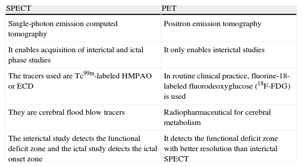
Neuroimaging studies play a fundamental role in the diagnosis and evaluation of epilepsy. Technological advances in neuroimaging techniques have made it possible to obtain functional as well as structural information. The pathophysiology of epilepsy consists of an abnormal increase in cerebral activity that can be appreciated on neuroimaging techniques such as functional magnetic resonance imaging (fMRI), positron emission tomography (PET), and single-photon emission computed tomography (SPECT). In patients with epilepsy that is refractory to drug therapy, the main aim of surgery is to control seizures and thus to improve the quality of life. In the preoperative workup of these patients, fMRI has an increasingly important role, evaluating the location of functional areas that must be safeguarded during surgery.

Keywords:
Epilepsy
Functional magnetic resonance imaging
Positron emission tomography
Single photon emission computed tomography
Resumen

Los estudios de neuroimagen tienen un papel fundamental en la evaluación y diagnóstico de la epilepsia. Los avances tecnológicos que se han producido en las técnicas de neuroimagen permiten en la actualidad obtener información no solo estructural, sino también funcional. La fisiopatología de la epilepsia consiste en un aumento anómalo de la actividad cerebral que puede llegar a apreciarse mediante técnicas de neuroimagen, como por ejemplo la RM funcional (RMf), la tomografía por emisión de positrones (PET) y la tomografía por emisión de fotón simple (SPECT). En pacientes con epilepsia farmacorresistente, la cirugía tiene como objetivo principal el control de las crisis, y por tanto mejorar la calidad de vida. La RMf juega un papel cada vez más relevante en la valoración prequirúrgica de estos pacientes, evaluando la localización de áreas funcionales que deben evitar lesionarse durante el acto quirúrgico.

Palabras clave:
Epilepsia
Resonancia magnética funcional
Tomografía por emisión de positrones
Tomografía por emisión de fotón simple

Article

These are the options to access the full texts of the publication Radiología (English Edition)
Subscriber
Subscriber

If you already have your login data, please click here .

If you have forgotten your password you can you can recover it by clicking here and selecting the option “I have forgotten my password”
Purchase
Purchase article

Purchasing article the PDF version will be downloaded

Price 19.34 €

Purchase now
Contact
Phone for subscriptions and reporting of errors
From Monday to Friday from 9 a.m. to 6 p.m. (GMT + 1) except for the months of July and August which will be from 9 a.m. to 3 p.m.
Calls from Spain
932 415 960
Calls from outside Spain
+34 932 415 960
E-mail
Article options
Tools
es en pt

¿Es usted profesional sanitario apto para prescribir o dispensar medicamentos?

Are you a health professional able to prescribe or dispense drugs?

Você é um profissional de saúde habilitado a prescrever ou dispensar medicamentos

Quizás le interese:
10.1016/j.rxeng.2020.03.006
No mostrar más