metricas
covid
Radiología (English Edition) Diagnostic value of contrast-enhanced mammography versus magnetic resonance imag...
Journal Information
Original articles
Available online 5 December 2025
Diagnostic value of contrast-enhanced mammography versus magnetic resonance imaging in invasive lobular carcinomas: Correlation with pathological assessment and detection of additional lesions
Valor diagnóstico de la mamografía con contraste frente a la RM en el carcinoma lobulillar invasivo: correlación con AP y detección de lesiones adicionales
E. Vila-Trias Jovera,
Corresponding author
, R. Alcántara Souzaa,b,c, A. Moncusí Armengold, M.M. Vernet Tomásb,c,e, L. Comerma Blesaf, J.M. Maiques Llácera, J. Azcona Sáenza,c
a Servicio de Radiología y Medicina Nuclear, DIBI-Hospital del Mar, Parc de Salut Mar, Barcelona, Spain
b Departamento de Medicina, Universidad Autónoma de Barcelona, Barcelona, Spain
c Departamento de Medicina y Ciencias de la Vida (MELIS), Universitat Pompeu Fabra, Barcelona, Spain
d Departamento de Epidemiología y Evaluación, Instituto de Investigación del Hospital del Mar, Barcelona, Spain
e Servicio de Obstetricia y Ginecología, Hospital del Mar, Parc de Salut Mar, Barcelona, Spain
f Servicio de Patología, Hospital del Mar, Parc de Salut Mar, Barcelona, Spain
Article information
Abstract
Full Text
Bibliography
Download PDF
Statistics
Figures (4)
Show moreShow less
Tables (6)
Table 1a. Findings of invasive lobular carcinoma on low-energy images.
Tables
Table 1b. Findings of invasive lobular carcinoma on recombinant images and magnetic resonance imaging.
Tables
Table 2. Correlation of the visibility of invasive lobular carcinoma (low, moderate, high) according to its histological tumour grade (G1, G2, G3) described in the post-surgical pathology report.
Tables
Table 3. Distribution of the estimation of tumour size of invasive lobular carcinoma in relation to the reference histological size (±1 cm), according to the imaging technique used: low energy, recombinant image and magnetic resonance.
Tables
Table 4a. Classification of additional lesions as false negatives, false positives, true negatives and true positives by contrast-enhanced mammography and magnetic resonance imaging.
Tables
Table 4b. Histology of additional lesions.
Tables
Show moreShow less
Abstract
Objective

To compare the diagnostic performance of contrast-enhanced mammography (CEM) with magnetic resonance imaging (MRI) in the preoperative assessment of invasive lobular carcinoma (ILC).

Materials and methods

This was a single-centre, retrospective observational study including women with histologically confirmed ILC who underwent both preoperative CEM and MRI between January 2018 and June 2022. The detection sensitivity of ILC was evaluated using low-energy images (LEI), recombined images (RCI), CEM (LEI + RCI), and MRI. We analysed the correlation of tumour size estimated by each imaging modality and pathology, and the correlation between lesion conspicuity and tumour grade. We also assessed the detection of additional lesions (AL).
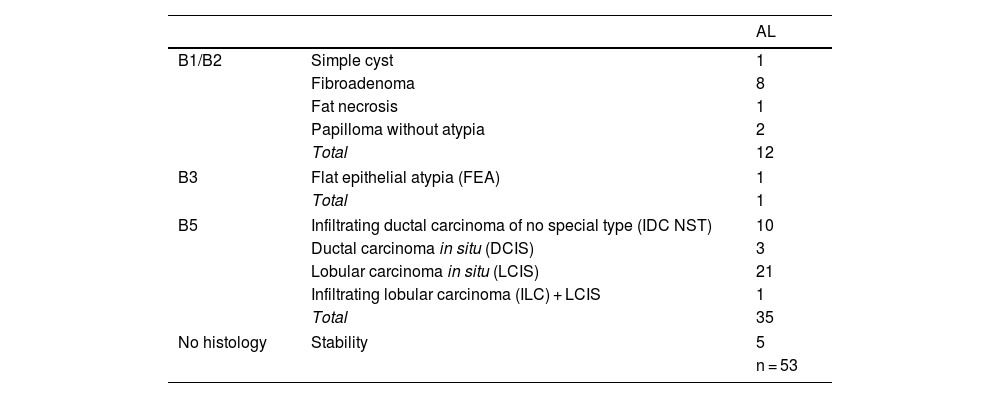
Results

Fifty-eight patients with 60 ILCs were included. LEIs showed a sensitivity of 83.3% compared to 100% for RCI, CEM and MRI. MRI demonstrated the strongest correlation with tumour size on HP (r = 0.792). High conspicuity was observed in 68% of ILCs and strongly correlated with MRI enhancement level (ρ = 0.81; P < .001). No significant association was found between conspicuity and tumour grade (P = .074). Of the 53 ALs detected, 39 were malignant. MRI identified all malignant lesions, whereas CEM failed to detect five.

Conclusion

CEM may be a comparable alternative to MRI in the detection and size estimation of ILC; however, MRI appears to be superior in identifying malignant additional lesions.

Keywords:
Lobular carcinoma
Mammography
Magnetic resonance imaging
Sensitivity
Neoplasm staging
Breast neoplasm
Diagnostic imaging
Resumen
Antecedentes y objetivos

Comparar el rendimiento diagnóstico de la mamografía con contraste (CEM) frente a la resonancia magnética (RM) en el estudio preoperatorio del carcinoma lobulillar invasivo (CLI).

Materiales y métodos

Estudio retrospectivo observacional, unicéntrico, que incluyó a aquellas mujeres con diagnóstico histológico de CLI, sometidas a CEM y RM preoperatorias entre enero de 2018 y junio de 2022. Se evaluó la capacidad de detección del CLI mediante imágenes de baja energía (LEI), recombinadas (RCI), CEM (LEI + RCI) y RM para el CLI. Se analizó la correlación entre el tamaño tumoral estimado por cada técnica respecto a la anatomía patológica (AP), así como la de la visibilidad con el grado tumoral. Asimismo, se valoró la detección de lesiones adicionales (LA).

Resultados

Fueron incluidas 58 pacientes con 60 CLI. La LEI mostró una sensibilidad del 83,3% vs. 100% de RCI, CEM, y RM. La RM mostró la mejor correlación con el tamaño tumoral en AP (r = 0,792). El 68% de los CLI presentaron alta visibilidad y una fuerte correlación con el nivel de realce en RM (ρ = 0,81; P < ,001). No se observó una asociación significativa entre la visibilidad y el grado tumoral (P = ,074). De las 53 LA detectadas, 39 resultaron malignas. La RM identificó todas las lesiones malignas, mientras que la CEM no logró detectar cinco de ellas.

Conclusión

La CEM puede ser una alternativa comparable a la RM en la detección y estimación del tamaño tumoral del CLI, aunque la RM parece ser superior en la detección de LA malignas.

Palabras clave:
Carcinoma lobulillar
Mamografía
Imagen por resonancia magnética
Sensibilidad
Estadificación de neoplasia
Neoplasia de mama
Diagnóstico por imagen

Article

These are the options to access the full texts of the publication Radiología (English Edition)
Subscriber
Subscriber

If you already have your login data, please click here .

If you have forgotten your password you can you can recover it by clicking here and selecting the option “I have forgotten my password”
Purchase
Purchase article

Purchasing article the PDF version will be downloaded

Purchase now
Contact
Phone for subscriptions and reporting of errors
From Monday to Friday from 9 a.m. to 6 p.m. (GMT + 1) except for the months of July and August which will be from 9 a.m. to 3 p.m.
Calls from Spain
932 415 960
Calls from outside Spain
+34 932 415 960
E-mail
Article options
Tools