


To explore the relationship between ventricular filling curves and the extent of late enhancement on cardiac magnetic resonance imaging (MRI) in patients with hypertrophic cardiomyopathy.
Material and methodsWe retrospectively included consecutive patients with suspected and/or confirmed hypertrophic cardiomyopathy and a control group of patients matched for age and sex who underwent cardiac MRI with evaluation of late enhancement. Among other determinations, we evaluated the following parameters on cine sequences: peak filling rate, time to the first peak filling rate, and filling rate normalized to the filling volume.
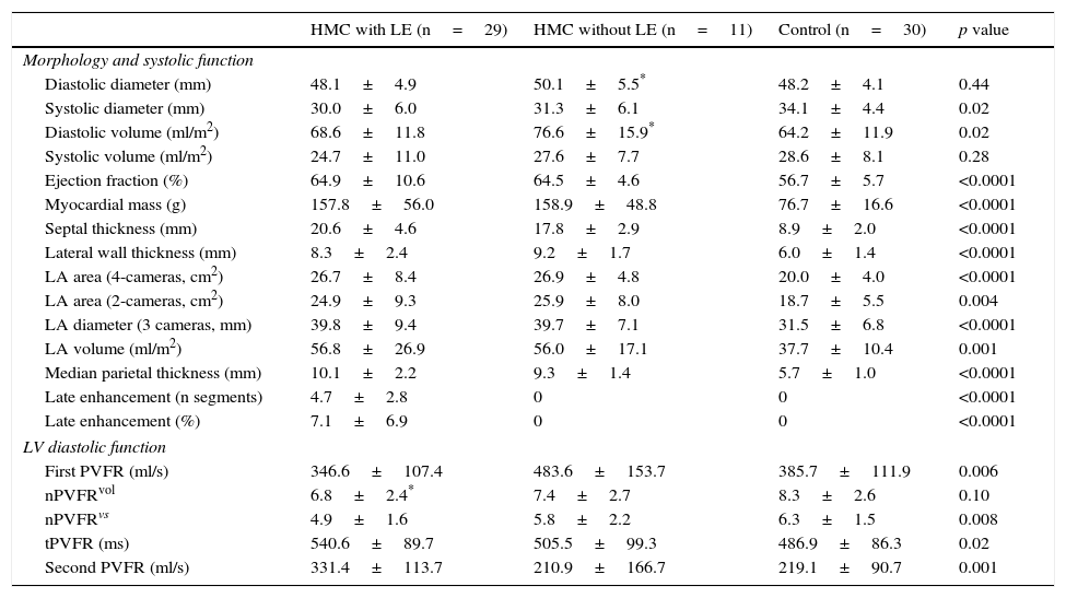
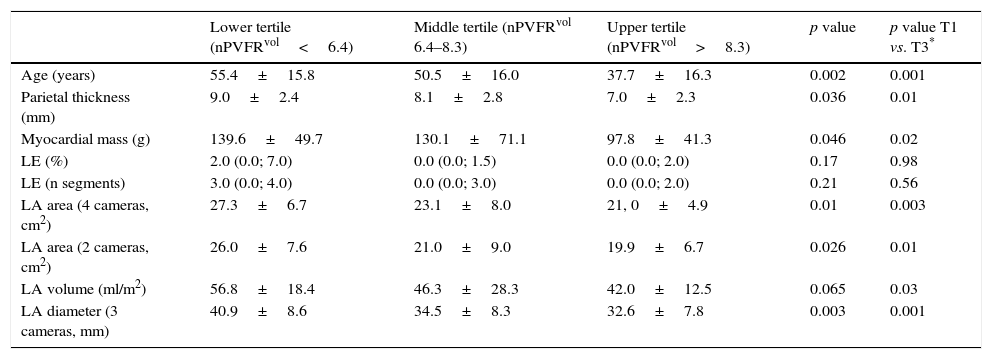
ResultsLate enhancement was observed in 29 (73%) of the 40 patients with hypertrophic cardiomyopathy. The normalized peak filling rate was significantly lower in patients with late enhancement (4.9±1.6 in those with hypertrophic cardiomyopathy positive for late enhancement vs. 5.8±2.2 in those with hypertrophic cardiomyopathy negative for late enhancement vs. 6.3±1.5 in controls, p=0.008) and the time to peak filling was longer in patients with late enhancement (540.6±89.7ms vs. 505.5±99.3ms in those with hypertrophic cardiomyopathy negative for late enhancement vs. 486.9±86.3ms in controls, p=0.02). When the population was stratified into three groups in function of the normalized peak filling rate, significant differences were observed among groups for age (p=0.002), mean wall thickness (p=0.036), and myocardial mass (p=0.046) and atrial dimensions, whereas no significant differences with respect to late enhancement were seen.
ConclusionsIn patients with hypertrophic cardiomyopathy, we found a significant association between ventricular filling patterns and age, wall thicknesses, and atrial dimensions, but not with the extent of late enhancement.
Explorar mediante resonancia magnética cardíaca la relación entre las curvas de llenado ventricular y la extensión del realce tardío (RT) en pacientes con miocardiopatía hipertrófica.
Material y métodosSe incluyeron de forma retrospectiva pacientes consecutivos con sospecha y/o diagnóstico de miocardiopatía hipertrófica, y un grupo control de pacientes pareados según sexo y edad en quienes se realizó una resonancia magnética cardíaca con valoración de RT. Entre otras determinaciones, se evaluaron mediante secuencias cine: tasa de llenado pico, tiempo a la primera tasa de llenado pico y tasa de llenado pico normalizada al volumen de llenado.
ResultadosDe los 40 pacientes con miocardiopatía hipertrófica, 29 (73%) presentaron RT. Se evidenciaron diferencias significativas respecto a la tasa de llenado pico normalizada (RT positivo 4,9±1,6, vs. RT negativo 5,8±2,2, vs. control 6,3±1,5, p=0,008) y al tiempo a la tasa de llenado pico (540,6±89,7ms, vs. 505,5±99,3ms, vs. 486,9±86,3ms, p=0,02). Al estratificar la población en tercios según la tasa de llenado pico normalizada al volumen de llenado se registraron diferencias significativas entre los grupos respecto a la edad (p=0,002), espesor parietal medio (p=0,036), masa miocárdica (p=0,046) y dimensiones auriculares, mientras que no se observaron diferencias significativas respecto al RT.
ConclusionesEn pacientes con miocardiopatía hipertrófica encontramos una asociación significativa entre patrones de llenado ventricular y edad, espesores parietales y dimensiones auriculares, mientras que no se identificó una relación significativa con la extensión del RT.

