Buscar en
Radiología (English Edition)
Toda la web
Inicio Radiología (English Edition) Non-traumatic spleen disorders in children. Assessment by imaging
Journal Information
Vol. 61. Issue 1.
Pages 16-25 (January - February 2019)
Share
Share
Download PDF
More article options
Visits
24
Vol. 61. Issue 1.
Pages 16-25 (January - February 2019)
Radiology through images
Non-traumatic spleen disorders in children. Assessment by imaging
El bazo pediátrico no traumático. Valoración por imagen
Visits
24
C. Sangüesa Nebota,
Corresponding author
sanguesa_cin@gva.es

Corresponding author.
, E. Carazo Palaciosb, R.L. Lorens Salvadora, S. Picó Aliagaa
a Área de Imagen Médica, Sección Pediatría, Hospital La Fe, Valencia, Spain
b Cirugía Pediátrica, Hospital La Fe, Valencia, Spain
This item has received
Article information
Abstract
Full Text
Bibliography
Download PDF
Statistics
Figures (15)
Show moreShow less
Tables (1)
Table 1. Non-traumatic splenic pathology in children.
Abstract

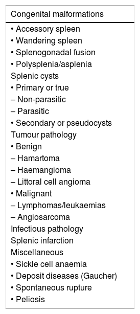
The spleen is considered a “forgotten organ” by most radiologists and paediatricians despite being affected in many clinical paediatric situations. While it is the organ most often affected in paediatric abdominal trauma, non-traumatic spleen disorders are less well known.

The spleen is well visualised by any imaging technique: ultrasound, computed tomography (CT) and magnetic resonance imaging (MRI); the former is used most often in children. Using imaging techniques to determine the features of splenic anomalies, both congenital and acquired, enables a correct diagnostic approach, avoids unnecessary surgical procedures or biopsies, and helps the clinician to prescribe appropriate treatment.

Our aim was to show the behaviour of the spleen in children using the different imaging techniques: its normal anatomy, the principal anatomical variants and the most common spleen disorder correlating with clinical symptoms, serology and histology.

Keywords:
Spleen
Ultrasound
Computed tomography
Magnetic resonance imaging
Resumen

Para la mayoría de radiólogos y pediatras, el bazo es el “órgano olvidado”, a pesar de estar afectado en múltiples situaciones clínicas de la infancia. Mientras que en el traumatismo abdominal pediátrico es el órgano más implicado, la patología esplénica no traumática es menos conocida.

El bazo se visualiza bien mediante cualquier técnica de imagen: ecografía, tomografía computarizada, resonancia magnética, y de ellas, la primera es la más utilizada en niños. Conocer las características por imagen de las anomalías esplénicas, tanto congénitas como adquiridas, permite realizar una aproximación diagnóstica correcta, evitar procedimientos quirúrgicos o biopsias innecesarias y guiar al clínico hacia un tratamiento adecuado.

Nuestro objetivo es mostrar el comportamiento del bazo en edad pediátrica con las diferentes técnicas de imagen: su anatomía normal, las principales variantes anatómicas y la patología esplénica no traumática más frecuente, correlacionando con clínica, serología o histología.

Palabras clave:
Bazo
Ecografía
Tomografía computarizada
Resonancia magnética

Article

These are the options to access the full texts of the publication Radiología (English Edition)
Subscriber
Subscriber

If you already have your login data, please click here .

If you have forgotten your password you can you can recover it by clicking here and selecting the option “I have forgotten my password”
Purchase
Purchase article

Purchasing article the PDF version will be downloaded

Price 19.34 €

Purchase now
Contact
Phone for subscriptions and reporting of errors
From Monday to Friday from 9 a.m. to 6 p.m. (GMT + 1) except for the months of July and August which will be from 9 a.m. to 3 p.m.
Calls from Spain
932 415 960
Calls from outside Spain
+34 932 415 960
E-mail
Article options
Tools
es en pt

¿Es usted profesional sanitario apto para prescribir o dispensar medicamentos?

Are you a health professional able to prescribe or dispense drugs?

Você é um profissional de saúde habilitado a prescrever ou dispensar medicamentos

Quizás le interese:
10.1016/j.rxeng.2023.02.001
No mostrar más