Buscar en
Radiología (English Edition)
Toda la web
Inicio Radiología (English Edition) Multidetector computed tomography in life-threatening hemoptysis
Journal Information
Vol. 55. Issue 6.
Pages 483-498 (November - December 2013)
Share
Share
Download PDF
More article options
Visits
3908
Vol. 55. Issue 6.
Pages 483-498 (November - December 2013)
Update in radiology
Multidetector computed tomography in life-threatening hemoptysis
La tomografía computarizada multidetector en la hemoptisis amenazante
Visits
3908
C. Spinu
Corresponding author
cspinu@tauli.cat

Corresponding author.
, E. Castañer, X. Gallardo, M. Andreu, A. Alguersuari
UDIAT-Centre Diagnòstic, Institut Universitari Parc Taulí-UAB, Corporació Sanitària Parc Taulí, Sabadell, Spain
This item has received
Article information
Abstract
Full Text
Bibliography
Download PDF
Statistics
Figures (11)
Show moreShow less
Tables (1)
Table 1. Causes of life-threatening hemoptysis.
Abstract

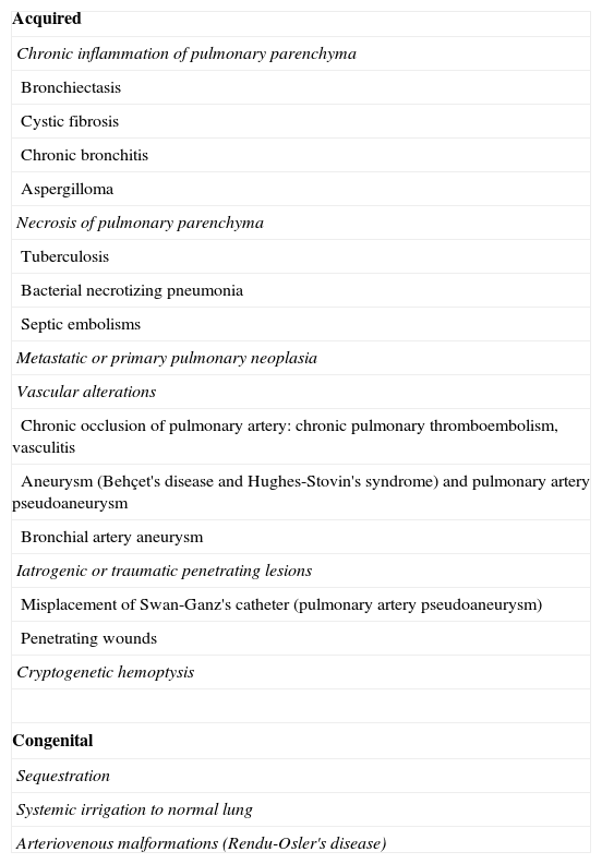
Life-threatening hemoptysis is a severe condition that requires rapid diagnosis and treatment. One of the treatments of choice is embolization. The initial assessment aims to locate the origin and cause of bleeding. The technological advance of the development of multidetector computed tomography (MDCT) has changed the management of patients with life-threatening hemoptysis. MDCT angiography makes it possible to evaluate the cause of bleeding and locate the vessels involved both rapidly and noninvasively; it is particularly useful for detecting ectopic bronchial arteries, nonbronchial systemic arteries, and pulmonary pseudoaneurysms. Performing MDCT angiography systematically before embolization enables better treatment planning. In this article, we review the pathophysiology and causes of life-threatening hemoptysis (including cryptogenic hemoptysis) and the MDCT angiography technique, and we review how to systematically evaluate the images (lung parenchyma, airways, and vascular structures).

Keywords:
Hemoptysis
Multidetector computed tomography
Bronchial arteries
Pulmonary artery
Resumen

La hemoptisis amenazante es una situación grave que precisa de un diagnóstico y tratamiento rápidos. Uno de los tratamientos de elección es la embolización. La evaluación inicial se dirige a localizar el origen y la causa del sangrado. El avance tecnológico de la TC multidetector (TCMD) ha supuesto un cambio en el manejo de estos pacientes. La angio-TCMD permite evaluar la causa rápida e incruentamente, y localizar los vasos implicados; es particularmente útil para detectar arterias bronquiales ectópicas, arterias sistémicas no bronquiales o seudoaneurismas pulmonares. Hacer sistemáticamente una angio-TCMD antes de la embolización permite planificar mejor el tratamiento. En este artículo revisamos la fisiopatología y las causas de la hemoptisis amenazante (incluyendo la hemoptisis criptogenética), la técnica del estudio de la angio-TCMD y describimos cómo evaluar sistemáticamente las imágenes (parénquima pulmonar, vía aérea y estructuras vasculares).

Palabras clave:
Hemoptisis
Tomografía computarizada multidetector
Arterias bronquiales
Arteria pulmonar

Article

These are the options to access the full texts of the publication Radiología (English Edition)
Subscriber
Subscriber

If you already have your login data, please click here .

If you have forgotten your password you can you can recover it by clicking here and selecting the option “I have forgotten my password”
Purchase
Purchase article

Purchasing article the PDF version will be downloaded

Price 19.34 €

Purchase now
Contact
Phone for subscriptions and reporting of errors
From Monday to Friday from 9 a.m. to 6 p.m. (GMT + 1) except for the months of July and August which will be from 9 a.m. to 3 p.m.
Calls from Spain
932 415 960
Calls from outside Spain
+34 932 415 960
E-mail
Article options
Tools
es en pt

¿Es usted profesional sanitario apto para prescribir o dispensar medicamentos?

Are you a health professional able to prescribe or dispense drugs?

Você é um profissional de saúde habilitado a prescrever ou dispensar medicamentos