metricas
covid
Radiología (English Edition) Magnetic resonance imaging of fetal cerebral ventriculomegaly
Journal Information
Serie: Fetal Magnetic Resonance Imaging
Available online 16 October 2025
Magnetic resonance imaging of fetal cerebral ventriculomegaly
Resonancia magnética de la ventriculomegalia cerebral fetal
P. Caro-Domíngueza,
Corresponding author
pablocaro82@hotmail.com

Corresponding author.
, L. García Díazb, G. Antiñolob, E. Millerc, M. Carvajoa, J.A. Sainz-Buenod
a Unidad de Radiología Pediátrica, Servicio de Radiología, Hospital Universitario Virgen del Rocío, Sevilla, Spain
b Departamento de Medicina Materno-Fetal, Genética y Reproducción y Departamento de Cirugía, Instituto de Biomedicina de Sevilla (IBIS), Hospital Universitario Virgen del Rocío, Universidad de Sevilla, Sevilla, Spain
c Department of Diagnostic and Interventional Radiology, The Hospital for Sick Children, University of Toronto, Toronto, Ontario, Canada
d Departamento de Obstetricia y Ginecología, Hospital Universitario Virgen de Valme y Facultad de Medicina, Universidad de Sevilla, Sevilla, Spain
Article information
Abstract
Full Text
Bibliography
Download PDF
Statistics
Figures (6)
Show moreShow less
Tables (2)
Table 1. Fetal MRI protocol for the assessment of ventriculomegaly using a 1.5 T Philips Ingenia scanner (Philips, The Netherlands).
Tables
Table 2. Standardised fetal MRI report of the central nervous system.
Tables
Show moreShow less
Abstract

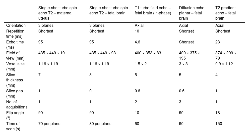
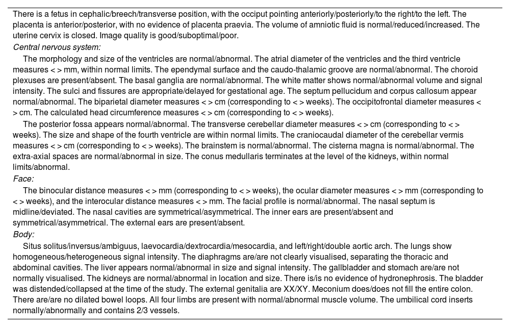
Fetal ventriculomegaly is one of the most common findings on prenatal ultrasound, and one of the most common indications for fetal magnetic resonance imaging (MRI). The aim of this article is to explain the different terminology used to describe the dilatation of the fetal cerebral ventricles, explain the impact of imaging (ultrasound and MRI) in this clinical scenario, illustrate common causes of ventriculomegaly and summarise the evidence regarding prognosis for these children, in order to be able to provide appropriate prenatal advice.

Keywords:
Brain
Central nervous system
Fetal
Magnetic resonance imaging
Ultrasound
Ventriculomegaly
Resumen

La ventriculomegalia fetal es uno de los hallazgos más frecuentes que se encuentran en la ecografía prenatal y una de las indicaciones más comunes de resonancia magnética fetal. El propósito de este artículo es definir la diferente terminología utilizada para describir la dilatación del sistema ventricular cerebral fetal, explicar el impacto de la imagen (ecografía y resonancia magnética fetal) en este escenario clínico, ilustrar con casos las causas más comunes y mostrar la evidencia que existe sobre el pronóstico de estos niños, para proporcionar un consejo prenatal adecuado.

Palabras clave:
Cerebro
Sistema nervioso central
Fetal
Resonancia magnética
Ecografía
Ventriculomegalia

Article

These are the options to access the full texts of the publication Radiología (English Edition)
Subscriber
Subscriber

If you already have your login data, please click here .

If you have forgotten your password you can you can recover it by clicking here and selecting the option “I have forgotten my password”
Purchase
Purchase article

Purchasing article the PDF version will be downloaded

Purchase now
Contact
Phone for subscriptions and reporting of errors
From Monday to Friday from 9 a.m. to 6 p.m. (GMT + 1) except for the months of July and August which will be from 9 a.m. to 3 p.m.
Calls from Spain
932 415 960
Calls from outside Spain
+34 932 415 960
E-mail
Article options
Tools