Buscar en
Radiología (English Edition)
Toda la web
Inicio Radiología (English Edition) Imaging findings in cardiac masses (Part i): Study protocol and benign tumors
Journal Information
Vol. 57. Issue 6.
Pages 480-488 (November - December 2015)
Share
Share
Download PDF
More article options
Visits
1793
Vol. 57. Issue 6.
Pages 480-488 (November - December 2015)
Radiology through images
Imaging findings in cardiac masses (Part i): Study protocol and benign tumors
Hallazgos de imagen de las masas cardíacas (parte i): protocolo de estudio y tumores benignos
Visits
1793
C. Díaz Angulo, C. Méndez Díaz, E. Rodríguez García, R. Soler Fernández
Corresponding author
rafaelasoler@telefonica.net

Corresponding author.
, A. Rois Siso, M. Marini Díaz
Servicio de Radiodiagnóstico, Complejo Hospitalario Universitario A Coruña (CHUAC), A Coruña, Spain
This item has received
Article information
Abstract
Full Text
Bibliography
Download PDF
Statistics
Figures (8)
Show moreShow less
Tables (2)
Table 1. Magnetic resonance (MR) protocol for the study of cardiac masses.
Table 2. More characteristic findings of benign cardiac tumors.
Show moreShow less
Abstract

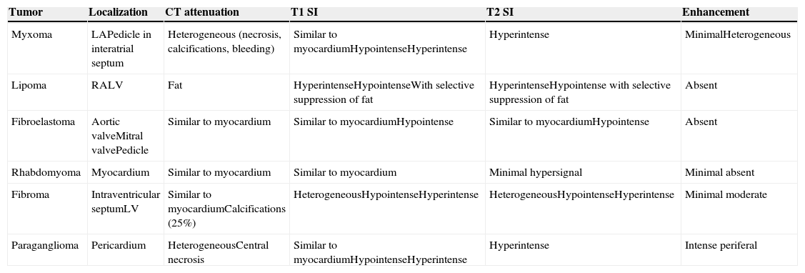
Cardiac masses represent a diagnostic challenge because decisions about treatment are based on imaging techniques. Echocardiography, magnetic resonance (MR) and computed tomography (CT) are fundamental for the detection, characterization, and staging of cardiac masses as well as for planning their treatment. Most primary cardiac tumors are benign; myxomas, papillary fibroelastomas, and lipomas are the most common. The location of the tumors and its characteristics on CT and MR orient the etiologic diagnosis in most cases.

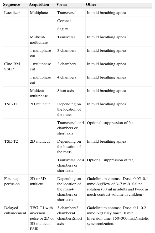
This article describes the protocols for CT and MR studies of cardiac masses as well as the morphologic findings, predominant locations, and most useful characteristics for characterizing benign cardiac masses and establishing the differential diagnosis with malignant cardiac tumors and non-neoplastic pseudotumors.

Keywords:
Cardiac masses
Myxoma
Fibroelastoma
Lipoma
Rhabdomyoma
Resumen

Las masas cardíacas son un reto diagnóstico porque las decisiones terapéuticas se basan en los hallazgos de las técnicas de imagen. La ecocardiografía, la resonancia magnética (RM) y la tomografía computarizada (TC) son fundamentales para la detección, caracterización, estadificación y planificación del tratamiento. La mayoría de los tumores primarios son benignos; los más frecuentes son el mixoma, el fibroelastoma papilar y el lipoma. La localización del tumor y sus características en la TC y la RM orientan el diagnóstico etiológico en la mayor parte de los casos.

Se describen los protocolos de estudio de TC y RM de las masas cardíacas, así como los hallazgos morfológicos, las localizaciones preferentes y las características más útiles para caracterizar las masas cardíacas benignas y establecer el diagnóstico diferencial con los tumores cardíacos malignos y las lesiones pseudotumorales no neoplásicas.

Palabras clave:
Masas cardíacas
Mixoma
Fibroelastoma
Lipoma
Rabdomioma

Article

These are the options to access the full texts of the publication Radiología (English Edition)
Subscriber
Subscriber

If you already have your login data, please click here .

If you have forgotten your password you can you can recover it by clicking here and selecting the option “I have forgotten my password”
Purchase
Purchase article

Purchasing article the PDF version will be downloaded

Price 19.34 €

Purchase now
Contact
Phone for subscriptions and reporting of errors
From Monday to Friday from 9 a.m. to 6 p.m. (GMT + 1) except for the months of July and August which will be from 9 a.m. to 3 p.m.
Calls from Spain
932 415 960
Calls from outside Spain
+34 932 415 960
E-mail
Article options
Tools
es en pt

¿Es usted profesional sanitario apto para prescribir o dispensar medicamentos?

Are you a health professional able to prescribe or dispense drugs?

Você é um profissional de saúde habilitado a prescrever ou dispensar medicamentos