Buscar en
Radiología (English Edition)
Toda la web
Inicio Radiología (English Edition) Imaging diagnosis of portal hypertension
Journal Information
Vol. 60. Issue 4.
Pages 290-300 (July - August 2018)
Share
Share
Download PDF
More article options
Visits
113
Vol. 60. Issue 4.
Pages 290-300 (July - August 2018)
Update in Radiology
Imaging diagnosis of portal hypertension
Diagnóstico por imagen de la hipertensión portal
Visits
113
M. Conangla-Planesa,
Corresponding author
marina.conangla@gmail.com

Corresponding author.
, X. Serresa, O. Persivaa, S. Augustínb,c
a Servicio de Radiodiagnóstico, Hospital Universitari Vall d’Hebron, Universitat Autònoma de Barcelona, Barcelona, Spain
b Servicio de Medicina Interna, Hospital Universitari Vall d’Hebron, Universitat Autònoma de Barcelona, Barcelona, Spain
c Centro de Investigación Biomédica en Red de Enfermedades Hepáticas y Digestivas (CIBEREHD), Spain
This item has received
Article information
Abstract
Full Text
Bibliography
Download PDF
Statistics
Figures (10)
Show moreShow less
Tables (2)
Table 1. Radiological findings of portal hypertension.
Table 2. Main causes of portal hypertension.
Show moreShow less
Abstract

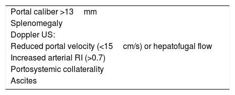
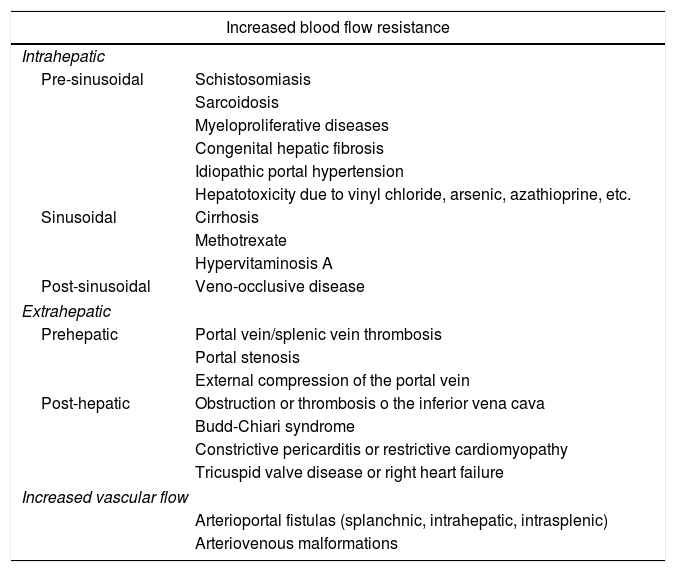
Portal hypertension (PHT) is a clinical entity defined when hydrostatic pressure >5mm Hg in the portal venous territory, being clinically significant if it reaches ≥10mm Hg. At this threshold, complications can develop, such as the bleeding of esophageal varices, the appearance of ascites, or hepatic encephalopathy. Imaging modalities play an important role here as non-invasive methods to determining whether PHT is present or not. This article analyzes various imaging findings that can be suggestive of PHT and contributes to define its etiology, severity, and possible complications.

Keywords:
Portal hypertension
Ultrasonography Doppler
Collateral circulation
Coronary veins
Resumen

La hipertensión portal (HTP) es una condición clínica definida por una presión hidrostática >5mmHg en el territorio venoso portal, siendo clínicamente significativa cuando es ≥10mmHg. A partir de este umbral pueden desarrollarse complicaciones, como sangrado de varices esofágicas, aparición de ascitis o encefalopatía hepática. Las técnicas de imagen tienen un papel importante como método no invasivo para determinar la presencia de HTP. En este artículo se analizan varios hallazgos radiológicos que pueden sugerir HTP y contribuir a definir su etiología, gravedad y posibles complicaciones.

Palabras clave:
Hipertensión portal
Ecografía Doppler
Circulación colateral
Venas coronarias

Article

These are the options to access the full texts of the publication Radiología (English Edition)
Subscriber
Subscriber

If you already have your login data, please click here .

If you have forgotten your password you can you can recover it by clicking here and selecting the option “I have forgotten my password”
Purchase
Purchase article

Purchasing article the PDF version will be downloaded

Price 19.34 €

Purchase now
Contact
Phone for subscriptions and reporting of errors
From Monday to Friday from 9 a.m. to 6 p.m. (GMT + 1) except for the months of July and August which will be from 9 a.m. to 3 p.m.
Calls from Spain
932 415 960
Calls from outside Spain
+34 932 415 960
E-mail
Article options
Tools
es en pt

¿Es usted profesional sanitario apto para prescribir o dispensar medicamentos?

Are you a health professional able to prescribe or dispense drugs?

Você é um profissional de saúde habilitado a prescrever ou dispensar medicamentos