


To assess the ability of urgent head computed tomography (CT) scan screening to detect patients who can evolve to brain death (BD).
Patients and methodPatients who underwent urgent head CT scan and meet the following criteria: midline shift greater than 5mm and/or decrease or absence of basal cisterns. A follow-up for 28 days of each patient was made. Epidemiological data (sex, age, cause of brain injury), clinical data (level of consciousness, severity index in the CT) and patient outcomes (death, BD, discharge or transfer) were recorded. This was a prospective observational study.
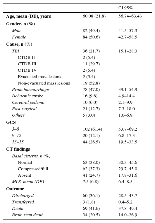
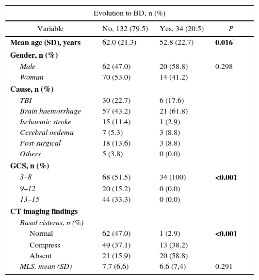
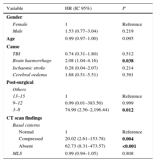
ResultsOne hundred and sixty-six patients were selected for study, with mean age 60.08 (SD 21.8) years. A percentage of 49.4 were men and the rest women. In the follow-up, 20.5% (n=34) had BD. In univariate analysis, intracerebral haemorrhage, Glasgow Coma Scale score less than 8 and alteration of basal cisterns were statistically significant in predicting BD (P<.05). Multivariate analysis showed that patients with compression of basal cisterns were 20 (95% confidence interval [95% CI] 2.61–153.78; P=.004] times more likely to progress to brain death, while the absence there of 62.6 (95% CI 13.1–738.8; P<.001] times more.
ConclusionsOur work shows that data as easy to interpret as compression/absence of basal cisterns can be a powerful tool for screening patients at risk for progression to BD.
Evaluar la capacidad de cribado de la tomografía computarizada (TC) de cráneo urgente para detectar a los pacientes que pueden evolucionar a muerte encefálica (ME).
Pacientes y métodoSe incluyeron los pacientes a los que se realiza TC de cráneo urgente y que cumplían los siguientes criterios: desplazamiento de línea media mayor de 5mm y/o disminución o ausencia de las cisternas de la base. Estos pacientes fueron sometidos a seguimiento durante 28 días. Se recogieron los datos epidemiológicos (sexo, edad, causa de la lesión encefálica), clínicos (nivel de consciencia, índices de gravedad tomográficos) y la evolución de los pacientes: fallecimiento, ME, alta o traslado.
ResultadosSe seleccionaron para su seguimiento 166 estudios, siendo la media (DE) de edad de 60,08 (21,8) años. El 49,4% fueron varones. Del total de casos estudiados, fallecieron por ME el 20,5% (n=34). En el análisis univariado, la hemorragia cerebral, la puntuación inferior a 8 de la Glasgow Coma Scale y la alteración de las cisternas de la base resultaron ser estadísticamente significativas para predecir ME (p<0,05). Mediante análisis multivariante observamos que la compresión de cisternas de la base suponía 20 (intervalo de confianza del 95% [IC 95%] 2,61–153,78; p=0,004) veces más posibilidades de evolucionar a ME, mientras que la ausencia de las mismas, hasta 62,6 (IC 95% 13,1–738,8; p<0,001) veces más.
ConclusionesNuestro trabajo demuestra que un dato tan sencillo de interpretar como la compresión/ausencia de las cisternas de la base puede ser una potente herramienta para el cribado de pacientes en riesgo de evolucionar a ME.
