metricas
Enfermedades Infecciosas y Microbiología Clínica (English Edition) Acute Q fever in Majorca island 2017–2022. An underestimated problem
Journal Information
Visits
675
Vol. 43. Issue 10.
Pages 629-724 (December 2025)
Original article
Full text access

Acute Q fever in Majorca island 2017–2022. An underestimated problem

Fiebre Q aguda en Mallorca, 2017−2022. Un problema infraestimado
Visits
675
Mercedes García-Gasallaa,b,j,k,1,
Corresponding author
, Sophia Pinecki Sociasb,1, Pablo A. Fraileb,c,k, Victoria Fernández-Bacad, Aroa Villosladab,e, Antonio Adrovere, Sandra Gregorioe, Marta González-Morenof, Alfredo Pinheiro Martinsg, Alexander Zidouha, Gemma Muth, Meritxell Gavaldài, Melchor Rieraa,b,j,k, Luisa Martín Penaa,b,k, Javier Murillas Angoitia,b,j,k
a Servicio de Medicina Interna, Hospital Universitario Son Espases, Palma, Balearic Islands, Spain
b Instituto de Investigación Sanitaria Illes Balears (IdISBa), Palma, Balearic Islands, Spain
c Servicio de Microbiología, Hospital Universitario Son Espases, Palma, Balearic Islands, Spain
d Servicio de Microbiología, Hospital Universitario Son Llàtzer, Palma, Balearic Islands, Spain
e Servicio de Medicina Interna, Hospital Universitario Son Llàtzer, Palma, Balearic Islands, Spain
f Servicio de Medicina Interna, Hospital de Manacor, Manacor, Balearic Islands, Spain
g Servicio de Medicina Interna, Hospital General de Mallorca, Palma, Balearic Islands, Spain
h Hospital Quirón Palma Planas, Palma, Balearic Islands, Spain
i Hospital de Inca, Inca, Balearic Islands, Spain
j Facultad de Medicina, Universidad de las Islas Baleares, Palma, Balearic Islands, Spain
k CIBER en Enfermedades Infecciosas, Instituto de Salud Carlos III (CIBERINFEC), Madrid, Spain
Ver más
This item has received
Article information
Abstract
Full Text
Bibliography
Download PDF
Statistics
Figures (1)
fig0005
Tables (3)
Table 1. Epidemiological data from the series of 223 patients diagnosed with AQF.
Tables
Table 2. Symptoms and main clinical diagnosis of the 223 cases of AQF.
Tables
Table 3. Laboratory test abnormalities detected in AQF cases (confirmed and highly probable/probable AQF).
Tables
Abstract
Introduction

Q fever (QF) is a zoonotic infection caused by Coxiella burnetii. Previous studies suggest that the incidence in the island of Majorca may be high. The objective was to know the epidemiological, clinical, diagnostic, and therapeutic characteristics of acute QF (AQF) on the island of Majorca during the years 2017–2022.

Methods

Retrospective analysis of a series of cases of AQF diagnosed in 3 out 4 hospitals of the Health Service of the island of Majorca. AQF was considered if a clinically compatible syndrome was present with one of the following serological criteria: confirmed AQF if IgG seroconversion phase II, very possible if only IgG ≥1/128 is available in addition to a positive IgM, and AQF possible if IgG≥ 1/512 with negative IgM or if IgM positive with negative IgG in a single serological determination.
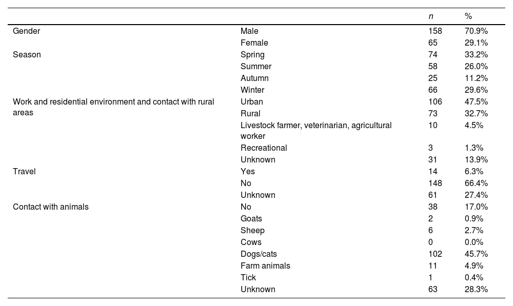
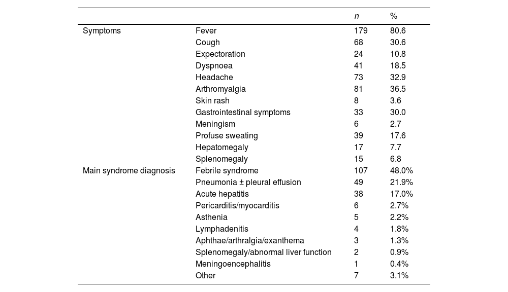
Results

223 cases of AQF were diagnosed, of which 102 (45.7%) were confirmed, 84 (37.7%) very probable and 37 (16.6%) probable AQF. Prolonged febrile syndrome was the most frequent clinical diagnosis (107, 48.0%), followed by pneumonia with/without pleural effusion (49, 21.9%), acute hepatitis (38, 17.0%), pericarditis and/or myocarditis (6, 2.6%). Three patients developed endocarditis (one in the acute phase, two others during follow-up). The median number of cases per year was 34 (p25–p75: 31.7–40.25). AQF-related mortality was 1.8% (4 patients).

Conclusion

AQF is a preventable and little recognized zoonosis, causing significant morbidity and mortality in Majorca.

Keywords:
Acute Q fever
Coxiella burneti
Febrile syndrome
Pneumonia
Hepatitis
Resumen
Introducción

La fiebre Q (FQ) es una zoonosis de distribución universal causada por Coxiella burnetii. Estudios previos sugieren que la incidencia en Mallorca puede ser elevada. El objetivo fue conocer las características epidemiológicas, clínicas, diagnósticas y terapéuticas de la FQ aguda (FQA) en la isla de Mallorca durante los años 2017 a 2022.

Métodos

Análisis retrospectivo de una serie de casos de FQA diagnosticados en 3 hospitales del Servicio de Salud de la isla de Mallorca. Se consideró FQA si el cuadro cínico era compatible y cumplían alguno de los siguientes criterios serológicos: FQA confirmada si seroconversión de IgG fase II, muy probable si solo se dispone de una determinación de IgG ≥1/128 además de IgM positiva y FQA probable si IgG ≥1/512 con IgM negativa o si IgM positiva con IgG negativa en una única determinación serológica.

Resultados

Se diagnosticaron en el periodo del estudio 223 casos de FQ aguda, de los cuales 102 (45,7%) FQA confirmada, 84 (37,7%) FQA muy posible y 37 (16,6%) FQA posible. El síndrome febril prolongado fue el diagnóstico clínico más frecuente (107, 48,0%), seguido de la neumonía con/sin derrame pleural (49, 21,9%), hepatitis aguda (38, 17,0%), pericarditis y/o miocarditis (6, 2,6%). Tres pacientes desarrollaron endocarditis (una en la fase aguda, otros dos durante el seguimiento). La mediana de casos por año fue de 34 (p25-p75: 31,7–40,25). Cuatro pacientes (1,4%) fallecieron en relación a la FQA.

Conclusión

La FQ es una zoonosis prevenible y poco reconocida, causante de una significativa morbimortalidad en Mallorca.

Palabras clave:
Fiebre Q aguda
Coxiella burnetii
Síndrome febril
Neumonía
Hepatitis
Full Text
Introduction

Q fever is a universally-distributed zoonosis caused by Coxiella burnetii. The reservoir of this bacterium is extensive and includes arthropods, in addition to wild and domestic mammals and birds, with humans being an accidental host.1,2C. burnetii is an extremely resistant microorganism in the environment. It is found in animal urine, faeces, milk, placenta and amniotic fluid.3 The most common source of infection in humans is domestic ruminants, especially goats and sheep,4 through airborne transmission by inhalation of spores, but oral and human-to-human transmission via the transplacental route during birth has also occasionally been reported. Transdermal transmission via vectors such as ticks has been demonstrated in animals but not in humans.4 Infection in animals is mostly subclinical and is associated with infertility or late abortions.3

In humans, the incubation period is estimated to be between seven and 32 days, with a median of 18 days.5 The spectrum of manifestations of the acute form (AQF) is variable, ranging from asymptomatic seroconversion (in 50-60%) to self-limiting febrile illness, or more severe manifestations, such as pneumonia, which can be critical, or acute hepatitis1,2; other clinical forms such as meningoencephalitis6 and myopericarditis7 have also been reported. It is estimated that 2–5% of symptomatic cases require hospital admission, and up to 20% in some series. The chronic form or localised persistent infection is more serious, with valvular heart disease and/or immunocompromised patients being more susceptible, and it often manifests as infected aneurysms, endocarditis or spondylodiscitis.8

Cases of Q fever have been reported on all continents except Antarctica. In Europe, an outbreak in 2007–2010 was described, with more than 4000 people affected in the Netherlands,9 but since 2016, Spain has been the country reporting the highest number of cases to the European Centre for Disease Prevention and Control (ECDC).10 In Spain, the Centro Nacional de Epidemiología [National Epidemiology Centre] has considered Q fever a notifiable disease (ND) since 2015 and recommends individualised and weekly notification of each diagnosed case to the epidemiological surveillance centre (RENAVE; Red Nacional de Vigilancia Epidemiológica [National Epidemiological Surveillance Network]).11 However, the Balearic Islands have not yet been incorporated into the national surveillance of Q fever as ND, as stipulated in Spanish Royal Decree (RD) 2210/1995. In 2022, 440 cases of C. burnetii infection were recorded in Spain, and 519 in 2023.12 Its prevalence was highest in the autonomous region of the Canary Islands. Case series have been published in practically all parts of Spain,13–19 including Majorca.20 A Spanish study reviewing hospital discharge diagnoses coded as Q fever between 1998 and 2015 estimated an incidence rate in that period of 0.53 cases/100,000 person-years, with the highest incidence recorded in the Canary Islands and the Balearic Islands (1.48 and 1.43 cases/100,000 person-years, respectively), and an overall estimated mortality rate of 1.5/100 cases.21

Our aim with this study was to increase our understanding of Q fever in Majorca by studying the clinical, epidemiological, diagnostic and therapeutic characteristics of the cases diagnosed at three of the four Balearic Islands Health Service (IB-Salut) hospitals over a six-year period.

Methods

This was a retrospective descriptive study in three hospitals on Majorca, which provide hospital care to 41 of the island’s 48 basic health areas (85.4% of the population). The study included the period from January 2017 to December 2022 at the hospitals of Manacor, Son Llàtzer and Son Espases, the latter being the reference hospital in the Balearic Islands.

In all cases the clinical signs and symptoms were consistent and there was no alternative diagnosis. Detection of C. burnetii phase II IgG was performed in all samples by indirect immunofluorescence assay (IFA) with Coxiella burnetii IFA IgG reagent (Vircell), while detection of IgM was performed by IFA (Coxiella burnetii IFA IgM, Vircell) or chemiluminescence (Coxiella burnetii VirClia IgM Monotest, Vircell) according to the protocol of the diagnostic laboratory. Sera with phase II IgG ≥1/512, or when requested by the clinician for patient follow-up, were also tested for phases I and II IgG and IgM with the Focus Coxiella phases reagent (Focus Diagnostics® Cypress, CA, USA). The serological criteria for diagnosis of the different conditions were based on the Centro Nacional de Epidemiología [National Epidemiology Centre] criteria11 with a modification to increase specificity in cases of probable AQF: the IgM result was considered positive, and in cases with a negative result, the titre considered positive for IgG was increased. The established criteria were therefore: 1) "confirmed AQF" when there was evidence of seroconversion of IgG titres to C. burnetii within 4–8 weeks; 2) in cases where a single serological determination was performed, "highly probable AQF" was defined if IgG was ≥1/128 with IgM also positive; and 3) a third group, "probable AQF", was considered when IgG serology was positive at a high titre (IgG ≥1/512) with negative IgM or when IgM was positive with negative IgG. For the analysis, we collected demographic and epidemiological data, risk factors (structural heart disease, HIV infection or other immunosuppression), clinical, analytical and radiological data, the need for hospital admission, treatment received and complications from the patient's medical records. An anonymised database was created in which the confidentiality of the data was guaranteed for the storage and analysis of the variables using the statistical package SPSS 12.0 for Windows. Descriptive statistical analysis was performed by calculating frequencies for each of the qualitative variables and the median and percentiles [p25–p75] for the quantitative variables. For comparison of qualitative variables, the Chi-square test with Yates correction was used where necessary, considering a difference to be statistically significant if p < 0.05. Incidence rates were calculated by dividing the number of cases per year/population in the health areas corresponding to the three hospitals in that year according to information from the Spanish Instituto Nacional de Estadística [National Institute of Statistics] and expressed as cases/100,000 person-years.

The study was approved by the Hospital Son Espases Research Committee (No. CI-562-21, and in its subsequent extension No. CI-922-24). In accordance with the applicable provisions of current regulations and the ethical principles for medical research on human subjects, Informed Consent was not requested from the patients as this was an anonymised retrospective study.

Results

A total of 223 cases of AQF were identified, 102 (45.7%) of which were confirmed AQF, 84 (37.7%) highly probable AQF and 37 (16.6%) probable AQF. The median age of the cases was 49 years (p25–p75: 39–62), and 158 (70.9%) of the 223 cases were male.

The number of cases diagnosed each year (total and per hospital) is shown in Fig. 1. There was a median of 34 cases per year (p25–p75: 31.7–40.25). There was a larger number of cases in 2018. The estimated annual incidence in the health areas covered by the three hospitals is shown in Fig. 1b.

Figure 1.

a. Cases of AQF diagnosed each year at each of the hospitals in the study. b. Estimated incidence of cases per year in the health area of the study (cases/100,000 person-years).

Table 1 shows the epidemiological characteristics of all the cases. The majority of cases occurred in spring and winter, but there was also a considerable number in summer. A large number of patients lived in urban areas and had no direct contact with animals. In terms of the comorbidities analysed, 27 patients (12.1%) had some form of immunosuppression and seven (3.1%) had advanced chronic liver disease. In total, 21 patients (9.4%) had structural or valvular heart disease, and six of them had a prosthetic valve. Three patients (1.3%) had aortic aneurysm and four patients (1.8%) had a prosthetic joint. The median number of days of symptoms prior to consultation/admission was seven (p25–p75: 3–10). A total of 184 cases (82.5%) required hospital admission, with a median length of stay of six days (p25–p75: 3–10); 74.4% of patients were admitted to general internal medicine and infectious diseases departments, and six required direct admission from Accident and Emergency to intensive care units. Prolonged febrile syndrome was the most common clinical diagnosis (109, 48.9%), followed by pneumonia with/without pleural effusion (50, 22.5%) and acute hepatitis (37, 16.6%). Acute hepatitis was significantly more common in the group of patients with confirmed AQF, with 29 cases (28.4%) vs 8 cases (9.5%) in the highly probable/probable AQF group (p < 0.001), and pneumonia was significantly more common in the highly probable/probable AQF group, with 40 cases (17.9%) vs 10 (4.5%) (p < 0.001). No other differences were detected between the three groups of confirmed, highly probable or probable AQF. Other clinical diagnoses were pericarditis/myocarditis, meningoencephalitis, lymphadenitis, pleural effusion, arthritis, exanthema and oral aphthae. One female patient was diagnosed with mitral endocarditis without microbiological isolation, with a serology consistent with AQF, but it was not possible to perform phase I and II serology (Table 2).

Table 1.

Epidemiological data from the series of 223 patients diagnosed with AQF.

    n 
GenderMale  158  70.9% 
Female  65  29.1% 
SeasonSpring  74  33.2% 
Summer  58  26.0% 
Autumn  25  11.2% 
Winter  66  29.6% 
Work and residential environment and contact with rural areasUrban  106  47.5% 
Rural  73  32.7% 
Livestock farmer, veterinarian, agricultural worker  10  4.5% 
Recreational  1.3% 
Unknown  31  13.9% 
TravelYes  14  6.3% 
No  148  66.4% 
Unknown  61  27.4% 
Contact with animalsNo  38  17.0% 
Goats  0.9% 
Sheep  2.7% 
Cows  0.0% 
Dogs/cats  102  45.7% 
Farm animals  11  4.9% 
Tick  0.4% 
Unknown  63  28.3% 
Table 2.

Symptoms and main clinical diagnosis of the 223 cases of AQF.

    n 
SymptomsFever  179  80.6 
Cough  68  30.6 
Expectoration  24  10.8 
Dyspnoea  41  18.5 
Headache  73  32.9 
Arthromyalgia  81  36.5 
Skin rash  3.6 
Gastrointestinal symptoms  33  30.0 
Meningism  2.7 
Profuse sweating  39  17.6 
Hepatomegaly  17  7.7 
Splenomegaly  15  6.8 
Main syndrome diagnosisFebrile syndrome  107  48.0% 
Pneumonia ± pleural effusion  49  21.9% 
Acute hepatitis  38  17.0% 
Pericarditis/myocarditis  2.7% 
Asthenia  2.2% 
Lymphadenitis  1.8% 
Aphthae/arthralgia/exanthema  1.3% 
Splenomegaly/abnormal liver function  0.9% 
Meningoencephalitis  0.4% 
Other  3.1% 

Abnormalities in laboratory test variables are shown in Table 3. Liver function tests were considered abnormal when values exceeded twice the upper limit of normal, which was found in 112 (50.2%) of the total number of patients diagnosed with AQF. All patients diagnosed with hepatitis had significantly abnormal liver function tests, as did 58 (54.2%) of those with prolonged febrile syndrome and 10 (20.8%) of those diagnosed with pneumonia. Anticardiolipin antibodies were not measured in any of the cases.

Table 3.

Laboratory test abnormalities detected in AQF cases (confirmed and highly probable/probable AQF).

  n 
Leukocytosis (leukocytes >11,500/mL) N = 219  46  21.0% 
Anaemia (haemoglobin <12 g/dl) N = 206  38  18.4% 
Thrombocytopaenia (platelets <120,000/mL) N = 218  62  28.4% 
Increased aspartate aminotransferase (AST > 68 U/l) N = 193  75  38.9% 
Increased alanine transaminase (ALT > 110 U/l) N = 210  71  33.8% 
Increased gamma-glutamyl transferase (GGT > 128 U/l) N = 210  82  39.0% 

In terms of imaging tests, chest X-rays were performed in 206 patients (92.4%) and abnormalities were found in 67 (32.5%). The most common radiological findings were pulmonary infiltrates in 46 cases and pleural effusion in nine cases. Transthoracic echocardiograms were performed in 43 patients (19.3%) at diagnosis or in the first few months of follow-up of the AQF episode (completed with transoesophageal echocardiogram in 10 cases), showing pericardial effusion in three cases, native valve endocarditis in two (in one case doubtful) and prosthetic valve endocarditis in another. Echocardiogram was performed if the patient had a risk factor for chronification (immunosuppression, prosthetic valve or endovascular prosthesis) or at the discretion of the treating physician. Abdominal imaging (ultrasound or computerised axial tomography [CT]) was performed in 98 cases (43.9%), and abnormalities were found in 42, most commonly hepatosplenomegaly (20 cases). Positron emission tomography (PET-CT) was performed in 13 cases, which was normal in all but one case, where a concomitant lung neoplasm was diagnosed.

At least 181 patients (77.7%) received some antibiotic therapy, the majority doxycycline (134 [60%]) and quinolones in 32 (14.3%). The mean length of antibiotic therapy was 12.5 days (SD: 5.05). Serological follow-up at six months was performed in 106/223 patients; 22 patients had elevated phase I antibody titres (>1/800), with one developing prosthetic aortic valve infective endocarditis, another possible native aortic valve endocarditis, and one persistent liver enzyme abnormalities. All three patients were treated with doxycycline and hydroxychloroquine. The remaining 19 patients were symptom-free; four of them were treated with doxycycline and hydroxychloroquine. Four patients died, three of them while in hospital: two from critical pneumonia and one from multiple-organ failure related to acute endocarditis. One patient died six months after the AQF episode due to prosthetic valve endocarditis.

Discussion

This study was carried out over a six-year period in three of the four public acute care hospitals on the island of Majorca which provide hospital care to 85.4% of the population. This allowed us to obtain a good perspective of the epidemiological situation and to estimate the incidence of acute infection. However, due to the retrospective nature of the study, this could be underestimated, as we only included cases diagnosed in public hospitals and not in primary care, and we were unable to include those diagnosed in the private medical sector. We followed strict diagnostic criteria for probable AQF by considering IgG titres ≥1/512, avoiding false positives, as we found persistent low titres in the asymptomatic population, suggesting a high seroprevalence, in line with reports from other regions.22 All of the above means we are able to affirm, as already suggested,20,21 the importance of this zoonosis in Spain. The number of cases varies from year to year, peaking at an incidence of seven cases/100,000 person-years in 2018, but we do not have the means to study whether or not this was an outbreak. The incidence in 2022 of five cases/100,000 population is much higher than that reported in the same year in other Autonomous Regions in Spain. In addition, cases were also reported in 2020 (25 cases between April and December 2020), when the wearing of masks to protect against airborne transmission was mandatory even in open spaces, suggesting that there may be transmission mechanisms other than airborne, or perhaps transmission by domestic animals. As in other series13 and in the RENAVE registry,12 the patients in our series were mostly middle-aged males. However, we found that they very often did not live in rural areas or have direct contact with livestock, as has been classically described.23 The highly resistant C. burnetii spores can travel up to 18 km on winds, with the highest infectivity rate considered to be within a radius of 5 km24; on Majorca, sheep are found across the island and frequently graze in the vicinity of towns and villages. The preliminary report of a recent study carried out on 165 small ruminant livestock farms, mostly with sheep, on the island of Majorca shows C. burnetii DNA to be present in 86.1% of the environmental samples.25 Environmental determinants such as wind, humidity and temperature could all affect the transmission of infection. Other animals, such as dogs and cats, and even pigeons, rodents and other wild animals26 can be carriers of C. burnetii. In our series, contact with dogs and cats was very common, and two patients recalled tick bites. Other alternative routes of transmission are still poorly understood, such as oral transmission through consumption of unpasteurised milk or dairy products or bites from ectoparasites such as ticks.4

There is great variability in the clinical presentation of AQF, which has been related to factors such as host susceptibility, inoculum size, route of infection and also geographical location.27 In our series of AQF cases, we found that prolonged febrile syndrome was the most common form of presentation, followed by hepatitis and pneumonia. These data differ from the series previously described in the Balearic Islands,20 in which pneumonia was the most common form of involvement, as has been reported in the north of the Iberian Peninsula,15,19 in Catalonia18 and also in Madrid.14 Prolonged fever was described as the most common symptom of AQF in Andalusia,16,21 associated with abnormal liver function tests. Endocarditis has very rarely been reported in AQF28 and could be related to an antiphospholipid syndrome (Libman-Sacks endocarditis),29 but in our case no anticardiolipin antibody test was performed to confirm this.

The six-month serological follow-up of patients diagnosed with AQF was not done routinely but at the discretion of the treating physician. There is debate in the literature regarding follow-up after AQF. Some authors suggest six-month follow-up serology for all patients after AQF,30 while others only for those with risk factors for chronification.2 The phase I antibody titre, which should be considered suggestive of QF complications and which should prompt further investigations, is also not well defined; 1/800 or 1/1,024.2,31 There is no agreement on whether or not to perform an echocardiogram and test for anticardiolipin antibodies in all cases of AQF, with Dr Raoult29 arguing they should be done, but others recommending echocardiogram only in cases with cardiovascular risk factors and in pregnancy.2 A recent study conducted at the Hospital de Valme in Seville has convincingly demonstrated the limited utility of serological follow-up for at least six months in cases of AQF for the diagnosis of persistent infection; 8.1% of patients diagnosed with AQF had phase I IgG titres >1/1024, but they were asymptomatic and no persistent infection was detected in any of the cases.32

Our study has important limitations, mainly due to its retrospective nature. Epidemiological data and serological follow-up were not available for all patients. In addition, it is possible that episodes of AQF may have gone unnoticed, especially in cases of pneumonia, but also in cases of acute hepatitis, as serological testing for QF is not routinely performed in these clinical situations, but is performed in cases of prolonged fever without a source, and this may have led to a bias in clinical diagnoses and an underestimation of the number of cases. In more than half of the cases, the diagnosis is probable AQF, as a second serological study was not performed after 2–3 weeks.

In conclusion, Q fever is a common disease on Majorca and the morbidity and mortality rates are considerable. Cases have been observed at all times of the year, in patients without the characteristic epidemiological factors of the disease, and even at the time of the COVID-19 pandemic, when the use of face masks in open spaces was mandatory. Under-diagnosis is also very likely, as serological studies for C. burnetii are not requested in all cases of pneumonia or acute hepatitis. We consider it necessary to establish a uniform QF surveillance system in each of Spain's Autonomous Regions, both in humans and in domestic animals and livestock, in order to achieve better characterisation of the clinical picture in our setting and its transmission mechanisms and reservoirs, and thus be able to introduce measures to prevent transmission.

Declaration of competing interest

The authors declare that they have no conflicts of interest.

References
[1]
M.T. Fariñas, C.M. Collado.
Infection by Coxiella burnetii (Q fever).
Enferm Infecc Microbiol Clin, 28 (2010), pp. 29-32
[2]
A. Anderson, H. Bijlmer, P.E. Fournier, S. Graves, J. Hartzell, G.J. Kersh, et al.
Diagnosis and management of Q fever—United States, 2013: recommendations from CDC and the Q Fever Working Group.
MMWR Recomm Rep, 62 (2013), pp. 1-30
[3]
S.R. Porter, G. Czaplicki, J. Mainil, R. Guattéo, C. Saegerman.
Q Fever: current state of knowledge and perspectives of research of a neglected zoonosis.
Int J Microbiol, 2011 (2011),
[4]
L.M. Koehler, B. Kloppert, H.P. Hamann, A. El-Sayed, M. Zschöck.
Comprehensive literature review of the sources of infection and transmission routes of Coxiella burnetii, with particular regard to the criteria of "evidence-based medicine".
Comp Immunol Microbiol Infect Dis, 64 (2019), pp. 67-72
[5]
D. Todkill, T. Fowler, J.I. Hawker.
Estimating the incubation period of acute Q fever, a systematic review.
Epidemiol Infect, 146 (2018), pp. 665-672
[6]
E. Bernit, J. Pouget, F. Janbon, H. Dutronc, P. Martinez, P. Brouqui, et al.
Neurological involvement in acute Q fever: a report of 29 cases and review of the literature.
Arch Intern Med, 162 (2002), pp. 693-700
[7]
P.E. Fournier, J. Etienne, J.R. Harle, G. Habib, D. Raoult.
Myocarditis, a rare but severe manifestation of Q fever: report of 8 cases and review of the literature.
Clin Infect Dis, 32 (2001), pp. 1440-1447
[8]
S.E. van Roeden, P.C. Wever, L.M. Kampschreur, P. Gruteke, W. van der Hoek, A.I.M. Hoepelman, et al.
Chronic Q fever-related complications and mortality: data from a nationwide cohort.
Clin Microbiol Infect, 25 (2019), pp. 1390-1398
[9]
M. Georgiev, A. Afonso, H. Neubauer, H. Needham, R. Thiery, A. Rodolakis, et al.
Q fever in humans and farm animals in four European countries, 1982 to 2010.
Euro Surveill, 18 (2013),
[10]
European Food Safety Authority and European Centre for Disease Prevention and Control (EFSA and ECDC).
The European Union One Health 2021 zoonoses report.
EFSA J, 20 (2022),
[11]
RNdVeR. Protocolo de vigilancia de la Fiebre Q [Accessed 30 July 2024]. Available from: https://cne.isciii.es/documents/d/cne/protocolo-20de-20vigilancia-20de-20fiebre-20q-pdf2018.
[12]
Informe epidemiológico sobre la situación de la Fiebre Q en España. Año 2024, Centro Nacional de Epidemiología. Instituto de Salud Carlos III, (2024),
[13]
J.L. Pérez-Arellano, C. Carranza Rodríguez, C. Gutierrez, M. Bolaños Rivero.
Epidemiology of Q fever in Spain (2018).
Rev Esp Quimioter, 31 (2018), pp. 386-405
[14]
M.P. Ruiz Seco, M. López Rodríguez, M. Estébanez Muñoz, B. Pagán, J.F. Gómez Cerezo, F.J. Barbado Hernández.
Q fever: 54 new cases from a tertiary hospital in Madrid.
Rev Clin Esp, 211 (2011), pp. 240-244
[15]
V. Alende-Castro, C. Macía-Rodríguez, I. Novo-Veleiro, X. García-Fernández, M. Treviño-Castellano, S. Rodríguez-Fernández, et al.
Q fever in Spain: description of a new series, and systematic review.
PLoS Negl Trop Dis, 12 (2018),
[16]
A. Alarcón, J.L. Villanueva, P. Viciana, L. López-Cortés, R. Torronteras, M. Bernabeu, et al.
Q fever: epidemiology, clinical features and prognosis. A study from 1983 to 1999 in the South of Spain.
J Infect, 47 (2003), pp. 110-116
[17]
M. Bolaños, O.E. Santana, J.L. Pérez-Arellano, A. Angel-Moreno, G. Moreno, J.L. Burgazzoli, et al.
Q fever in Gran Canaria: 40 new cases.
Enferm Infecc Microbiol Clin, 21 (2003), pp. 20-23
[18]
P. Domingo, C. Muñoz, T. Franquet, M. Gurguí, F. Sancho, G. Vazquez.
Acute Q fever in adult patients: report on 63 sporadic cases in an urban area.
Clin Infect Dis, 29 (1999), pp. 874-879
[19]
M. Montes, G. Cilla, D. Vicente, V. Nieto, M. Ercibengoa, E. Perez-Trallero, et al.
Gipuzkoa, Basque Country, Spain (1984-2004): a hyperendemic area of Q fever.
Ann N Y Acad Sci, 1078 (2006), pp. 129-132
[20]
M. Raya Cruz, C. Gállego Lezaún, M. García Gasalla, C. Cifuentes Luna, T. Forteza Forteza, V. Fernández-Baca, et al.
Symptomatic acute Q fever: a series of 87 cases in an area of Mallorca.
Enferm Infecc Microbiol Clin, 32 (2014), pp. 213-218
[21]
B. Rodríguez-Alonso, H. Almeida, M. Alonso-Sardón, A. López-Bernus, J. Pardo-Lledias, V. Velasco-Tirado, et al.
Epidemiological scenario of Q fever hospitalized patients in the Spanish Health System: what’s new.
Int J Infect Dis, 90 (2020), pp. 226-233
[22]
J.M. Sanzo, M.A. Garcia-Calabuig, A. Audicana, V. Dehesa.
Q fever: prevalence of antibodies to Coxiella burnetii in the Basque country.
Int J Epidemiol, 22 (1993), pp. 1183-1188
[23]
T. Groten, K. Kuenzer, U. Moog, B. Hermann, K. Maier, K. Boden.
Who is at risk of occupational Q fever: new insights from a multi-profession cross-sectional study.
BMJ Open, 10 (2020),
[24]
N.J. Clark, R.J. Soares Magalhães.
Airborne geographical dispersal of Q fever from livestock holdings to human communities: a systematic review and critical appraisal of evidence.
BMC Infect Dis, 18 (2018), pp. 218
[25]
Zendoia JB, Cevidanes A, García-Pérez AL. NEIKER-Instituto Vasco de Investigación y Desarrollo Agrario. Departamento de Sanidad Animal, D Ferrer CR, R Vera, T Martí, Pujol.IRFAP (Institut de Recerca i, - FAiPdlIBLdSA, IRFAP DdSAd. Prevalencia y distribución de la infección por Coxiella burnetii en las explotaciones de pequeños rumiantes de las Islas Baleares- BIA03/20-2 [Accessed 30 July 2024]. Available from: https://neiker.eus/es/2023.
[26]
D. González-Barrio, F. Ruiz-Fons.
Coxiella burnetii in wild mammals: a systematic review..
Transbound Emerg Dis, 66 (2019), pp. 662-671
[27]
D. Raoult, T. Marrie, J. Mege.
Natural history and pathophysiology of Q fever.
Lancet Infect Dis, 5 (2005), pp. 219-226
[28]
T. Moreira Marques, L. Cruz, R. Soares, S. Eusébio.
The diagnostic challenge of acute Q fever endocarditis.
BMJ Case Rep, 14 (2021),
[29]
M. Million, D. Raoult.
Recent advances in the study of Q fever epidemiology, diagnosis and management.
J Infect, 71 (2015), pp. S2-S9
[30]
C. Eldin, C. Mélenotte, O. Mediannikov, E. Ghigo, M. Million, S. Edouard, et al.
From Q fever to Coxiella burnetii infection: a paradigm change.
Clin Microbiol Rev, 30 (2017), pp. 115-190
[31]
L.M. Kampschreur, M.C. Wegdam-Blans, P.C. Wever, N.H. Renders, C.E. Delsing, T. Sprong, et al.
Chronic Q fever diagnosis— consensus guideline versus expert opinion.
Emerg Infect Dis, 21 (2015), pp. 1183-1188
[32]
M. Rodríguez-Fernández, R. Espíndola Gómez, M. Trigo-Rodríguez, C. Castro, P. Martínez Pérez-Crespo, R. Herrero, et al.
High incidence of asymptomatic phase I IgG seroconversion after an acute Q fever episode: implications for chronic Q fever diagnosis.
Clin Infect Dis, 74 (2022), pp. 2122-2128

These authors share first authorship.

Copyright © 2025. Sociedad Española de Enfermedades Infecciosas y Microbiología Clínica
Article options
Tools