Buscar en
Cirugía Española (English Edition)
Toda la web
Inicio Cirugía Española (English Edition) Osteolytic Lesions on SPECT-CT
Journal Information
Vol. 91. Issue 1.
Pages 53 (January 2013)
Image of the Month
Full text access
Osteolytic Lesions on SPECT-CT
Lesiones osteolíticas en SPECT-TAC
Visits
2333
M. Rubio-Manzanares-Dorado
Corresponding author
mercedesrmd@gmail.com

Corresponding author.
, J. Montes-Carmona, J.M. Martos-Martínez, J. Padillo-Ruiz
Cirugía General y Digestiva, Hospital Universitario Virgen del Rocío, Sevilla, Spain
This item has received
Article information
Full Text
Download PDF
Statistics
Figures (2)
Full Text

A 52-year-old woman with a prior history of haemodialysis and a failed renal transplant was studied for hyperparathyroidism.

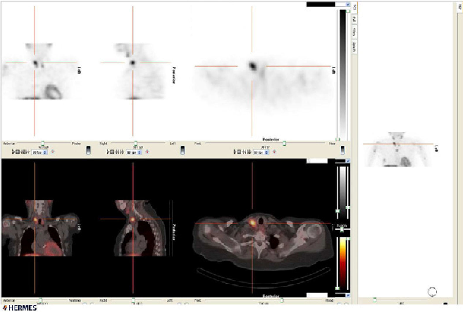
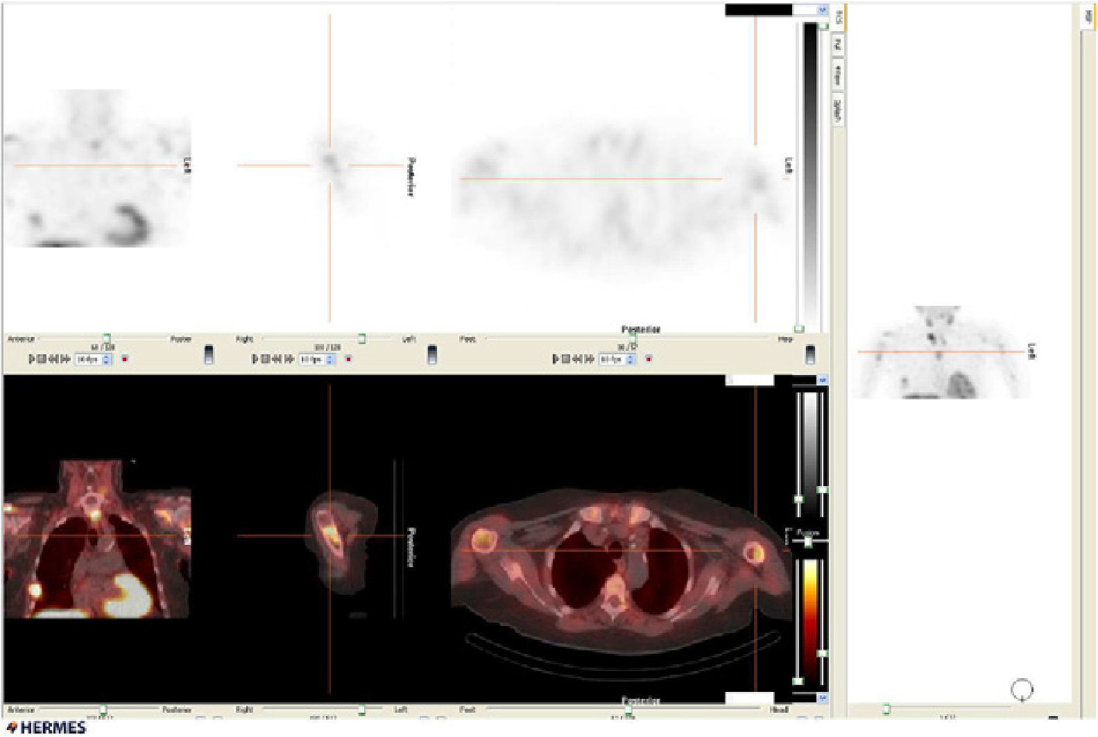
A SPECT (single-photon emission computed tomography)-CT scan was performed that revealed intense-tracer uptake in the right superior thyroid lobe (Fig. 1), and several extra-thyroid locations, in the sternum, right acromium, proximal third of both humerus bones, and in the middle-third of the 6th and 7th right ribs (Fig. 2), compatible with osteitis fibrosa cystica (brown tumours), although bone metastasis could not be ruled out. Biopsy confirmed brown tumours.

Fig. 1
(0.13MB).
Fig. 2
(0.14MB).

A parathyroidectomy was performed and PTH and calcium levels returned to normal.

At present this presentation of hyperparathyroidism is very uncommon.

Diagnosis: Brown tumours secondary to parathyroid adenoma.

Please, cite this article as: Rubio-Manzanares-Dorado M, et al. Lesiones osteolíticas en SPECT-TAC. Cir Esp. 2013;91:53.

Copyright © 2011. AEC
Article options
Tools
es en pt

¿Es usted profesional sanitario apto para prescribir o dispensar medicamentos?

Are you a health professional able to prescribe or dispense drugs?

Você é um profissional de saúde habilitado a prescrever ou dispensar medicamentos