


metricas
Cirugía Española (English Edition) Giant Cell Tumor of the Superior Maxilla
Journal Information
Vol. 97. Issue 5.
Pages 245-302 (May 2019)
Cite
Cite
Share
Download PDF
More article options
Vol. 97. Issue 5.
Pages 245-302 (May 2019)
Image of the Month
Full text access

Giant Cell Tumor of the Superior Maxilla

Tumor de células gigantes de maxilar superior
Visits
2788
Jorge Luna-Abantoa,b,
Corresponding author
Jorgelunaabanto@gmail.com

Corresponding author.
, Erick Gonzales Laguadoc
a Departamento de Cirugía Oncológica, Instituto Nacional de Enfermedades Neoplásicas, Lima, Peru
b Escuela de Posgrado, Universidad Peruana Cayetano Heredia, Lima, Peru
c Departamento de Cirugía de Cabeza y Cuello, Instituto Nacional de Enfermedades Neoplásicas, Lima, Peru
This item has received
Article information
Full Text
Download PDF
Statistics
Figures (2)
fig0005
fig0010
Full Text

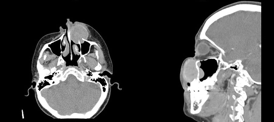
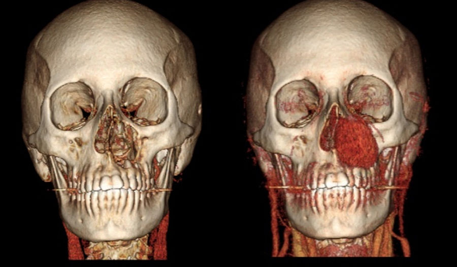
A 37-year-old woman presented with a fast-growing mass in the left nasolabial region that had been developing over the previous 9 months. Computed tomography scan with contrast of the facial bones showed a solid, homogeneous lesion with partially defined margins, dependent on the left upper maxilla (Figs. 1 and 2).
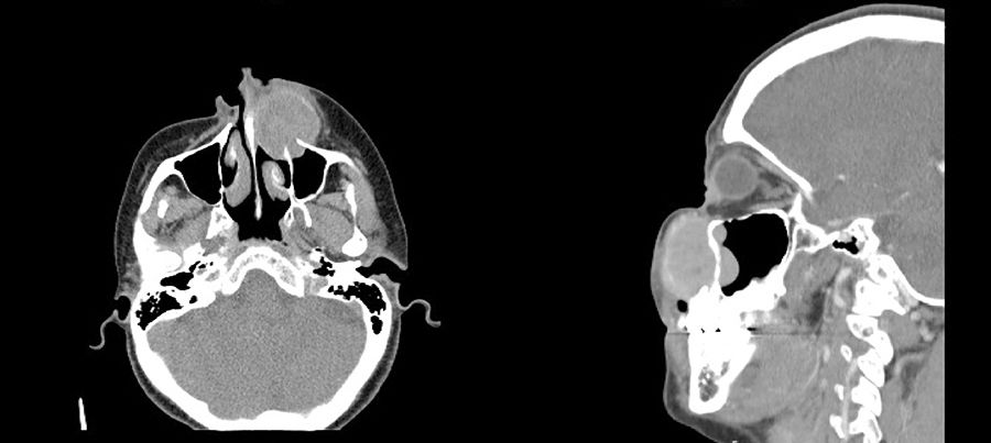
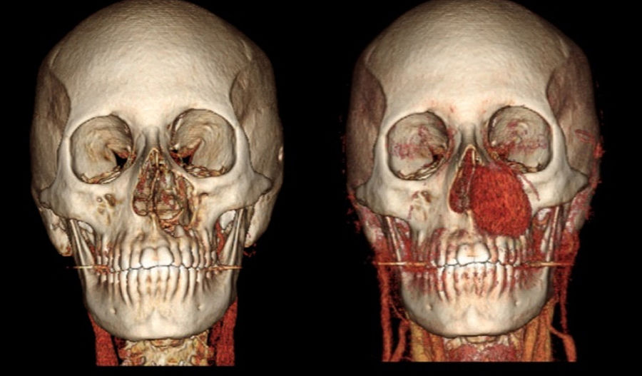
Fig. 1
Fig. 2

Biopsy of the tumor identified the mass as a giant cell tumor (GCT). GCT of the bone are lesions characterized by the presence of multinucleated giant cells and represent 4% of all bone tumors. Only 2% originate in the craniofacial skeleton; maxillary tumors are exceptional. Standard treatment is surgical resection.

Please cite this article as: Luna-Abanto J, Gonzales Laguado E. Tumor de células gigantes de maxilar superior. Cir Esp. 2019;97:289.


Copyright © 2018. AEC
asdasdasd
Article options
Tools