Buscar en
Cirugía Española (English Edition)
Toda la web
Inicio Cirugía Española (English Edition) Extrapleural Solitary Fibrous Tumor
Journal Information
Vol. 92. Issue 10.
Pages e65 (December 2014)
Vol. 92. Issue 10.
Pages e65 (December 2014)
Image of the month
Full text access
Extrapleural Solitary Fibrous Tumor
Tumor solitario fibroso extrapleural
Visits
2125
Francisco B. de Lacy
Corresponding author
borjalacy@gmail.com

Corresponding author.
, Luis Flores, Ramón Rull, Juan Carlos García-Valdecasas
Servicio de Cirugía General y del Aparato Digestivo, Hospital Clínico, Barcelona, Spain
This item has received
Article information
Full Text
Download PDF
Statistics
Figures (1)
Full Text

A 70-year-old patient presented an incidental finding on CT of a well-defined retroperitoneal mass measuring 7.2cm in the caudate lobe of the liver that was displacing adjacent organs (Fig. 1). Epithelial markers were negative. Endoscopic ultrasound-guided fine-needle aspiration collected insufficient material for diagnosis, so the patient was treated surgically with laparotomy, dissecting the mass from the right pillar of the diaphragm, the gastro-esophageal junction and the splenic vein.

Fig. 1
(0.13MB).

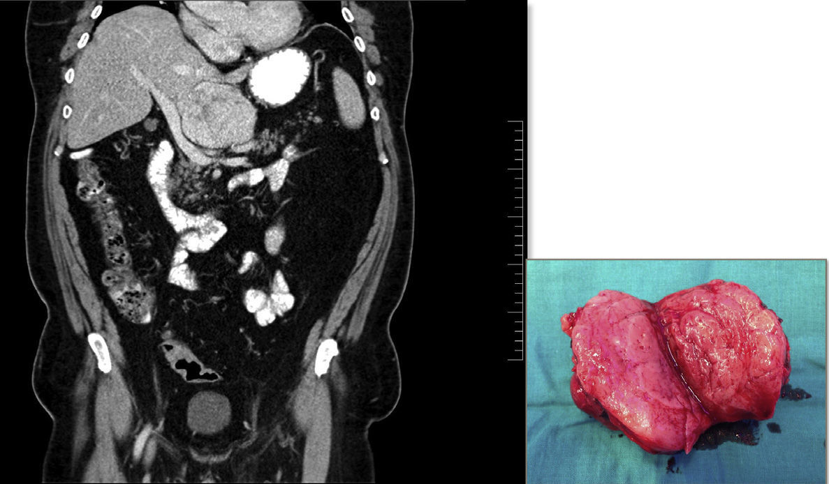
The pathology study confirmed the diagnosis of extrapleural solitary fibrous tumor (CD34+). The resection margins were free of neoplasm and the Ki67 proliferative rate was 10%, indicating a low risk of local recurrence and malignancy.

Please cite this article as: de Lacy FB, Flores L, Rull R, García-Valdecasas JC. Tumor solitario fibroso extrapleural. Cir Esp. 2014;92:e65.

Copyright © 2014. AEC
Article options
Tools
es en pt

¿Es usted profesional sanitario apto para prescribir o dispensar medicamentos?

Are you a health professional able to prescribe or dispense drugs?

Você é um profissional de saúde habilitado a prescrever ou dispensar medicamentos