Buscar en
Archivos de la Sociedad Española de Oftalmología (English Edition)
Toda la web
Inicio Archivos de la Sociedad Española de Oftalmología (English Edition) SD-OCT findings in polypoidal choroidal vasculopathy
Journal Information
Vol. 91. Issue 1.
Pages 23-26 (January 2016)
Share
Share
Download PDF
More article options
Visits
677
Vol. 91. Issue 1.
Pages 23-26 (January 2016)
Original article
SD-OCT findings in polypoidal choroidal vasculopathy
Hallazgos con la SD-OCT en vasculopatía coroidea polipoidea
Visits
677
V.M. Asensio-Sánchez
Servicio de Oftalmología, Hospital Clínico Universitario, Valladolid, Spain
This item has received
Article information
Abstract
Full Text
Bibliography
Download PDF
Statistics
Figures (3)
Show moreShow less
Tables (2)
Table 1. Characteristics of group (n=10).
Table 2. Diagnostic and inclusion criteria (n=15).
Show moreShow less
Abstract
Objective

To examine patients with polypoidal choroidal vasculopathy (PCV), using spectral-domain optical coherence tomography (SD-OCT) to characterize and locate the PCV lesions.
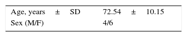
Patients and methods

A series of 15 eyes of 10 patients diagnosed with PCV were examined. All eyes were imaged with macular SD-OCT.
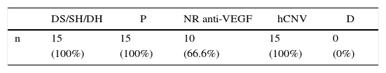
Results

SD-OCT cross-sectional scan findings included atypical and typical pigment epithelial detachments (PEDs). Polyps and neovascularisation were located above Bruch's membrane. All 15 eyes (100%) showed sub-retinal fluid (SRF) in association with PEDs.

Conclusion

These SD-OCT findings located the vascular lesions of PCV in the sub-retinal pigment epithelium (RPE) space, and strongly suggest that PCV is a variant of type 1 neovascularization.

Keywords:
SD-OCT
PED
Choroidal neovascularisation type 1
Polyps
Bruch's membrane
Resumen
Objetivo

Examinar a los pacientes con vasculopatía coroidea polipoidea (VCP), usando la tomografía de coherencia óptica de dominio espectral (SD-OCT) para caracterizar y localizar las lesiones en la VCP.

Pacientes y métodos

Se estudió una serie de 15 ojos de 10 pacientes diagnosticados de VCP. Todos los ojos fueron explorados con SD-OCT.

Resultados

Con cortes transversales de la SD-OCT se encontraron desprendimientos del epitelio pigmentario (DEP) típicos y atípicos. Los pólipos y la neovascularización se localizaron encima de la membrana de Bruch. Los 15 ojos (100%) mostraron líquido subretiniano (LSR) en asociación con los DEP.

Conclusión

Los hallazgos de la SD-OCT localizan las lesiones vasculares de la VCP en el espacio sub-EPR e indican fuertemente que la VCP es una variante de neovascularización tipo 1.

Palabras clave:
SD-OCT
DEP
Neovascularización coroidea tipo 1
Pólipos
Membrana de Bruch

Article

These are the options to access the full texts of the publication Archivos de la Sociedad Española de Oftalmología (English Edition)
Subscriber
Subscriber

If you already have your login data, please click here .

If you have forgotten your password you can you can recover it by clicking here and selecting the option “I have forgotten my password”
Subscribe
Subscribe to

Archivos de la Sociedad Española de Oftalmología (English Edition)

Purchase
Purchase article

Purchasing article the PDF version will be downloaded

Price 19.34 €

Purchase now
Contact
Phone for subscriptions and reporting of errors
From Monday to Friday from 9 a.m. to 6 p.m. (GMT + 1) except for the months of July and August which will be from 9 a.m. to 3 p.m.
Calls from Spain
932 415 960
Calls from outside Spain
+34 932 415 960
E-mail
Article options
Tools
es en pt

¿Es usted profesional sanitario apto para prescribir o dispensar medicamentos?

Are you a health professional able to prescribe or dispense drugs?

Você é um profissional de saúde habilitado a prescrever ou dispensar medicamentos