


To provide a qualitative analysis of filtering blebs after being surgically repaired due to late blebs leaks.
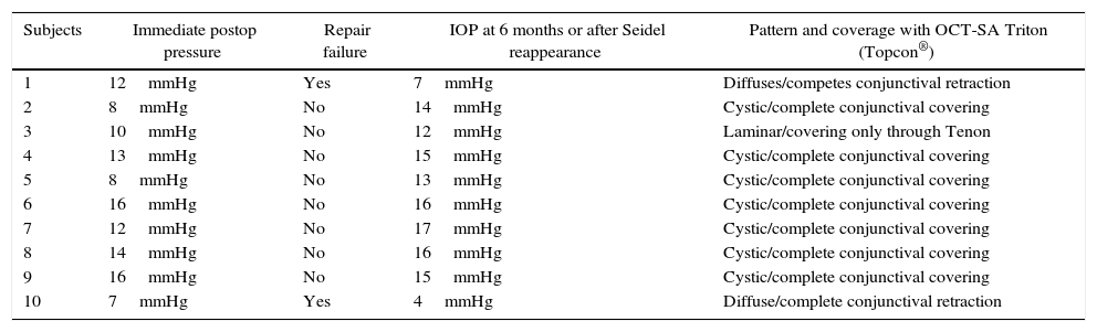
MethodsBlebs were studied 6 months after surgical reparation using AS-OCT Triton (Topcon®). An analysis was made of the morphological pattern and internal structures of blebs, including the covering, in 10 patients. The images were obtained using OCTs at a wavelength of 1050nm.
ResultsAccording to the Hirooka classification, three different patterns were found in the structure of blebs, which made it possible to correlate them with their functionality. A full covering was observed in 70% of the cases, and they showed sub-epithelial cysts (cystoid pattern). Two cases showed a full conjunctival retraction without Tenon's covering. The walls were thin, with a de-structured bleb (diffuse pattern) being visualized. In the third group, the image showed a partial conjunctival retraction with Tenon's covering. There were some sub-epithelial diffuse cysts with walls following a laminar pattern.
ConclusionUsing AS-OCT, it is possible to study the bleb's characteristics in detail, as well as the cover, in the case of blebs requiring repair due to late leaks, using conjunctival advancement. It allows for the early visualization of conjunctival retractions that were not visible in a slit lamp, and to predict the functionality of the blebs by their morphology.
Realizar un análisis cualitativo de las ampollas de filtración que han sido reparadas quirúrgicamente por presentar Seidel tardío.
MétodosEstudio de 10 ampollas de filtración que requirieron reparación quirúrgica mediante la OCT-SA Triton (Topcon®). Se analizó, a los 6 meses de la cirugía, el patrón morfológico y las estructuras internas de las mismas, así como el estado del recubrimiento. La obtención de las imágenes fue mediante longitudes de onda de 1.050nm.
ResultadosSegún la clasificación de Hirooka, encontramos 3 patrones diferentes en la morfología de la ampolla que pudimos relacionar con la funcionalidad de la misma. En un 70% de los casos el recubrimiento fue completo, presentando quistes subepiteliales difusos (tipo quístico). Dos casos mostraron una retracción conjuntival completa, sin cobertura por Tenon. Las paredes estaban adelgazadas, mostrando una desestructuración de la ampolla (patrón difuso). En un tercer grupo, la imagen obtenida mostraba una retracción conjuntival parcial con cobertura por Tenon. Presentaba algún quiste subepitelial difuso y con paredes siguiendo un patrón laminar.
ConclusiónMediante la OCT-SA es posible estudiar de forma detallada las características de la ampolla y las de su cobertura en el caso de reparación con avance conjuntival por Seidel tardío. Permite visualizar precozmente la retracción de la conjuntiva que en la lámpara de hendidura no sería visible y predecir mediante la morfología de la ampolla la funcionalidad de la misma.
