


Photic maculopathy is a retinal condition caused by exposure to intense light sources, resulting in varying degrees of visual impairment. Optical coherence tomography angiography (OCT-A) offers high-resolution, non-invasive imaging of the retinal microvasculature, facilitating detailed assessment of vascular alterations linked to this pathology. This study aims to characterize the OCT-A features of photic maculopathy and to emphasize the utility of OCT-A in evaluating retinal damage and microvascular changes.
Materials and methodsThis retrospective descriptive study included patients diagnosed with PM between September 2021 and September 2023. Inclusion required a history of intense or prolonged light exposure temporally related to visual symptoms, characteristic fundus and clinical findings, and multimodal imaging confirming focal outer retinal damage at the fovea. Patients with alternative diagnoses, poor-quality imaging, or incomplete records were excluded. Clinical evaluation included structural optical coherence tomography (OCT), OCT-A, fluorescein angiography, and visual function tests. Demographic and ophthalmic data were collected, and a literature review was conducted to support interpretation.
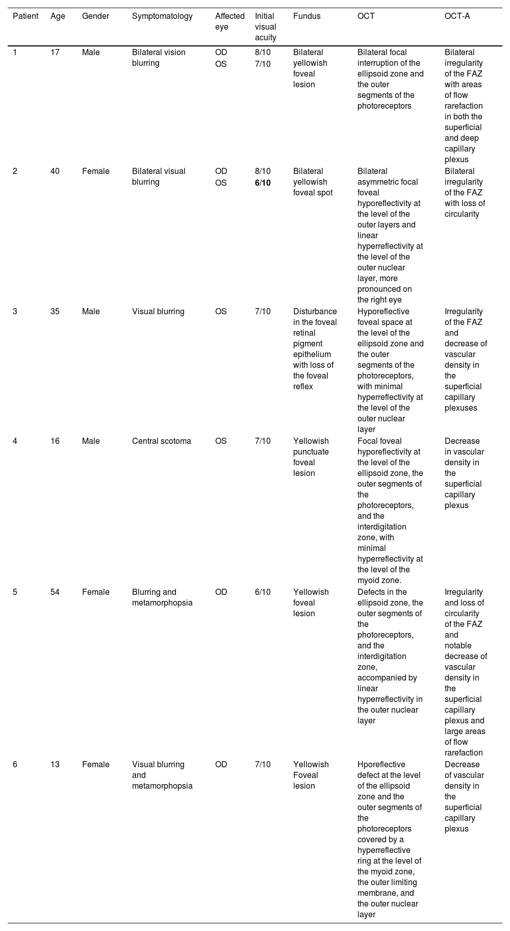
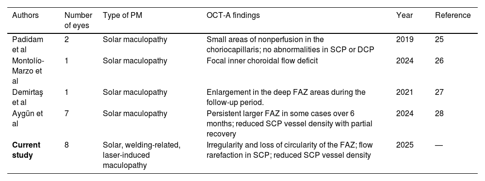
ResultsEight eyes from six patients were analyzed: two bilateral cases of solar maculopathy, one unilateral welder’s maculopathy, and three unilateral laser-induced maculopathies. All patients presented with reduced visual acuity. Fundus examination revealed a yellowish foveal lesion in seven eyes and retinal pigment epithelium changes in one. OCT showed consistent disruption of the outer retinal layers. OCT-A demonstrated an irregular foveal avascular zone in six eyes and reduced vessel density in the superficial capillary plexus in four eyes.
ConclusionsOCT-A plays a critical role in detecting structural retinal damage and associated microvascular alterations, offering key insights into the underlying pathophysiology. This study emphasizes the added value of OCT-A in assessing retinal integrity and vascular changes, which may enhance diagnostic accuracy and guide the clinical management of photic maculopathy.
La maculopatía fótica es una afección retiniana causada por la exposición a fuentes de luz intensa, que resulta en diversos grados de deterioro visual. La angiografía por tomografía de coherencia óptica (OCT-A) ofrece imágenes de alta resolución y no invasivas de la microvasculatura retiniana, facilitando la evaluación detallada de las alteraciones vasculares asociadas a esta patología. El presente estudio tiene como objetivo caracterizar las características de la OCT-A en la maculopatía fótica y destacar la utilidad de esta técnica en la evaluación del daño retiniano y los cambios microvasculares.
Materiales y métodosEste estudio descriptivo retrospectivo incluyó pacientes diagnosticados con MF entre septiembre de 2021 y septiembre de 2023. Los criterios de inclusión requerían antecedentes de exposición intensa o prolongada a la luz relacionados temporalmente con los síntomas visuales, hallazgos clínicos y del fondo de ojo característicos, y estudios de imagen multimodal que confirmaran daño focal en la retina externa a nivel de la fóvea. Se excluyeron pacientes con diagnósticos alternativos, imágenes de mala calidad o historiales médicos incompletos. La evaluación clínica incluyó OCT estructural, OCT-A, angiografía con fluoresceína y pruebas de función visual. Se recopilaron datos demográficos y oftalmológicos, y se realizó una revisión de la literatura para apoyar la interpretación de los hallazgos.
ResultadosSe analizaron ocho ojos de seis pacientes: dos casos bilaterales de maculopatía solar, un caso unilateral de maculopatía por soldadura y tres casos unilaterales de maculopatía inducida por láser. Todos los pacientes presentaron disminución de la agudeza visual. El examen del fondo de ojo reveló una lesión amarillenta en la fóvea en siete ojos y alteraciones del epitelio pigmentario de la retina en uno. La OCT mostró una alteración consistente de las capas externas de la retina. La OCT-A evidenció una zona avascular foveal irregular en seis ojos y una disminución de la densidad vascular en el plexo capilar superficial en cuatro ojos.
ConclusionesLa OCT-A desempeña un papel fundamental en la detección del daño estructural retiniano y las alteraciones microvasculares asociadas, proporcionando información clave sobre la fisiopatología subyacente. Este estudio resalta el valor añadido de la OCT-A en la evaluación de la integridad retiniana y los cambios vasculares, lo que puede mejorar la precisión diagnóstica y orientar el manejo clínico de la maculopatía fótica.
