


Uveal melanoma is the most common primary malignant intraocular tumour in the adult population, with a survival rate of 50% despite advances in treatment and knowledge of this disease. The presence of extraocular extension (EE) worsens the prognosis of these patients, so its proper identification can ensure its management and early intervention. Ophthalmological ultrasound is the technique of choice for the diagnosis and follow-up of these patients, both of the anterior EE using ultrasonic biomicroscopy (UBM), and the posterior EE using A and B ultrasound. The aim of this study is to describe the ultrasound characteristics of the BMU and the A and B ultrasound.
Material and methodsA descriptive and retrospective study is carried out on patients diagnosed with uveal melanoma (UM) and EE from 2003 to 2019. The ultrasound characteristics of the local disease and the follow-up after treatment were recorded completely and at each visit. In the case of anterior EE, photographs of the anterior segment and UBM were taken, while those involving the posterior segment were explored under A and B mode ultrasound. All enucleated eyes were sent for anatomopathological study.
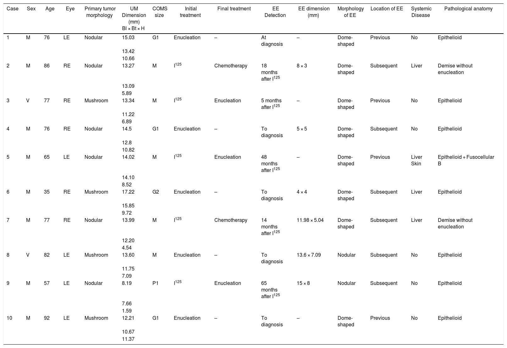
ResultsTen patients with an average age of 72.3 years were included. The largest proportion of them were medium-sized tumours, followed by large and small ones. The most frequent morphology of the primary tumour was cupuliform. All the EE presented lower internal reflectivity compared to the primary tumour. No trans-scleral connection bridges were found between the primary tumour and the EE in the ultrasound studies. 50% of patients underwent primary enucleation at the time of diagnosis of intraocular MU due to the presence of the EE, and the remaining 50% presented the EE after initial treatment of the primary tumour with I125 brachytherapy. Sixty percent of the patients presented with posterior EE, and were therefore diagnosed with ultrasound A and B. The most frequent histopathological pattern with 87.5% of patients was the epithelioid pattern.
DiscussionUltrasound scanning in patients with MU is mandatory for diagnosis and follow-up of EE. BMU and A and B ultrasound are the test of choice for anterior and posterior EE, respectively. EE have particular ultrasound characteristics such as low internal reflectivity, regularity of their contour and their location usually adjacent to the base of the primary intraocular tumor.
El melanoma uveal es el tumor primario maligno intraocular más frecuente en la población adulta, con una tasa de supervivencia del 50% a pesar de los avances en el tratamiento y conocimiento de esta enfermedad. La presencia de extensión extraocular (EE) empeora el pronóstico de estos pacientes, por lo que su correcta identificación puede asegurar su manejo e intervención temprana. La ecografía oftalmológica es la técnica de elección para el diagnóstico y seguimiento de estos pacientes, tanto de la EE anterior mediante biomicroscopía ultrasónica (UBM), como de la EE posterior mediante ecografía A y B. El objetivo de este estudio es describir las características ecográficas de la UBM y de la ecografía A y B.
Material y métodosSe realiza un estudio descriptivo y retrospectivo de los pacientes diagnosticados de melanoma uveal (UM) y EE desde 2003 hasta 2019. Las características ecográficas de la enfermedad local y el seguimiento luego del tratamiento se registraron de manera completa y en cada visita. En caso de EE anteriores se realizaron fotografías de segmento anterior y BMU, por el contrario las que involucran el segmento posterior se exploraron bajo ecografía modo A y B. Todos los ojos enucleados se enviaron para su estudio anatomopatológico.
ResultadosSe incluyeron 10 pacientes con una edad media de 72,3 años. La mayor proporción de ellos eran tumores de tamaño medio, seguidos de los grandes y pequeños. La morfología más frecuente del tumor primario fue la cupuliforme. Todos los EE presentaban una reflectividad interna inferior a la del tumor primario. No se encontraron puentes de conexión transescleral entre el tumor primario y el EE en los estudios ecográficos. El 50% de los pacientes fueron sometidos a enucleación primaria en el momento del diagnóstico del MU intraocular debido a la presencia de la EE, y el 50% restante presentó la EE tras el tratamiento inicial del tumor primario con braquiterapia I125. El 60% de los pacientes presentaron EE posterior, por lo que fueron diagnosticados con ecografía A y B. El patrón histopatológico más frecuente con el 87,5% de los pacientes fue el patrón epitelioide.
DiscusiónLa ecografía en pacientes con MU es obligatoria para el diagnóstico y seguimiento de la EE. La BMU y la ecografía A y B son las pruebas de elección para las EE anteriores y posteriores, respectivamente. Las EE tienen características ecográficas particulares como la baja reflectividad interna, la regularidad de su contorno y su localización generalmente adyacente a la base del tumor intraocular primario.
