Buscar en
Archivos de la Sociedad Española de Oftalmología (English Edition)
Toda la web
Inicio Archivos de la Sociedad Española de Oftalmología (English Edition) Evaluation of non-exudative tomographic signs in cases of exudative age-related ...
Journal Information
Vol. 98. Issue 5.
Pages 276-280 (May 2023)
Share
Share
Download PDF
More article options
Visits
4
Vol. 98. Issue 5.
Pages 276-280 (May 2023)
Original article
Evaluation of non-exudative tomographic signs in cases of exudative age-related macular degeneration
Evaluación de signos tomográficos no exudativos en casos de degeneración macular asociada a la edad de tipo exudativo
Visits
4
L. Monje-Fernándeza,
Corresponding author
lmonje@saludcastillayleon.es

Corresponding author.
, R. Gallego-Pinazob, M. Cordero-Comac, J. Donate-Lópezd, R.M. Coco-Martíne, R. Dolz-Marcob
a Complejo Asistencial Universitario de León, León, Spain
b Unidad de Mácula, Clínica Oftalvist Valencia, Valencia, Spain
c Área de Oftalmología, HM Hospitales, Málaga, Spain
d Clínica la Luz, Madrid, Spain
e Instituto de Oftalmo Biología Aplicada (IOBA), Valladolid, Spain
This item has received
Article information
Abstract
Full Text
Bibliography
Download PDF
Statistics
Tables (2)
Table 1. Relationship between visual acuity (VA) and the different variables. Result of beta linear regression, in which each of the levels of the variable is compared to a base level, the measure of which results in the row called the overall mean; within this measure, type 1 NV was taken as this type of NV had the lowest logMAR (highest VA), so each p-value of the other types is telling us whether that type of NV is different from type 1.
Table 2. Shows associations between covariates (rows) and signs (columns). Pseudocysts and subretinal clefts are increasing. ORT increases with follow-up and is more frequent in type 3 and mixed NV. The onion sign increases with age. The probability of pseudoswelling increases with both age and time of evolution. Macular atrophy increases with age and follow-up and is more present in NV types 2 and 3 (results with respect to NV-1).
Show moreShow less
Abstract
Objective

To analyse the prevalence of non-exudative tomographic signs (onion sign, pseudoswelling, external retinal tubulation, pseudocysts, subretinal clefts and macular atrophy) in patients with neovascular age-related macular degeneration.

Material and methods

A total of 174 eyes of patients with neovascular age-related macular degeneration who had not received previous treatment were included in the study. Visual acuity, neovascularization activity, and the appearance or not of the different signs under study were assessed at times 0 (initial visit), 4 months, one year, year and a half, and at 2 and 3 years of follow-up. The following were also evaluated: age, sex, affected eye and type of neovascularization (1, 2, 3, polypoid or mixed). The analysis were performed using the statistical software R (version 3.3.2) and the glmmADMB package (version 0.8.3.3).
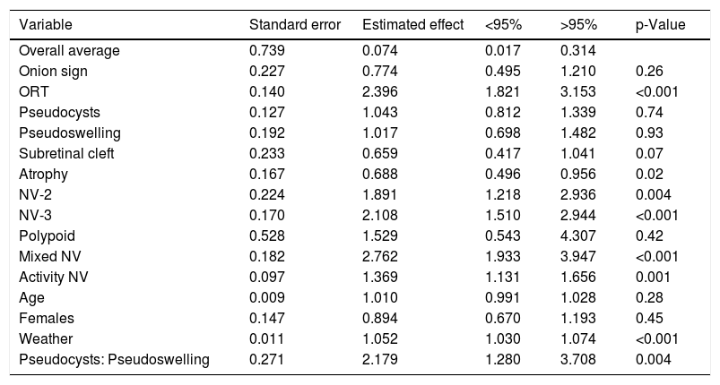
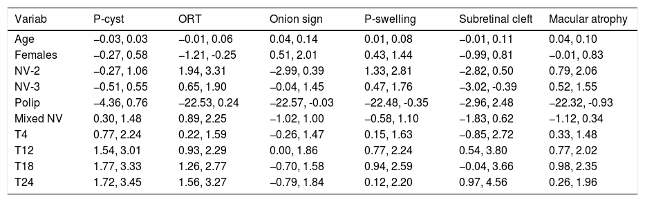
Results

The presence of pseudocysts and external retinal tubulation increases throughout the follow-up. The onion sign begins with an ascending frequency up to 12 months, then decreases at 18 months and increases again at 24 months. Regarding pseudowelling, it maintains an increase until 18 months to finally decrease. Subretinal clefts is the rarest sign, presenting in 1.1% on the first visit. Finally, macular atrophy, present in 12.6% of the eyes initially, is found in 25% after 2 years.

Conclusion

Pseudocysts, external retinal tubulation and macular atrophy were the most prevalent signs, while subretinal clefts were the most infrequent.

Keywords:
Age macular degeneration
Neovascularization
Pseudocyst
Outer retinal tabulation
Optical coherence tomography
Pseudoswelling
Subretinal cleft
Onion sign
Macular atrophy
Resumen
Objetivo

Analizar la prevalencia de signos tomográficos no exudativos (signo de cebolla, seudoedema, tubulación de la retina externa, seudoquistes, hendiduras subretinianas y atrofia macular) en pacientes con degeneración macular asociada a la edad neovascular.

Material y métodos

Un total de 174 ojos de pacientes con degeneración macular asociada a la edad neovascular que no habían recibido tratamiento previo fueron incluidos en el estudio. Se valoró la agudeza visual, la actividad de la neovascularización y la aparición o no de los distintos signos objeto de estudio en los tiempos 0 (visita inicial), 4 meses, un año, año y medio y a los 2 y 3 años de seguimiento. Se evaluaron también: la edad, el sexo, el ojo afecto y el tipo de neovascularización (1, 2, 3, polipoidea o mixta). Los análisis se han realizado mediante el software estadístico R (versión 3.3.2) y el paquete glmmADMB (versión 0.8.3.3).

Resultados

La presencia de seudoquistes y tubulación de la retina externa va en aumento a lo largo del seguimiento. El signo de cebolla comienza con una frecuencia ascendente hasta los 12 meses, posteriormente desciende a los 18 meses y vuelve a incrementarse a los 24 meses. En cuanto al seudoedema, mantiene un incremento hasta los 18 meses para finalmente descender. Las hendiduras subretinianas son el signo más raro, presentándose en el 1,1% en la primera visita. Finalmente, la atrofia macular, presente en el 12,6% de los ojos inicialmente, se encuentra en el 25% a los 2 años.

Conclusión

Los seudoquistes, la tubulación de la retina externa y la atrofia macular fueron los signos más prevalentes, mientras que las hendiduras subretinianas fueron los más infrecuentes.

Palabras clave:
Degeneración macular asociada a la edad
Neovascularización
Seudoquiste
Tubulación de la retina externa
Tomografía de coherencia óptica
Seudoedema
Hendidura subretiniana
Signo de cebolla
Atrofia macular

Article

These are the options to access the full texts of the publication Archivos de la Sociedad Española de Oftalmología (English Edition)
Subscriber
Subscriber

If you already have your login data, please click here .

If you have forgotten your password you can you can recover it by clicking here and selecting the option “I have forgotten my password”
Subscribe
Subscribe to

Archivos de la Sociedad Española de Oftalmología (English Edition)

Purchase
Purchase article

Purchasing article the PDF version will be downloaded

Price 19.34 €

Purchase now
Contact
Phone for subscriptions and reporting of errors
From Monday to Friday from 9 a.m. to 6 p.m. (GMT + 1) except for the months of July and August which will be from 9 a.m. to 3 p.m.
Calls from Spain
932 415 960
Calls from outside Spain
+34 932 415 960
E-mail
Article options
Tools
es en pt

¿Es usted profesional sanitario apto para prescribir o dispensar medicamentos?

Are you a health professional able to prescribe or dispense drugs?

Você é um profissional de saúde habilitado a prescrever ou dispensar medicamentos