


To study clinical and pathological variables leading to a poor prognosis in a sample of uveal malignant melanoma patients who required eyeball enucleation as final treatment approach. All patients were seen and treated in the same public tertiary hospital in Madrid (Spain) within a 6-year time-period.
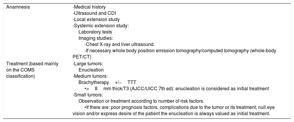
MethodsLongitudinal observational retrospective study. The presence of clinical and pathologic factors known to be linked to poor prognosis – as well as other features – was assessed in 30 malignant melanoma: 20 de novo-enucleated malignant melanoma eyes (group A), and 10 in eyes that received radiotherapy prior to enucleation (group B). The diagnostic reliability of magnetic resonance imaging was assessed by comparing it with the histology results – “gold standard” – as a means to detect scleral and extra-scleral extension.
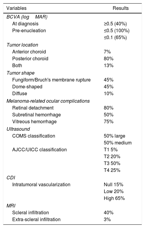
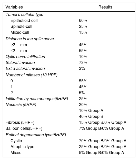
ResultsTumor size, Bruch's membrane rupture, scleral infiltration, and distance to the optic nerve were the most decisive factors for a poor prognosis in the study sample. In 93% of cases the condition was under control, with a 6% incidence rate of metastatic spread and a 100% rate of overall survival for a mean follow-up period of 3±1.5 (range 1.2–6) years. In the study population, the sensitivity of the magnetic resonance imaging to detect scleral infiltration was 27%, which increased to 100% for identifying extra-scleral involvement.
ConclusionsThe analyses of the clinical and pathological data collected within the framework of this study justify enucleation as the treatment of choice for the patients of this study. Magnetic resonance imaging was not found to be an optimum screening method to detect scleral infiltration in this study sample.
Estudiar las variables clínicas y anatomopatológicas de mal pronóstico en una muestra de pacientes con melanoma maligno de úvea que requirieron enucleación como tratamiento definitivo en un hospital terciario en Madrid (España) durante un período de tiempo de 6 años.
Material y métodosEstudio retrospectivo, observacional, longitudinal. Se analiza la presencia de factores clínicos y anatomopatológicos conocidos de mal pronóstico y otros en 30 melanomas malignos: 20 enucleados de novo (grupo A) y 10 tras recibir tratamiento con radioterapia (grupo B). Se estudia el grado de fiabilidad diagnóstica de la resonancia magnética nuclear, comparándola con la histología (gold standard) para predecir la presencia de invasión escleral y extraescleral.
ResultadosEl tamaño tumoral, la rotura de la membrana de Bruch, la invasión escleral y la proximidad al nervio óptico fueron los factores de mal pronóstico más determinantes. Se logró controlar la enfermedad en el 93% de los casos, con una incidencia de diseminación metastásica del 6% y una supervivencia del 100% a una media de seguimiento de 3±1,5 (rango 1,2-6) años. La sensibilidad de la resonancia magnética nuclear, en nuestra población, para detectar infiltración escleral fue del 27%, y extraescleral, del 100%.
ConclusionesEl análisis de los datos clínicos e histopatológicos recogidos justifican la enucleación como tratamiento final en los pacientes estudiados. La resonancia magnética nuclear no resultó un buen método de cribado para detectar la extensión escleral.
